Downloaded 146 times

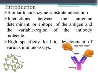
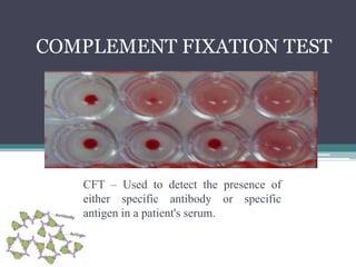
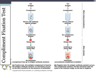
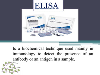
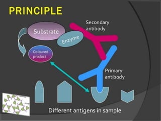
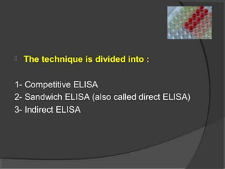
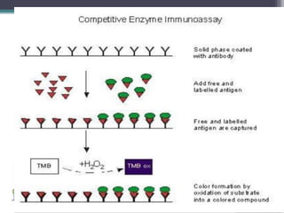
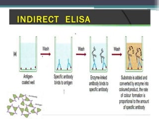
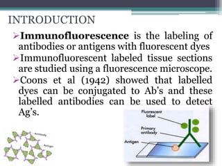
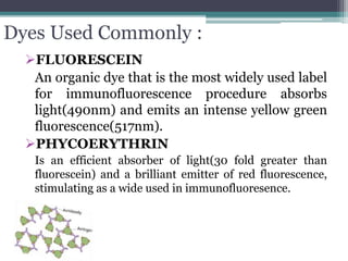
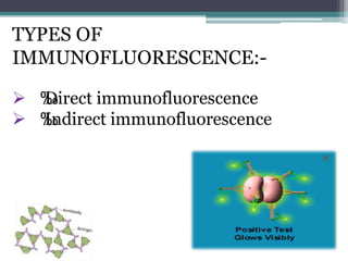
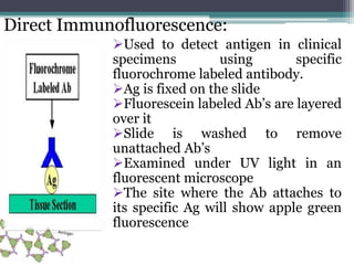
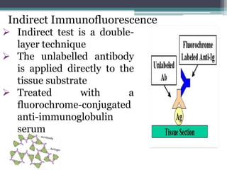
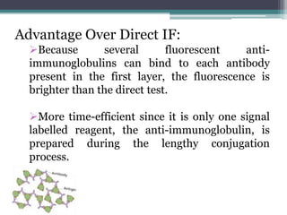

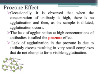
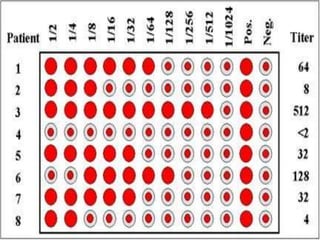
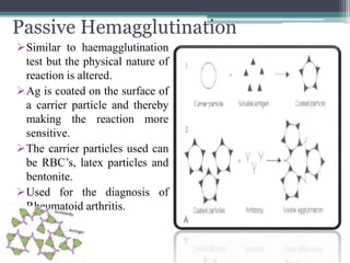
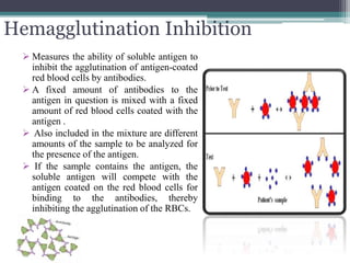

The document outlines various antigen-antibody interactions and their significance in immunoassays. It discusses several techniques, including the complement fixation test, ELISA, immunofluorescence, and agglutination tests, detailing how they work and their applications. Different types of immunofluorescence methods and agglutination reactions, including qualitative and quantitative tests, along with concepts like the prozone effect and hemagglutination inhibition, are also explored.


![Antibodies [Immunoglobulins]](https://cdn.slidesharecdn.com/ss_thumbnails/presentation11-201227140050-thumbnail.jpg?width=640&height=640&fit=bounds)