Downloaded 129 times


![Applications
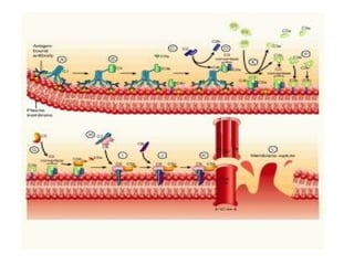
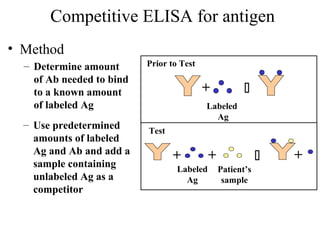
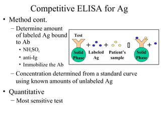
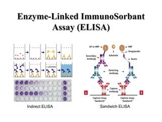
• Because the ELISA can be performed to evaluate either the presence of
antigen or the presence of antibody in a sample, it is a useful tool both for
determining serum antibody concentrations (such as with the HIV test [1] or
West Nile Virus) and also for detecting the presence of antigen. It has also
found applications in the food industry in detecting potential food allergens
such as milk,peanuts,walnuts,almonds, and eggs [2]The ELISA test, or the
enzyme immunoassay (EIA), was the first screening test commonly employed
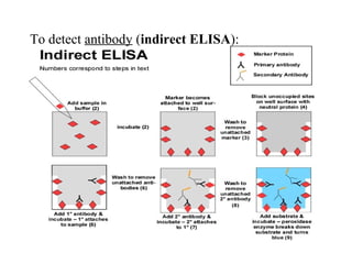
for HIV. It has a high sensitivity.In an ELISA test, a person's serum is diluted
400-fold and applied to a plate to which HIV antigens have been attached. If
antibodies to HIV are present in the serum, they may bind to these HIV
antigens. The plate is then washed to remove all other components of the
serum. A specially prepared "secondary antibody" — an antibody that binds to
human antibodies — is then applied to the plate, followed by another wash.
This secondary antibody is chemically linked in advance to an enzyme. Thus
the plate will contain enzyme in proportion to the amount of secondary
antibody bound to the plate. A substrate for the enzyme is applied, and
catalysis by the enzyme leads to a change in color or fluorescence. ELISA
results are reported as a number; the most controversial aspect of this test is
determining the "cut-off" point between a positive and negative result.](https://image.slidesharecdn.com/antigenantibodyinteraction-151031153315-lva1-app6892/85/Antigen-antibody-interaction-66-320.jpg)

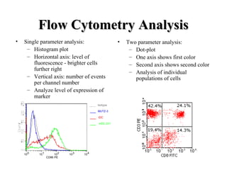
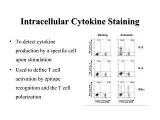
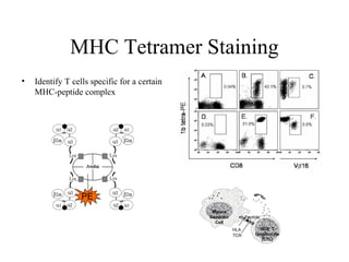

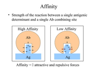
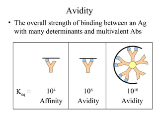
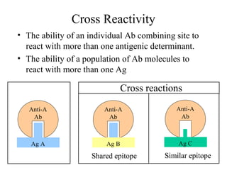
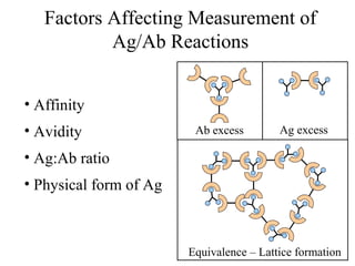
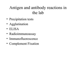
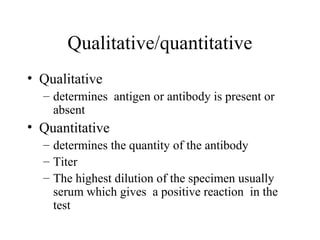
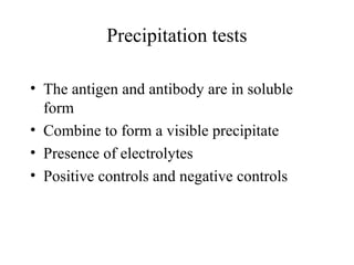
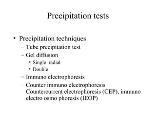
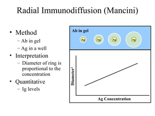
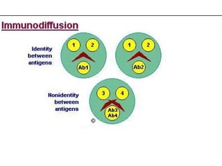
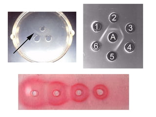
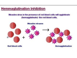
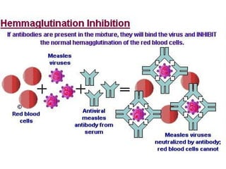
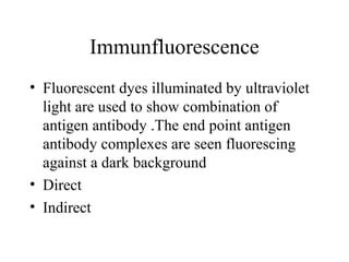
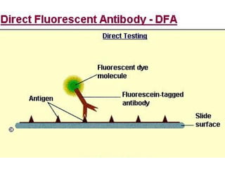
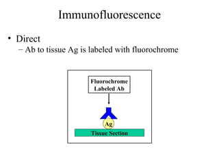
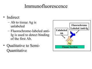
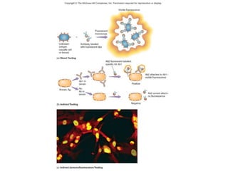
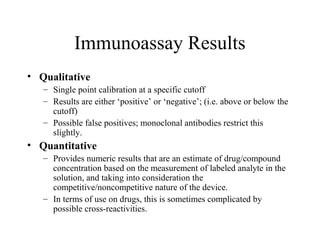
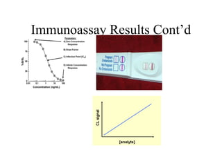
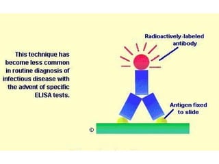
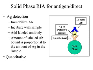
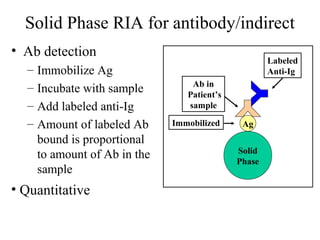
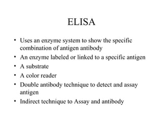
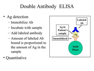
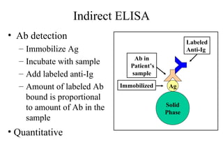
This document discusses antigen-antibody reactions including factors that affect their measurement and techniques used to measure them in the lab. Key points covered include: affinity and avidity being measures of strength of antigen-antibody binding; specificity and cross-reactivity relating to reaction with single or multiple antigens; and techniques including precipitation tests, agglutination, ELISA, radioimmunoassay, immunofluorescence, and complement fixation. Both qualitative and quantitative applications are discussed.