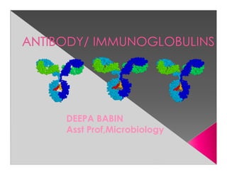
DEEPA BABIN
Asst Prof,Microbiology
Ab are glycoprotein molecule produced
by plasma cells in response to an Ag
and react specifically in an observable
manner
FUNCTIONS-Ag binding
Effector functions- Complement
fixtn,other cells fixation
› Soluble: secreted in blood and tissue

› Membrane-bound: found on surface of B-
  cell, also known as a B-cell receptor (BCR)

- BCR binds circulating antigen, activating the
  B-cell and forming plasma cells or memory
  B-cells
- Epitope-Ag
- Paratope-Ab
- Idiotype-Antigenic determinant on paratope
V   heavy
                                  Fab

HV                                  Variable
                 S-S   Fab

         C

                             Constant


                       Fc
Monomer: A flexible Y-shaped molecule with
four protein chains:
    2 identical light chains
     2 identical heavy chains
Each heavy and light chain has a constant
and variable region
The variable region binds the antigen in a
“lock-and-key” manner
L chains : 2 forms – kappa (κ) & lambda (λ)
Each molecule of Ig can have either κ or λ,
but never both.
Antibodies can also be divided into two
regions based on their function
 › Fab (fragment, antigen binding) region.
    Tip of the antibody
    Binds the antigen
 › Fc (fragment, crystallizable) region
 › Determines biological properties of Ig
   molecule.
    Base of the antibody
    Can bind cell receptors, complement
    proteins and other molecules
H chain designated by Greek letter.

– 5 different types: IgA, IgD, IgE, IgG, IgM
 › IgM      µ      (mu)
 › IgD      δ      (delta)
 › IgG      γ      (gamma)
 › IgA      α      (alpha)
 › IgE      ε      (epsilon)
H chain also divided into VH & CH regions; the CH region
is further divided into CH1, CH2 & CH3.
Regions also called as DOMAINS :
 - globular in shape
 - stabilized by intrachain disulphide bonds
Ag binding sites are located in the variable domains.
IgG- PROTECTS BODY FLUIDS
IgA- PROTECTS BODY SURFACE
IgM- PROTECTS BLOOD STREAM
IgE- MEDIATES REAGINIC HYPERSENSITIVITY
IgD- Recognisation receptor for Ag
Structure: Monomer
Percentage serum antibodies: 80%
Location: Blood, lymph, intestine
Half-life in serum: 23 days
Complement Fixation: Yes
Placental Transfer: Yes only Ab
Major Ab of secondary response, found both in
serum & body fluids.
4 subclasses found in humans – IgG1, IgG2,
IgG3 & IgG4, each having a distinct type of
gamma chain
Functions: Enhances phagocytosis,
neutralizes toxins and viruses, protects fetus
and newborn.
Structure: Pentamer
Percentage serum antibodies: 5-10%
Location: Blood, lymph, B cell surface
(monomer)
Half-life in serum: 5 days
Complement Fixation: Yes
Placental Transfer: No
primary immune response.
Functions: First antibodies produced during
an infection. Effective against microbes and
agglutinating antigens. Useful in the diagnosis
of congenital infections like syphilis, rubella, HIV,
dengue,toxoplasmosis etc.
Structure: Dimer second most abundunt Ab
Location: Secretions (colostrum,tears, saliva,
intestine, milk), blood and lymph.
Half-life in serum: 6 days
Complement Fixation: No
Placental Transfer: No
Occur in 2 forms : IgA1 & IgA2
Secretory IgA is always in dimeric form – composed
of 2 basic chain units, a J chain & the secretory
component.
Secretory component helps to transport the dimer
from the submucosa to the mucosal cell surface
Functions: Localized protection of mucosal
surfaces. Provides immunity to infant digestive
tract.
Structure: Monomer resemble Ig G
Percentage serum antibodies: 0.2%
Location: B-cell surface, blood, and
lymph
Half-life in serum: 3 days
Complement Fixation: No
Placental Transfer: No
Functions: In serum function is unknown.
Occurs along with Ig M on the surface of B
cell- initiate immune response.
Structure: Monomer LOW LEVEL IN SERUM
Percentage serum antibodies: 0.002%
Location: linings of respiratory & intestinal
tracts.Bound to mast cells and basophils
throughout body. Blood.
Half-life in serum: 2 days
Complement Fixation: No
Placental Transfer: No
Functions: anaphylactic type of
hypersensitivity ,Allergic reactions.
Possibly lysis of worms.
› B cells develop from stem cells in the bone
  marrow of adults (liver of fetuses).
› After maturation B cells migrate to lymphoid
  organs (lymph node or spleen).
› Clonal Selection: When a B cell encounters
 an antigen it recognizes, it is stimulated and
 divides into many clones called plasma
 cells, which actively secrete antibodies.
› Each B cell produces antibodies that will
  recognize only one antigenic determinant.
Programmed cell death (“Falling away”).
  › Human body makes 100 million lymphocytes
    every day. If an equivalent number doesn’t
    die, will develop leukemia.
  › B cells that do not encounter stimulating
    antigen will self-destruct and send signals to
    phagocytes to dispose of their remains.
  › Many virus infected cells will undergo
    apoptosis, to help prevent spread of the
    infection.
Structurally similar proteins in serum seen
in certain pathological conditions.

Bence Jones protein in multiple myeloma
– light chains of Igs.

Cryoglobulinemia – formation of gel or ppt
on cooling the serum which redissolves
on warming – in myelomas, SLE etc.
An individual produces a large number of
Abs to cope with the vast number of different
Ags.

This Ab diversity is due to the Ig genes.

Genes coding for the variable & constant
portions of the chains are separate

One or only few genes code for C region
whereas many genes code for the V region.
Multiple V- region genes.

     V-J & V-D-J recombination.

     Junctional diversity
1.   Nucleotide addition – extra nucleotides may
     get inserted between VH & D, and between
     D & JH segments
     Somatic mutation – point mutation in the
     genes for V domain.
THANK U

Antibody deepa babin

  • 1.
  • 2.
    Ab are glycoproteinmolecule produced by plasma cells in response to an Ag and react specifically in an observable manner FUNCTIONS-Ag binding Effector functions- Complement fixtn,other cells fixation
  • 3.
    › Soluble: secretedin blood and tissue › Membrane-bound: found on surface of B- cell, also known as a B-cell receptor (BCR) - BCR binds circulating antigen, activating the B-cell and forming plasma cells or memory B-cells - Epitope-Ag - Paratope-Ab - Idiotype-Antigenic determinant on paratope
  • 5.
    V heavy Fab HV Variable S-S Fab C Constant Fc
  • 6.
    Monomer: A flexibleY-shaped molecule with four protein chains: 2 identical light chains 2 identical heavy chains Each heavy and light chain has a constant and variable region The variable region binds the antigen in a “lock-and-key” manner L chains : 2 forms – kappa (κ) & lambda (λ) Each molecule of Ig can have either κ or λ, but never both.
  • 7.
    Antibodies can alsobe divided into two regions based on their function › Fab (fragment, antigen binding) region. Tip of the antibody Binds the antigen › Fc (fragment, crystallizable) region › Determines biological properties of Ig molecule. Base of the antibody Can bind cell receptors, complement proteins and other molecules
  • 9.
    H chain designatedby Greek letter. – 5 different types: IgA, IgD, IgE, IgG, IgM › IgM µ (mu) › IgD δ (delta) › IgG γ (gamma) › IgA α (alpha) › IgE ε (epsilon) H chain also divided into VH & CH regions; the CH region is further divided into CH1, CH2 & CH3. Regions also called as DOMAINS : - globular in shape - stabilized by intrachain disulphide bonds Ag binding sites are located in the variable domains.
  • 11.
    IgG- PROTECTS BODYFLUIDS IgA- PROTECTS BODY SURFACE IgM- PROTECTS BLOOD STREAM IgE- MEDIATES REAGINIC HYPERSENSITIVITY IgD- Recognisation receptor for Ag
  • 12.
    Structure: Monomer Percentage serumantibodies: 80% Location: Blood, lymph, intestine Half-life in serum: 23 days Complement Fixation: Yes Placental Transfer: Yes only Ab Major Ab of secondary response, found both in serum & body fluids. 4 subclasses found in humans – IgG1, IgG2, IgG3 & IgG4, each having a distinct type of gamma chain Functions: Enhances phagocytosis, neutralizes toxins and viruses, protects fetus and newborn.
  • 13.
    Structure: Pentamer Percentage serumantibodies: 5-10% Location: Blood, lymph, B cell surface (monomer) Half-life in serum: 5 days Complement Fixation: Yes Placental Transfer: No primary immune response. Functions: First antibodies produced during an infection. Effective against microbes and agglutinating antigens. Useful in the diagnosis of congenital infections like syphilis, rubella, HIV, dengue,toxoplasmosis etc.
  • 14.
    Structure: Dimer secondmost abundunt Ab Location: Secretions (colostrum,tears, saliva, intestine, milk), blood and lymph. Half-life in serum: 6 days Complement Fixation: No Placental Transfer: No Occur in 2 forms : IgA1 & IgA2 Secretory IgA is always in dimeric form – composed of 2 basic chain units, a J chain & the secretory component. Secretory component helps to transport the dimer from the submucosa to the mucosal cell surface Functions: Localized protection of mucosal surfaces. Provides immunity to infant digestive tract.
  • 15.
    Structure: Monomer resembleIg G Percentage serum antibodies: 0.2% Location: B-cell surface, blood, and lymph Half-life in serum: 3 days Complement Fixation: No Placental Transfer: No Functions: In serum function is unknown. Occurs along with Ig M on the surface of B cell- initiate immune response.
  • 16.
    Structure: Monomer LOWLEVEL IN SERUM Percentage serum antibodies: 0.002% Location: linings of respiratory & intestinal tracts.Bound to mast cells and basophils throughout body. Blood. Half-life in serum: 2 days Complement Fixation: No Placental Transfer: No Functions: anaphylactic type of hypersensitivity ,Allergic reactions. Possibly lysis of worms.
  • 17.
    › B cellsdevelop from stem cells in the bone marrow of adults (liver of fetuses). › After maturation B cells migrate to lymphoid organs (lymph node or spleen). › Clonal Selection: When a B cell encounters an antigen it recognizes, it is stimulated and divides into many clones called plasma cells, which actively secrete antibodies. › Each B cell produces antibodies that will recognize only one antigenic determinant.
  • 18.
    Programmed cell death(“Falling away”). › Human body makes 100 million lymphocytes every day. If an equivalent number doesn’t die, will develop leukemia. › B cells that do not encounter stimulating antigen will self-destruct and send signals to phagocytes to dispose of their remains. › Many virus infected cells will undergo apoptosis, to help prevent spread of the infection.
  • 19.
    Structurally similar proteinsin serum seen in certain pathological conditions. Bence Jones protein in multiple myeloma – light chains of Igs. Cryoglobulinemia – formation of gel or ppt on cooling the serum which redissolves on warming – in myelomas, SLE etc.
  • 20.
    An individual producesa large number of Abs to cope with the vast number of different Ags. This Ab diversity is due to the Ig genes. Genes coding for the variable & constant portions of the chains are separate One or only few genes code for C region whereas many genes code for the V region.
  • 21.
    Multiple V- regiongenes. V-J & V-D-J recombination. Junctional diversity 1. Nucleotide addition – extra nucleotides may get inserted between VH & D, and between D & JH segments Somatic mutation – point mutation in the genes for V domain.
  • 22.