Antibody Customer Review for Anti-Survivin Monoclonal Antibody (STJ97436)
•
0 likes•157 views

The document summarizes an experiment validating an anti-Survivin antibody for use in mass cytometry analysis. MCF7, SKBR3, MDA-MB-231, and T47D breast cancer cell lines were cultured and fixed with paraformaldehyde. The fixed cells were stained with the primary STJ97436 Anti-Survivin Antibody at 4 μg/ml, followed by a 165Ho-labeled secondary antibody, and analyzed by mass cytometry. The results showed the antibody labeled Survivin expression in the cell lines.
Recommended

Recommended
More Related Content
What's hot
What's hot (20)
Viewers also liked
Viewers also liked (13)
Similar to Antibody Customer Review for Anti-Survivin Monoclonal Antibody (STJ97436)
Similar to Antibody Customer Review for Anti-Survivin Monoclonal Antibody (STJ97436) (20)
More from St John's Laboratory Ltd
More from St John's Laboratory Ltd (20)
Recently uploaded
Recently uploaded (20)
Antibody Customer Review for Anti-Survivin Monoclonal Antibody (STJ97436)
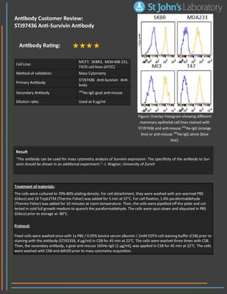
- 1. Cell Line: MCF7, SKBR3, MDA-MB-231, T47D cell lines (ATCC) Method of validation: Mass Cytometry Primary Antibody: STJ97436 Anti-Survivin Anti- body Secondary Antibody 165 Ho-IgG goat anti-mouse Dilution ratio: Used at 4 µg/ml Result “The antibody can be used for mass cytometry analysis of Survivin expression. The specificity of the antibody to Sur- vivin should be shown in an additional experiment.”- J. Wagner, University of Zurich Treatment of materials: The cells were cultured to 70%-80% plating density. For cell detachment, they were washed with pre-warmed PBS (Gibco) and 1X TrypLETM (Thermo Fisher) was added for 5 min at 37°C. For cell fixation, 1.6% paraformaldehyde (Thermo Fisher) was added for 10 minutes at room temperature. Then, the cells were pipetted off the plate and col- lected in cold full growth medium to quench the paraformaldehyde. The cells were spun down and aliquoted in PBS (Gibco) prior to storage at -80°C. Protocol: Fixed cells were washed once with 1x PBS / 0.05% bovine serum albumin / 2mM EDTA cell staining buffer (CSB) prior to staining with the antibody (STJ92356, 4 µg/ml) in CSB for 45 min at 22°C. The cells were washed three times with CSB. Then, the secondary antibody, a goat anti-mouse 165Ho-IgG (1 µg/ml), was applied in CSB for 45 min at 22°C. The cells were washed with CSB and ddH20 prior to mass cytometry acquisition. Antibody Customer Review: STJ97436 Anti-Survivin Antibody Antibody Rating: Figure: Overlay histogram showing different mammary epithelial cell lines stained with STJ97436 and anti-mouse 165 Ho-IgG (orange line) or anti-mouse 165 Ho-IgG alone (blue line).