Antibody Customer Review for CCND1 Polyclonal Antibody (STJ31335)
•
0 likes•336 views

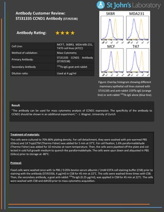
The document summarizes an experiment analyzing CCND1 expression in four breast cancer cell lines (MCF7, SKBR3, MDA-MB-231, T47D) using mass cytometry. The primary antibody STJ31335 CCND1 Antibody was used at 4 μg/ml along with a secondary antibody 159Tb-IgG goat anti-rabbit. The cells were cultured, fixed with paraformaldehyde, and stained according to the described protocol before mass cytometry acquisition. A reviewer commented that additional experiments are needed to demonstrate the specificity of the CCND1 antibody.
Report
Share
Report
Share
Download to read offline
Recommended
Regulatory component of the cyclin D1-CDK4 (DC) complex that phosphorylates and inhibits members of the retinoblastoma (RB) protein family including RB1 and regulates the cell-cycle during G1/S transition. Phosphorylation of RB1 allows dissociation of the transcription factor E2F from the RB/E2F complex and the subsequent transcription of E2F target genes which are responsible for the progression through the G1 phase. Hypophosphorylates RB1 in early G1 phase. Cyclin D-CDK4 complexes are major integrators of various mitogenenic and antimitogenic signals. Also substrate for SMAD3, phosphorylating SMAD3 in a cell-cycle-dependent manner and repressing its transcriptional activity. Component of the ternary complex, cyclin D1/CDK4/CDKN1B, required for nuclear translocation and activity of the cyclin D-CDK4 complex. Exhibits transcriptional corepressor activity with INSM1 on the NEUROD1 and INS promoters in a cell cycle-independent manner.
Anti-Cyclin D1-http://www.stjohnslabs.com/cyclin-d1-antibody-p-91890
Join our Antibody Validation Project - http://www.stjohnslabs.com/services/antibody-validation
Immunohistochemistry Antibody Validation Report for Anti-Cyclin D1 Antibody (...
Immunohistochemistry Antibody Validation Report for Anti-Cyclin D1 Antibody (...St John's Laboratory Ltd
Regulatory component of the cyclin D1-CDK4 (DC) complex that phosphorylates and inhibits members of the retinoblastoma (RB) protein family including RB1 and regulates the cell-cycle during G1/S transition. Phosphorylation of RB1 allows dissociation of the transcription factor E2F from the RB/E2F complex and the subsequent transcription of E2F target genes which are responsible for the progression through the G1 phase.
Anti-Cyclin D1 -http://www.stjohnslabs.com/cyclin-d1-antibody-p-91890
Join our Antibody Validation Project - http://www.stjohnslabs.com/services/antibody-validationImmunofluorescence Antibody Validation Report for Anti-Cyclin D1 Antibody (ST...
Immunofluorescence Antibody Validation Report for Anti-Cyclin D1 Antibody (ST...St John's Laboratory Ltd
NF-kappa-B is a pleiotropic transcription factor present in almost all cell types and is the endpoint of a series of signal transduction events that are initiated by a vast array of stimuli related to many biological processes such as inflammation, immunity, differentiation, cell growth, tumorigenesis and apoptosis. NF-kappa-B is a homo- or heterodimeric complex formed by the Rel-like domain-containing proteins RELA/p65, RELB, NFKB1/p105, NFKB1/p50, REL and NFKB2/p52 and the heterodimeric p65-p50 complex appears to be most abundant one. The dimers bind at kappa-B sites in the DNA of their target genes and the individual dimers have distinct preferences for different kappa-B sites that they can bind with distinguishable affinity and specificity. Different dimer combinations act as transcriptional activators or repressors, respectively. NF-kappa-B is controlled by various mechanisms of post-translational modification and subcellular compartmentalization as well as by interactions with other cofactors or corepressors.
Anti-NFkB p65-http://www.stjohnslabs.com/nfkb-p65-antibody-p-98648
Join our Antibody Validation Project - http://www.stjohnslabs.com/services/antibody-validationImmunohistochemistry Antibody Validation Report for Anti-NFkB p65 Antibody (S...
Immunohistochemistry Antibody Validation Report for Anti-NFkB p65 Antibody (S...St John's Laboratory Ltd
May play an essential role in local proteolysis of the extracellular matrix and in leukocyte migration. Could play a role in bone osteoclastic resorption. Cleaves KiSS1 at a Gly-|-Leu bond. Cleaves type IV and type V collagen into large C-terminal three quarter fragments and shorter N-terminal one quarter fragments. Degrades fibronectin but not laminin or Pz-peptide. / Cleavage of gelatin types I and V and collagen types IV and V.
Anti-MMP-9-http://www.stjohnslabs.com/mmp-9-antibody-p-
95079
Join our Antibody Validation Project - http://www.stjohnslabs.com/services/antibody-validationImmunohistochemistry Antibody Validation Report for Anti-MMP-9 Antibody (STJ9...
Immunohistochemistry Antibody Validation Report for Anti-MMP-9 Antibody (STJ9...St John's Laboratory Ltd
Antigen KI-67 also known as Ki-67 or MKI67 is a protein that in humans is encoded by the MKI67 gene (antigen identified by monoclonal antibody Ki-67). Antigen KI-67 is a nuclear protein that is associated with and may be necessary for cellular proliferation. Furthermore, it is associated with ribosomal RNA transcription.[5] Inactivation of antigen KI-67 leads to inhibition of ribosomal RNA synthesis.
Anti-Ki67 -http://www.stjohnslabs.com/ki-67-antibody-p-92897
Join our Antibody Validation Project - http://www.stjohnslabs.com/services/antibody-validation
Immunohistochemistry Antibody Validation Report for Anti-Ki67 Antibody (STJ93...
Immunohistochemistry Antibody Validation Report for Anti-Ki67 Antibody (STJ93...St John's Laboratory Ltd
NF-kappa-B is a pleiotropic transcription factor present in almost all cell types and is the endpoint of a series of signal transduction events that are initiated by a vast array of stimuli related to many biological processes such as inflammation, immunity, differentiation, cell growth, tumorigenesis and apoptosis. NF-kappa-B is a homo- or heterodimeric complex formed by the Rel-like domain-containing proteins RELA/p65, RELB, NFKB1/p105, NFKB1/p50, REL and NFKB2/p52 and the heterodimeric p65-p50 complex appears to be most abundant one. The dimers bind at kappa-B sites in the DNA of their target genes and the individual dimers have distinct preferences for different kappa-B sites that they can bind with distinguishable affinity and specificity. Different dimer combinations act as transcriptional activators or repressors, respectively. NF-kappa-B is controlled by various mechanisms of post-translational modification and subcellular compartmentalization as well as by interactions with other cofactors or corepressors. NF-kappa-B complexes are held in the cytoplasm in an inactive state complexed with members of the NF-kappa-B inhibitor (I-kappa-B) family. In a conventional activation pathway, I-kappa-B is phosphorylated by I-kappa-B kinases (IKKs) in response to different activators, subsequently degraded thus liberating the active NF-kappa-B complex which translocates to the nucleus. NF-kappa-B heterodimeric p65-p50 and p65-c-Rel complexes are transcriptional activators. The NF-kappa-B p65-p65 complex appears to be involved in invasin-mediated activation of IL-8 expression. The inhibitory effect of I-kappa-B upon NF-kappa-B the cytoplasm is exerted primarily through the interaction with p65. p65 shows a weak DNA-binding site which could contribute directly to DNA binding in the NF-kappa-B complex. Associates with chromatin at the NF-kappa-B promoter region via association with DDX1. Essential for cytokine gene expression in T-cells.
Anti-NFκB-p65-http://www.stjohnslabs.com/nfkb-p65-antibody-p-93372
Join our Antibody Validation Project - http://www.stjohnslabs.com/services/antibody-validationImmunohistochemistry Antibody Validation Report for Anti-NFκB-p65 Antibody (S...
Immunohistochemistry Antibody Validation Report for Anti-NFκB-p65 Antibody (S...St John's Laboratory Ltd
Tyrosine-protein kinase that acts as a cell-surface receptor for PDGFA, PDGFB and PDGFC and plays an essential role in the regulation of embryonic development, cell proliferation, survival and chemotaxis. Depending on the context, promotes or inhibits cell proliferation and cell migration. Plays an important role in the differentiation of bone marrow-derived mesenchymal stem cells. Required for normal skeleton development and cephalic closure during embryonic development. Required for normal development of the mucosa lining the gastrointestinal tract, and for recruitment of mesenchymal cells and normal development of intestinal villi. Plays a role in cell migration and chemotaxis in wound healing. Plays a role in platelet activation, secretion of agonists from platelet granules, and in thrombin-induced platelet aggregation.
Anti-PDGFRα-http://www.stjohnslabs.com/pdgfra-antibody-2
Join our Antibody Validation Project - http://www.stjohnslabs.com/services/antibody-validationImmunohistochemistry Antibody Validation Report for Anti-PDGFRα Antibody (STJ...
Immunohistochemistry Antibody Validation Report for Anti-PDGFRα Antibody (STJ...St John's Laboratory Ltd
Receptor-regulated SMAD (R-SMAD) that is an intracellular signal transducer and transcriptional modulator activated by TGF-beta (transforming growth factor) and activin type 1 receptor kinases. Binds the TRE element in the promoter region of many genes that are regulated by TGF-beta and, on formation of the SMAD3/SMAD4 complex, activates transcription. Also can form a SMAD3/SMAD4/JUN/FOS complex at the AP-1/SMAD site to regulate TGF-beta-mediated transcription. Has an inhibitory effect on wound healing probably by modulating both growth and migration of primary keratinocytes and by altering the TGF-mediated chemotaxis of monocytes. This effect on wound healing appears to be hormone-sensitive. Regulator of chondrogenesis and osteogenesis and inhibits early healing of bone fractures. Positively regulates PDPK1 kinase activity by stimulating its dissociation from the 14-3-3 protein YWHAQ which acts as a negative regulator.
Anti-Phospho-Smad3 (S425)-http://www.stjohnslabs.com/phospho-smad3-s425-antibody
Join our Antibody Validation Project - http://www.stjohnslabs.com/services/antibody-validationImmunohistochemistry Antibody Validation Report for Anti-Phospho-Smad3 (S425)...
Immunohistochemistry Antibody Validation Report for Anti-Phospho-Smad3 (S425)...St John's Laboratory Ltd
Nuclear phosphoprotein which forms a tight but non-covalently linked complex with the JUN/AP-1 transcription factor. In the heterodimer, FOS and JUN/AP-1 basic regions each seems to interact with symmetrical DNA half sites. On TGF-beta activation, forms a multimeric SMAD3/SMAD4/JUN/FOS complex at the AP1/SMAD-binding site to regulate TGF-beta-mediated signaling. Has a critical function in regulating the development of cells destined to form and maintain the skeleton. It is thought to have an important role in signal transduction, cell proliferation and differentiation. In growing cells, activates phospholipid synthesis, possibly by activating CDS1 and PI4K2A. This activity requires Tyr-dephosphorylation and association with the endoplasmic reticulum.
Anti-c-Fos-http://www.stjohnslabs.com/c-fos-antibody-p-91669
Join our Antibody Validation Project - http://www.stjohnslabs.com/services/antibody-validationImmunohistochemistry Antibody Validation Report for Anti-c-Fos Antibody (STJ9...
Immunohistochemistry Antibody Validation Report for Anti-c-Fos Antibody (STJ9...St John's Laboratory Ltd
Important regulator of cell cycle progression. Involved in G1 arrest. Potent inhibitor of cyclin E- and cyclin A-CDK2 complexes. Forms a complex with cyclin type D-CDK4 complexes and is involved in the assembly, stability, and modulation of CCND1-CDK4 complex activation. Acts either as an inhibitor or an activator of cyclin type D-CDK4 complexes depending on its phosphorylation state and/or stoichometry.
Anti-p27-http://www.stjohnslabs.com/p27-antibody-p-93722
Join our Antibody Validation Project - http://www.stjohnslabs.com/services/antibody-validationImmunohistochemistry Antibody Validation Report for Anti-p27 Antibody (STJ94866)
Immunohistochemistry Antibody Validation Report for Anti-p27 Antibody (STJ94866)St John's Laboratory Ltd
Telomerase is a ribonucleoprotein enzyme essential for the replication of chromosome termini in most eukaryotes. Active in progenitor and cancer cells. Inactive, or very low activity, in normal somatic cells. Catalytic component of the teleromerase holoenzyme complex whose main activity is the elongation of telomeres by acting as a reverse transcriptase that adds simple sequence repeats to chromosome ends by copying a template sequence within the RNA component of the enzyme. Catalyzes the RNA-dependent extension of 3'-chromosomal termini with the 6-nucleotide telomeric repeat unit, 5'-TTAGGG-3'. The catalytic cycle involves primer binding, primer extension and release of product once the template boundary has been reached or nascent product translocation followed by further extension. More active on substrates containing 2 or 3 telomeric repeats. Telomerase activity is regulated by a number of factors including telomerase complex-associated proteins, chaperones and polypeptide modifiers. Modulates Wnt signaling. Plays important roles in aging and antiapoptosis.
Anti-TERT-http://www.stjohnslabs.com/anti-tert-antibody?filter_name=STJ98964
Join our Antibody Validation Project - http://www.stjohnslabs.com/services/antibody-validationImmunohistochemistry Antibody Validation Report for Anti-TERT Antibody (STJ98...
Immunohistochemistry Antibody Validation Report for Anti-TERT Antibody (STJ98...St John's Laboratory Ltd
Receptor for TNFSF6/FASLG. The adapter molecule FADD recruits caspase-8 to the activated receptor. The resulting death-inducing signaling complex (DISC) performs caspase-8 proteolytic activation which initiates the subsequent cascade of caspases (aspartate-specific cysteine proteases) mediating apoptosis. FAS-mediated apoptosis may have a role in the induction of peripheral tolerance, in the antigen-stimulated suicide of mature T-cells, or both. The secreted isoforms 2 to 6 block apoptosis (in vitro).
Anti-FAS-http://www.stjohnslabs.com/fas-antibody-p-92276
Join our Antibody Validation Project - http://www.stjohnslabs.com/services/antibody-validation
Immunohistochemistry Antibody Validation Report for Anti-FAS Antibody (STJ93041)
Immunohistochemistry Antibody Validation Report for Anti-FAS Antibody (STJ93041)St John's Laboratory Ltd
Suppresses apoptosis in a variety of cell systems including factor-dependent lymphohematopoietic and neural cells. Regulates cell death by controlling the mitochondrial membrane permeability. Appears to function in a feedback loop system with caspases. Inhibits caspase activity either by preventing the release of cytochrome c from the mitochondria and/or by binding to the apoptosis-activating factor (APAF-1). May attenuate inflammation by impairing NLRP1-inflammasome activation, hence CASP1 activation and IL1B release .
Anti-Bcl-2-http://www.stjohnslabs.com/bcl-2-antibody-1
Join our Antibody Validation Project - http://www.stjohnslabs.com/services/antibody-validationImmunohistochemistry Antibody Validation Report for Anti-Bcl-2 Antibody (STJ9...
Immunohistochemistry Antibody Validation Report for Anti-Bcl-2 Antibody (STJ9...St John's Laboratory Ltd
Non-receptor tyrosine kinase involved in various processes such as cell growth, development, or differentiation. Mediates essential signaling events in both innate and adaptive immunity and plays a crucial role in hematopoiesis during T-cells development. In the cytoplasm, plays a pivotal role in signal transduction via its association with type I receptors sharing the common subunit gamma such as IL2R, IL4R, IL7R, IL9R, IL15R and IL21R. Following ligand binding to cell surface receptors, phosphorylates specific tyrosine residues on the cytoplasmic tails of the receptor, creating docking sites for STATs proteins.
Anti-JAK3-http://www.stjohnslabs.com/jak3-antibody-p-92869
Join our Antibody Validation Project - http://www.stjohnslabs.com/services/antibody-validation
Immunohistochemistry Antibody Validation Report for Anti-JAK3 Antibody (STJ93...
Immunohistochemistry Antibody Validation Report for Anti-JAK3 Antibody (STJ93...St John's Laboratory Ltd
Ubiquitin-like modifier involved in formation of autophagosomal vacuoles (autophagosomes) . Whereas LC3s are involved in elongation of the phagophore membrane, the GABARAP/GATE-16 subfamily is essential for a later stage in autophagosome maturation.
Anti-LC3A -http://www.stjohnslabs.com/lc3a-antibody
Join our Antibody Validation Project - http://www.stjohnslabs.com/services/antibody-validationImmunofluorescence Antibody Validation Report for Anti-LC3A Antibody (STJ97755)
Immunofluorescence Antibody Validation Report for Anti-LC3A Antibody (STJ97755)St John's Laboratory Ltd
Required for genome-wide de novo methylation and is essential for the establishment of DNA methylation patterns during development. DNA methylation is coordinated with methylation of histones. May preferentially methylates nucleosomal DNA within the nucleosome core region. May function as transcriptional co-repressor by associating with CBX4 and independently of DNA methylation. Seems to be involved in gene silencing (By similarity). In association with DNMT1 and via the recruitment of CTCFL/BORIS, involved in activation of BAG1 gene expression by modulating dimethylation of promoter histone H3 at H3K4 and H3K9. Isoforms 4 and 5 are probably not functional due to the deletion of two conserved methyltransferase motifs. Function as transcriptional corepressor by associating with ZHX1. / S-adenosyl-L-methionine + DNA = S-adenosyl-L-homocysteine + DNA containing 5-methylcytosine.
Anti-Dnmt3b-http://www.stjohnslabs.com/dnmt3b-antibody-p-92052
Join our Antibody Validation Project - http://www.stjohnslabs.com/services/antibody-validation
Immunohistochemistry Antibody Validation Report for Anti-Dnmt3b Antibody (STJ...
Immunohistochemistry Antibody Validation Report for Anti-Dnmt3b Antibody (STJ...St John's Laboratory Ltd
Toll-like receptor 4 is a protein that in humans is encoded by the TLR4 gene. TLR4 is a transmembrane protein, member of the toll-like receptor family, which belongs to the Pattern Recognition Receptor (PRRs) family. Its activation leads to an intracellular signaling pathway NF-κB and inflammatory cytokine production which is responsible for activating the innate immune system. It is most well known for recognizing lipopolysaccharide (LPS), a component present in many Gram-negative bacteria (e.g. Neisseria spp) and select Gram-positive bacteria. Its ligands also include several viral proteins, polysaccharide, and a variety of endogenous proteins such as low-density lipoprotein, beta-defensins, and heat shock protein.
Anti-CD284 -http://www.stjohnslabs.com/cd284-antibody
Join our Antibody Validation Project - http://www.stjohnslabs.com/services/antibody-validation
Immunohistochemistry Antibody Validation Report for Anti-CD284 Antibody (STJ9...
Immunohistochemistry Antibody Validation Report for Anti-CD284 Antibody (STJ9...St John's Laboratory Ltd
Signal transducer and transcription activator that mediates cellular responses to interleukins, KITLG/SCF, LEP and other growth factors. Once activated, recruits coactivators, such as NCOA1 or MED1, to the promoter region of the target gene . May mediate cellular responses to activated FGFR1, FGFR2, FGFR3 and FGFR4. Binds to the interleukin-6 (IL-6)-responsive elements identified in the promoters of various acute-phase protein genes. Activated by IL31 through IL31RA. Involved in cell cycle regulation by inducing the expression of key genes for the progression from G1 to S phase, such as CCND1 . Mediates the effects of LEP on melanocortin production, body energy homeostasis and lactation (By similarity). May play an apoptotic role by transctivating BIRC5 expression under LEP activation . Cytoplasmic STAT3 represses macroautophagy by inhibiting EIF2AK2/PKR activity.
Anti-Stat3-http://www.stjohnslabs.com/stat3-antibody-p-94446
Join our Antibody Validation Project - http://www.stjohnslabs.com/services/antibody-validationImmunohistochemistry Antibody Validation Report for Anti-Stat3 Antibody (STJ9...
Immunohistochemistry Antibody Validation Report for Anti-Stat3 Antibody (STJ9...St John's Laboratory Ltd
Facilitative glucose transporter. This isoform may be responsible for constitutive or basal glucose uptake. Has a very broad substrate specificity; can transport a wide range of aldoses including both pentoses and hexoses.
Anti-Glut1-http://www.stjohnslabs.com/glut1-antibody-p-92472
Join our Antibody Validation Project - http://www.stjohnslabs.com/services/antibody-validation
Immunohistochemistry Antibody Validation Report for Anti-Glut1 Antibody (STJ9...
Immunohistochemistry Antibody Validation Report for Anti-Glut1 Antibody (STJ9...St John's Laboratory Ltd
Produces nitric oxide (NO) which is a messenger molecule with diverse functions throughout the body . In macrophages, NO mediates tumoricidal and bactericidal actions. Also has nitrosylase activity and mediates cysteine S-nitrosylation of cytoplasmic target proteins such PTGS2/COX2 (By similarity). As component of the iNOS-S100A8/9 transnitrosylase complex involved in the selective inflammatory stimulus-dependent S-nitrosylation of GAPDH on 'Cys-247' implicated in regulation of the GAIT complex activity and probably multiple targets including ANXA5, EZR, MSN and VIM . Involved in inflammation, enhances the synthesis of proinflammatory mediators such as IL6 and IL8 .
Anti-NOS2-http://www.stjohnslabs.com/nos2-antibody-p-93419
Join our Antibody Validation Project - http://www.stjohnslabs.com/services/antibody-validationImmunohistochemistry Antibody Validation Report for Anti-NOS2 Antibody (STJ94...
Immunohistochemistry Antibody Validation Report for Anti-NOS2 Antibody (STJ94...St John's Laboratory Ltd
Receptor for the Fc region of IgG. Binds complexed or aggregated IgG and also monomeric IgG. Mediates antibody-dependent cellular cytotoxicity (ADCC) and other antibody-dependent responses, such as phagocytosis.
Anti-CD16-http://www.stjohnslabs.com/cd16-antibody-p-98627
Join our Antibody Validation Project - http://www.stjohnslabs.com/services/antibody-validationImmunohistochemistry Antibody Validation Report for Anti-CD16 Antibody (STJ96...
Immunohistochemistry Antibody Validation Report for Anti-CD16 Antibody (STJ96...St John's Laboratory Ltd
Collagen alpha-2(I) chain is a protein that in humans is encoded by the COL1A2 gene.
This gene encodes one of the chains for type I collagen, the fibrillar collagen found in most connective tissues. Mutations in this gene are associated with osteogenesis imperfecta, Ehlers-Danlos syndrome, idiopathic osteoporosis, and atypical Marfan syndrome. Symptoms associated with mutations in this gene, however, tend to be less severe than mutations in the gene for alpha-1 type I collagen since alpha-2 is less abundant. Multiple messages for this gene result from multiple polyadenylation signals, a feature shared by most of the other collagen genes.
Anti-COL1A2 -http://www.stjohnslabs.com/col1a2-antibody
Join our Antibody Validation Project - http://www.stjohnslabs.com/services/antibody-validationImmunohistochemistry Antibody Validation Report for Anti-COL1A2 Antibody (STJ...
Immunohistochemistry Antibody Validation Report for Anti-COL1A2 Antibody (STJ...St John's Laboratory Ltd
Signal transducer and transcription activator that mediates cellular responses to interleukins, KITLG/SCF, LEP and other growth factors. Once activated, recruits coactivators, such as NCOA1 or MED1, to the promoter region of the target gene . May mediate cellular responses to activated FGFR1, FGFR2, FGFR3 and FGFR4. Binds to the interleukin-6 (IL-6)-responsive elements identified in the promoters of various acute-phase protein genes. Activated by IL31 through IL31RA. Involved in cell cycle regulation by inducing the expression of key genes for the progression from G1 to S phase, such as CCND1 . Mediates the effects of LEP on melanocortin production, body energy homeostasis and lactation (By similarity). May play an apoptotic role by transctivating BIRC5 expression under LEP activation.
Anti-Stat3 -http://www.stjohnslabs.com/stat3-antibody-p-94446
Join our Antibody Validation Project - http://www.stjohnslabs.com/services/antibody-validationImmunofluorescence Antibody Validation Report for Anti-Stat3 Antibody (STJ95808)
Immunofluorescence Antibody Validation Report for Anti-Stat3 Antibody (STJ95808)St John's Laboratory Ltd
Tyrosine-protein kinase that acts as a cell-surface receptor for VEGFA, VEGFC and VEGFD. Plays an essential role in the regulation of angiogenesis, vascular development, vascular permeability, and embryonic hematopoiesis. Promotes proliferation, survival, migration and differentiation of endothelial cells. Promotes reorganization of the actin cytoskeleton. Isoforms lacking a transmembrane domain, such as isoform 2 and isoform 3, may function as decoy receptors for VEGFA, VEGFC and/or VEGFD. Isoform 2 plays an important role as negative regulator of VEGFA- and VEGFC-mediated lymphangiogenesis by limiting the amount of free VEGFA and/or VEGFC and preventing their binding to FLT4.
Anti-Flk-1-http://www.stjohnslabs.com/flk-1-antibody-p-92317
Join our Antibody Validation Project - http://www.stjohnslabs.com/services/antibody-validation
Immunohistochemistry Antibody Validation Report for Anti-Flk-1 Antibody (STJ9...
Immunohistochemistry Antibody Validation Report for Anti-Flk-1 Antibody (STJ9...St John's Laboratory Ltd
Suppresses apoptosis in a variety of cell systems including factor-dependent lymphohematopoietic and neural cells. Regulates cell death by controlling the mitochondrial membrane permeability. Appears to function in a feedback loop system with caspases. Inhibits caspase activity either by preventing the release of cytochrome c from the mitochondria and/or by binding to the apoptosis-activating factor (APAF-1). May attenuate inflammation by impairing NLRP1-inflammasome activation, hence CASP1 activation and IL1B release .
Anti-Bcl-2-http://www.stjohnslabs.com/bcl-2-antibody-p-91340
Join our Antibody Validation Project - http://www.stjohnslabs.com/services/antibody-validationImmunohistochemistry Antibody Validation Report for Anti-Bcl-2 Antibody (STJ9...
Immunohistochemistry Antibody Validation Report for Anti-Bcl-2 Antibody (STJ9...St John's Laboratory Ltd
Accelerates programmed cell death by binding to, and antagonizing the apoptosis repressor BCL2 or its adenovirus homolog E1B 19k protein. Under stress conditions, undergoes a conformation change that causes translocation to the mitochondrion membrane, leading to the release of cytochrome c that then triggers apoptosis. Promotes activation of CASP3, and thereby apoptosis.
Anti-Bax-http://www.stjohnslabs.com/bax-antibody-p-91328
Join our Antibody Validation Project - http://www.stjohnslabs.com/services/antibody-validationImmunohistochemistry Antibody Validation Report for Anti-Bax Antibody (STJ91820)
Immunohistochemistry Antibody Validation Report for Anti-Bax Antibody (STJ91820)St John's Laboratory Ltd
Produces nitric oxide (NO) which is a messenger molecule with diverse functions throughout the body . In macrophages, NO mediates tumoricidal and bactericidal actions. Also has nitrosylase activity and mediates cysteine S-nitrosylation of cytoplasmic target proteins such PTGS2/COX2 (By similarity). As component of the iNOS-S100A8/9 transnitrosylase complex involved in the selective inflammatory stimulus-dependent S-nitrosylation of GAPDH on 'Cys-247' implicated in regulation of the GAIT complex activity and probably multiple targets including ANXA5, EZR, MSN and VIM. Involved in inflammation, enhances the synthesis of proinflammatory mediators such as IL6 and IL8.
Anti-NOS2 -http://www.stjohnslabs.com/nos2-antibody-p-93419
Join our Antibody Validation Project - http://www.stjohnslabs.com/services/antibody-validationImmunofluorescence Antibody Validation Report for Anti-NOS2 Antibody (STJ94534)
Immunofluorescence Antibody Validation Report for Anti-NOS2 Antibody (STJ94534)St John's Laboratory Ltd
Antigen KI-67 also known as Ki-67 or MKI67 is a protein that in humans is encoded by the MKI67 gene (antigen identified by monoclonal antibody Ki-67). Antigen KI-67 is a nuclear protein that is associated with and may be necessary for cellular proliferation. Furthermore, it is associated with ribosomal RNA transcription.[5] Inactivation of antigen KI-67 leads to inhibition of ribosomal RNA synthesis.
Anti-Ki67 -http://www.stjohnslabs.com/ki-67-antibody-p-92897
Join our Antibody Validation Project - http://www.stjohnslabs.com/services/antibody-validation
Immunofluorescence Antibody Validation Report for Anti-Ki67 Antibody (STJ93832)
Immunofluorescence Antibody Validation Report for Anti-Ki67 Antibody (STJ93832)St John's Laboratory Ltd
Transcription factor that binds DNA in a non-specific manner, yet also specifically recognizes the core sequence 5'-CAC[GA]TG-3'. Activates the transcription of growth-related genes.
Anti-c-Myc -http://www.stjohnslabs.com/c-myc-antibody-p-91755
Join our Antibody Validation Project - http://www.stjohnslabs.com/services/antibody-validationImmunofluorescence Antibody Validation Report for Anti-c-Myc Antibody (STJ92356)
Immunofluorescence Antibody Validation Report for Anti-c-Myc Antibody (STJ92356)St John's Laboratory Ltd
More Related Content
What's hot
Nuclear phosphoprotein which forms a tight but non-covalently linked complex with the JUN/AP-1 transcription factor. In the heterodimer, FOS and JUN/AP-1 basic regions each seems to interact with symmetrical DNA half sites. On TGF-beta activation, forms a multimeric SMAD3/SMAD4/JUN/FOS complex at the AP1/SMAD-binding site to regulate TGF-beta-mediated signaling. Has a critical function in regulating the development of cells destined to form and maintain the skeleton. It is thought to have an important role in signal transduction, cell proliferation and differentiation. In growing cells, activates phospholipid synthesis, possibly by activating CDS1 and PI4K2A. This activity requires Tyr-dephosphorylation and association with the endoplasmic reticulum.
Anti-c-Fos-http://www.stjohnslabs.com/c-fos-antibody-p-91669
Join our Antibody Validation Project - http://www.stjohnslabs.com/services/antibody-validationImmunohistochemistry Antibody Validation Report for Anti-c-Fos Antibody (STJ9...
Immunohistochemistry Antibody Validation Report for Anti-c-Fos Antibody (STJ9...St John's Laboratory Ltd
Important regulator of cell cycle progression. Involved in G1 arrest. Potent inhibitor of cyclin E- and cyclin A-CDK2 complexes. Forms a complex with cyclin type D-CDK4 complexes and is involved in the assembly, stability, and modulation of CCND1-CDK4 complex activation. Acts either as an inhibitor or an activator of cyclin type D-CDK4 complexes depending on its phosphorylation state and/or stoichometry.
Anti-p27-http://www.stjohnslabs.com/p27-antibody-p-93722
Join our Antibody Validation Project - http://www.stjohnslabs.com/services/antibody-validationImmunohistochemistry Antibody Validation Report for Anti-p27 Antibody (STJ94866)
Immunohistochemistry Antibody Validation Report for Anti-p27 Antibody (STJ94866)St John's Laboratory Ltd
Telomerase is a ribonucleoprotein enzyme essential for the replication of chromosome termini in most eukaryotes. Active in progenitor and cancer cells. Inactive, or very low activity, in normal somatic cells. Catalytic component of the teleromerase holoenzyme complex whose main activity is the elongation of telomeres by acting as a reverse transcriptase that adds simple sequence repeats to chromosome ends by copying a template sequence within the RNA component of the enzyme. Catalyzes the RNA-dependent extension of 3'-chromosomal termini with the 6-nucleotide telomeric repeat unit, 5'-TTAGGG-3'. The catalytic cycle involves primer binding, primer extension and release of product once the template boundary has been reached or nascent product translocation followed by further extension. More active on substrates containing 2 or 3 telomeric repeats. Telomerase activity is regulated by a number of factors including telomerase complex-associated proteins, chaperones and polypeptide modifiers. Modulates Wnt signaling. Plays important roles in aging and antiapoptosis.
Anti-TERT-http://www.stjohnslabs.com/anti-tert-antibody?filter_name=STJ98964
Join our Antibody Validation Project - http://www.stjohnslabs.com/services/antibody-validationImmunohistochemistry Antibody Validation Report for Anti-TERT Antibody (STJ98...
Immunohistochemistry Antibody Validation Report for Anti-TERT Antibody (STJ98...St John's Laboratory Ltd
Receptor for TNFSF6/FASLG. The adapter molecule FADD recruits caspase-8 to the activated receptor. The resulting death-inducing signaling complex (DISC) performs caspase-8 proteolytic activation which initiates the subsequent cascade of caspases (aspartate-specific cysteine proteases) mediating apoptosis. FAS-mediated apoptosis may have a role in the induction of peripheral tolerance, in the antigen-stimulated suicide of mature T-cells, or both. The secreted isoforms 2 to 6 block apoptosis (in vitro).
Anti-FAS-http://www.stjohnslabs.com/fas-antibody-p-92276
Join our Antibody Validation Project - http://www.stjohnslabs.com/services/antibody-validation
Immunohistochemistry Antibody Validation Report for Anti-FAS Antibody (STJ93041)
Immunohistochemistry Antibody Validation Report for Anti-FAS Antibody (STJ93041)St John's Laboratory Ltd
Suppresses apoptosis in a variety of cell systems including factor-dependent lymphohematopoietic and neural cells. Regulates cell death by controlling the mitochondrial membrane permeability. Appears to function in a feedback loop system with caspases. Inhibits caspase activity either by preventing the release of cytochrome c from the mitochondria and/or by binding to the apoptosis-activating factor (APAF-1). May attenuate inflammation by impairing NLRP1-inflammasome activation, hence CASP1 activation and IL1B release .
Anti-Bcl-2-http://www.stjohnslabs.com/bcl-2-antibody-1
Join our Antibody Validation Project - http://www.stjohnslabs.com/services/antibody-validationImmunohistochemistry Antibody Validation Report for Anti-Bcl-2 Antibody (STJ9...
Immunohistochemistry Antibody Validation Report for Anti-Bcl-2 Antibody (STJ9...St John's Laboratory Ltd
Non-receptor tyrosine kinase involved in various processes such as cell growth, development, or differentiation. Mediates essential signaling events in both innate and adaptive immunity and plays a crucial role in hematopoiesis during T-cells development. In the cytoplasm, plays a pivotal role in signal transduction via its association with type I receptors sharing the common subunit gamma such as IL2R, IL4R, IL7R, IL9R, IL15R and IL21R. Following ligand binding to cell surface receptors, phosphorylates specific tyrosine residues on the cytoplasmic tails of the receptor, creating docking sites for STATs proteins.
Anti-JAK3-http://www.stjohnslabs.com/jak3-antibody-p-92869
Join our Antibody Validation Project - http://www.stjohnslabs.com/services/antibody-validation
Immunohistochemistry Antibody Validation Report for Anti-JAK3 Antibody (STJ93...
Immunohistochemistry Antibody Validation Report for Anti-JAK3 Antibody (STJ93...St John's Laboratory Ltd
Ubiquitin-like modifier involved in formation of autophagosomal vacuoles (autophagosomes) . Whereas LC3s are involved in elongation of the phagophore membrane, the GABARAP/GATE-16 subfamily is essential for a later stage in autophagosome maturation.
Anti-LC3A -http://www.stjohnslabs.com/lc3a-antibody
Join our Antibody Validation Project - http://www.stjohnslabs.com/services/antibody-validationImmunofluorescence Antibody Validation Report for Anti-LC3A Antibody (STJ97755)
Immunofluorescence Antibody Validation Report for Anti-LC3A Antibody (STJ97755)St John's Laboratory Ltd
Required for genome-wide de novo methylation and is essential for the establishment of DNA methylation patterns during development. DNA methylation is coordinated with methylation of histones. May preferentially methylates nucleosomal DNA within the nucleosome core region. May function as transcriptional co-repressor by associating with CBX4 and independently of DNA methylation. Seems to be involved in gene silencing (By similarity). In association with DNMT1 and via the recruitment of CTCFL/BORIS, involved in activation of BAG1 gene expression by modulating dimethylation of promoter histone H3 at H3K4 and H3K9. Isoforms 4 and 5 are probably not functional due to the deletion of two conserved methyltransferase motifs. Function as transcriptional corepressor by associating with ZHX1. / S-adenosyl-L-methionine + DNA = S-adenosyl-L-homocysteine + DNA containing 5-methylcytosine.
Anti-Dnmt3b-http://www.stjohnslabs.com/dnmt3b-antibody-p-92052
Join our Antibody Validation Project - http://www.stjohnslabs.com/services/antibody-validation
Immunohistochemistry Antibody Validation Report for Anti-Dnmt3b Antibody (STJ...
Immunohistochemistry Antibody Validation Report for Anti-Dnmt3b Antibody (STJ...St John's Laboratory Ltd
Toll-like receptor 4 is a protein that in humans is encoded by the TLR4 gene. TLR4 is a transmembrane protein, member of the toll-like receptor family, which belongs to the Pattern Recognition Receptor (PRRs) family. Its activation leads to an intracellular signaling pathway NF-κB and inflammatory cytokine production which is responsible for activating the innate immune system. It is most well known for recognizing lipopolysaccharide (LPS), a component present in many Gram-negative bacteria (e.g. Neisseria spp) and select Gram-positive bacteria. Its ligands also include several viral proteins, polysaccharide, and a variety of endogenous proteins such as low-density lipoprotein, beta-defensins, and heat shock protein.
Anti-CD284 -http://www.stjohnslabs.com/cd284-antibody
Join our Antibody Validation Project - http://www.stjohnslabs.com/services/antibody-validation
Immunohistochemistry Antibody Validation Report for Anti-CD284 Antibody (STJ9...
Immunohistochemistry Antibody Validation Report for Anti-CD284 Antibody (STJ9...St John's Laboratory Ltd
Signal transducer and transcription activator that mediates cellular responses to interleukins, KITLG/SCF, LEP and other growth factors. Once activated, recruits coactivators, such as NCOA1 or MED1, to the promoter region of the target gene . May mediate cellular responses to activated FGFR1, FGFR2, FGFR3 and FGFR4. Binds to the interleukin-6 (IL-6)-responsive elements identified in the promoters of various acute-phase protein genes. Activated by IL31 through IL31RA. Involved in cell cycle regulation by inducing the expression of key genes for the progression from G1 to S phase, such as CCND1 . Mediates the effects of LEP on melanocortin production, body energy homeostasis and lactation (By similarity). May play an apoptotic role by transctivating BIRC5 expression under LEP activation . Cytoplasmic STAT3 represses macroautophagy by inhibiting EIF2AK2/PKR activity.
Anti-Stat3-http://www.stjohnslabs.com/stat3-antibody-p-94446
Join our Antibody Validation Project - http://www.stjohnslabs.com/services/antibody-validationImmunohistochemistry Antibody Validation Report for Anti-Stat3 Antibody (STJ9...
Immunohistochemistry Antibody Validation Report for Anti-Stat3 Antibody (STJ9...St John's Laboratory Ltd
Facilitative glucose transporter. This isoform may be responsible for constitutive or basal glucose uptake. Has a very broad substrate specificity; can transport a wide range of aldoses including both pentoses and hexoses.
Anti-Glut1-http://www.stjohnslabs.com/glut1-antibody-p-92472
Join our Antibody Validation Project - http://www.stjohnslabs.com/services/antibody-validation
Immunohistochemistry Antibody Validation Report for Anti-Glut1 Antibody (STJ9...
Immunohistochemistry Antibody Validation Report for Anti-Glut1 Antibody (STJ9...St John's Laboratory Ltd
Produces nitric oxide (NO) which is a messenger molecule with diverse functions throughout the body . In macrophages, NO mediates tumoricidal and bactericidal actions. Also has nitrosylase activity and mediates cysteine S-nitrosylation of cytoplasmic target proteins such PTGS2/COX2 (By similarity). As component of the iNOS-S100A8/9 transnitrosylase complex involved in the selective inflammatory stimulus-dependent S-nitrosylation of GAPDH on 'Cys-247' implicated in regulation of the GAIT complex activity and probably multiple targets including ANXA5, EZR, MSN and VIM . Involved in inflammation, enhances the synthesis of proinflammatory mediators such as IL6 and IL8 .
Anti-NOS2-http://www.stjohnslabs.com/nos2-antibody-p-93419
Join our Antibody Validation Project - http://www.stjohnslabs.com/services/antibody-validationImmunohistochemistry Antibody Validation Report for Anti-NOS2 Antibody (STJ94...
Immunohistochemistry Antibody Validation Report for Anti-NOS2 Antibody (STJ94...St John's Laboratory Ltd
Receptor for the Fc region of IgG. Binds complexed or aggregated IgG and also monomeric IgG. Mediates antibody-dependent cellular cytotoxicity (ADCC) and other antibody-dependent responses, such as phagocytosis.
Anti-CD16-http://www.stjohnslabs.com/cd16-antibody-p-98627
Join our Antibody Validation Project - http://www.stjohnslabs.com/services/antibody-validationImmunohistochemistry Antibody Validation Report for Anti-CD16 Antibody (STJ96...
Immunohistochemistry Antibody Validation Report for Anti-CD16 Antibody (STJ96...St John's Laboratory Ltd
Collagen alpha-2(I) chain is a protein that in humans is encoded by the COL1A2 gene.
This gene encodes one of the chains for type I collagen, the fibrillar collagen found in most connective tissues. Mutations in this gene are associated with osteogenesis imperfecta, Ehlers-Danlos syndrome, idiopathic osteoporosis, and atypical Marfan syndrome. Symptoms associated with mutations in this gene, however, tend to be less severe than mutations in the gene for alpha-1 type I collagen since alpha-2 is less abundant. Multiple messages for this gene result from multiple polyadenylation signals, a feature shared by most of the other collagen genes.
Anti-COL1A2 -http://www.stjohnslabs.com/col1a2-antibody
Join our Antibody Validation Project - http://www.stjohnslabs.com/services/antibody-validationImmunohistochemistry Antibody Validation Report for Anti-COL1A2 Antibody (STJ...
Immunohistochemistry Antibody Validation Report for Anti-COL1A2 Antibody (STJ...St John's Laboratory Ltd
Signal transducer and transcription activator that mediates cellular responses to interleukins, KITLG/SCF, LEP and other growth factors. Once activated, recruits coactivators, such as NCOA1 or MED1, to the promoter region of the target gene . May mediate cellular responses to activated FGFR1, FGFR2, FGFR3 and FGFR4. Binds to the interleukin-6 (IL-6)-responsive elements identified in the promoters of various acute-phase protein genes. Activated by IL31 through IL31RA. Involved in cell cycle regulation by inducing the expression of key genes for the progression from G1 to S phase, such as CCND1 . Mediates the effects of LEP on melanocortin production, body energy homeostasis and lactation (By similarity). May play an apoptotic role by transctivating BIRC5 expression under LEP activation.
Anti-Stat3 -http://www.stjohnslabs.com/stat3-antibody-p-94446
Join our Antibody Validation Project - http://www.stjohnslabs.com/services/antibody-validationImmunofluorescence Antibody Validation Report for Anti-Stat3 Antibody (STJ95808)
Immunofluorescence Antibody Validation Report for Anti-Stat3 Antibody (STJ95808)St John's Laboratory Ltd
Tyrosine-protein kinase that acts as a cell-surface receptor for VEGFA, VEGFC and VEGFD. Plays an essential role in the regulation of angiogenesis, vascular development, vascular permeability, and embryonic hematopoiesis. Promotes proliferation, survival, migration and differentiation of endothelial cells. Promotes reorganization of the actin cytoskeleton. Isoforms lacking a transmembrane domain, such as isoform 2 and isoform 3, may function as decoy receptors for VEGFA, VEGFC and/or VEGFD. Isoform 2 plays an important role as negative regulator of VEGFA- and VEGFC-mediated lymphangiogenesis by limiting the amount of free VEGFA and/or VEGFC and preventing their binding to FLT4.
Anti-Flk-1-http://www.stjohnslabs.com/flk-1-antibody-p-92317
Join our Antibody Validation Project - http://www.stjohnslabs.com/services/antibody-validation
Immunohistochemistry Antibody Validation Report for Anti-Flk-1 Antibody (STJ9...
Immunohistochemistry Antibody Validation Report for Anti-Flk-1 Antibody (STJ9...St John's Laboratory Ltd
Suppresses apoptosis in a variety of cell systems including factor-dependent lymphohematopoietic and neural cells. Regulates cell death by controlling the mitochondrial membrane permeability. Appears to function in a feedback loop system with caspases. Inhibits caspase activity either by preventing the release of cytochrome c from the mitochondria and/or by binding to the apoptosis-activating factor (APAF-1). May attenuate inflammation by impairing NLRP1-inflammasome activation, hence CASP1 activation and IL1B release .
Anti-Bcl-2-http://www.stjohnslabs.com/bcl-2-antibody-p-91340
Join our Antibody Validation Project - http://www.stjohnslabs.com/services/antibody-validationImmunohistochemistry Antibody Validation Report for Anti-Bcl-2 Antibody (STJ9...
Immunohistochemistry Antibody Validation Report for Anti-Bcl-2 Antibody (STJ9...St John's Laboratory Ltd
Accelerates programmed cell death by binding to, and antagonizing the apoptosis repressor BCL2 or its adenovirus homolog E1B 19k protein. Under stress conditions, undergoes a conformation change that causes translocation to the mitochondrion membrane, leading to the release of cytochrome c that then triggers apoptosis. Promotes activation of CASP3, and thereby apoptosis.
Anti-Bax-http://www.stjohnslabs.com/bax-antibody-p-91328
Join our Antibody Validation Project - http://www.stjohnslabs.com/services/antibody-validationImmunohistochemistry Antibody Validation Report for Anti-Bax Antibody (STJ91820)
Immunohistochemistry Antibody Validation Report for Anti-Bax Antibody (STJ91820)St John's Laboratory Ltd
Produces nitric oxide (NO) which is a messenger molecule with diverse functions throughout the body . In macrophages, NO mediates tumoricidal and bactericidal actions. Also has nitrosylase activity and mediates cysteine S-nitrosylation of cytoplasmic target proteins such PTGS2/COX2 (By similarity). As component of the iNOS-S100A8/9 transnitrosylase complex involved in the selective inflammatory stimulus-dependent S-nitrosylation of GAPDH on 'Cys-247' implicated in regulation of the GAIT complex activity and probably multiple targets including ANXA5, EZR, MSN and VIM. Involved in inflammation, enhances the synthesis of proinflammatory mediators such as IL6 and IL8.
Anti-NOS2 -http://www.stjohnslabs.com/nos2-antibody-p-93419
Join our Antibody Validation Project - http://www.stjohnslabs.com/services/antibody-validationImmunofluorescence Antibody Validation Report for Anti-NOS2 Antibody (STJ94534)
Immunofluorescence Antibody Validation Report for Anti-NOS2 Antibody (STJ94534)St John's Laboratory Ltd
What's hot (20)
Immunohistochemistry Antibody Validation Report for Anti-c-Fos Antibody (STJ9...
Immunohistochemistry Antibody Validation Report for Anti-c-Fos Antibody (STJ9...
Immunohistochemistry Antibody Validation Report for Anti-p27 Antibody (STJ94866)
Immunohistochemistry Antibody Validation Report for Anti-p27 Antibody (STJ94866)
Immunohistochemistry Antibody Validation Report for Anti-TERT Antibody (STJ98...
Immunohistochemistry Antibody Validation Report for Anti-TERT Antibody (STJ98...
Immunohistochemistry Antibody Validation Report for Anti-FAS Antibody (STJ93041)
Immunohistochemistry Antibody Validation Report for Anti-FAS Antibody (STJ93041)
In Cell-Western Antibody Customer Review for Anti-LC3A Antibody (STJ97755)
In Cell-Western Antibody Customer Review for Anti-LC3A Antibody (STJ97755)
Immunohistochemistry Antibody Validation Report for Anti-Bcl-2 Antibody (STJ9...
Immunohistochemistry Antibody Validation Report for Anti-Bcl-2 Antibody (STJ9...
Immunohistochemistry Antibody Validation Report for Anti-JAK3 Antibody (STJ93...
Immunohistochemistry Antibody Validation Report for Anti-JAK3 Antibody (STJ93...
Immunofluorescence Antibody Validation Report for Anti-LC3A Antibody (STJ97755)
Immunofluorescence Antibody Validation Report for Anti-LC3A Antibody (STJ97755)
Immunohistochemistry Antibody Validation Report for Anti-Dnmt3b Antibody (STJ...
Immunohistochemistry Antibody Validation Report for Anti-Dnmt3b Antibody (STJ...
Immunohistochemistry Antibody Validation Report for Anti-CD284 Antibody (STJ9...
Immunohistochemistry Antibody Validation Report for Anti-CD284 Antibody (STJ9...
Immunohistochemistry Antibody Validation Report for Anti-Stat3 Antibody (STJ9...
Immunohistochemistry Antibody Validation Report for Anti-Stat3 Antibody (STJ9...
Immunohistochemistry Antibody Validation Report for Anti-Glut1 Antibody (STJ9...
Immunohistochemistry Antibody Validation Report for Anti-Glut1 Antibody (STJ9...
Immunohistochemistry Antibody Validation Report for Anti-NOS2 Antibody (STJ94...
Immunohistochemistry Antibody Validation Report for Anti-NOS2 Antibody (STJ94...
Immunohistochemistry Antibody Validation Report for Anti-CD16 Antibody (STJ96...
Immunohistochemistry Antibody Validation Report for Anti-CD16 Antibody (STJ96...
Immunohistochemistry Antibody Validation Report for Anti-COL1A2 Antibody (STJ...
Immunohistochemistry Antibody Validation Report for Anti-COL1A2 Antibody (STJ...
Immunofluorescence Antibody Validation Report for Anti-Stat3 Antibody (STJ95808)
Immunofluorescence Antibody Validation Report for Anti-Stat3 Antibody (STJ95808)
Immunohistochemistry Antibody Validation Report for Anti-Flk-1 Antibody (STJ9...
Immunohistochemistry Antibody Validation Report for Anti-Flk-1 Antibody (STJ9...
Immunohistochemistry Antibody Validation Report for Anti-Bcl-2 Antibody (STJ9...
Immunohistochemistry Antibody Validation Report for Anti-Bcl-2 Antibody (STJ9...
Immunohistochemistry Antibody Validation Report for Anti-Bax Antibody (STJ91820)
Immunohistochemistry Antibody Validation Report for Anti-Bax Antibody (STJ91820)
Immunofluorescence Antibody Validation Report for Anti-NOS2 Antibody (STJ94534)
Immunofluorescence Antibody Validation Report for Anti-NOS2 Antibody (STJ94534)
Viewers also liked
Antigen KI-67 also known as Ki-67 or MKI67 is a protein that in humans is encoded by the MKI67 gene (antigen identified by monoclonal antibody Ki-67). Antigen KI-67 is a nuclear protein that is associated with and may be necessary for cellular proliferation. Furthermore, it is associated with ribosomal RNA transcription.[5] Inactivation of antigen KI-67 leads to inhibition of ribosomal RNA synthesis.
Anti-Ki67 -http://www.stjohnslabs.com/ki-67-antibody-p-92897
Join our Antibody Validation Project - http://www.stjohnslabs.com/services/antibody-validation
Immunofluorescence Antibody Validation Report for Anti-Ki67 Antibody (STJ93832)
Immunofluorescence Antibody Validation Report for Anti-Ki67 Antibody (STJ93832)St John's Laboratory Ltd
Transcription factor that binds DNA in a non-specific manner, yet also specifically recognizes the core sequence 5'-CAC[GA]TG-3'. Activates the transcription of growth-related genes.
Anti-c-Myc -http://www.stjohnslabs.com/c-myc-antibody-p-91755
Join our Antibody Validation Project - http://www.stjohnslabs.com/services/antibody-validationImmunofluorescence Antibody Validation Report for Anti-c-Myc Antibody (STJ92356)
Immunofluorescence Antibody Validation Report for Anti-c-Myc Antibody (STJ92356)St John's Laboratory Ltd
Core component of nucleosome. Nucleosomes wrap and compact DNA into chromatin, limiting DNA accessibility to the cellular machineries which require DNA as a template. Histones thereby play a central role in transcription regulation, DNA repair, DNA replication and chromosomal stability. DNA accessibility is regulated via a complex set of post-translational modifications of histones, also called histone code, and nucleosome remodeling.
Anti-Histone H2B -http://www.stjohnslabs.com/histone-h2b-
antibody-p-98699
Join our Antibody Validation Project - http://www.stjohnslabs.com/services/antibody-validationImmunofluorescence Antibody Validation Report for Anti-Histone H2B Antibody (...
Immunofluorescence Antibody Validation Report for Anti-Histone H2B Antibody (...St John's Laboratory Ltd
Tyrosine phosphorylated upon stimulation with EGF. Tyrosine phosphorylated in response to constitutively activated FGFR1, FGFR2, FGFR3 and FGFR4 (By similarity). Activated through tyrosine phosphorylation by BMX. Tyrosine phosphorylated in response to IL6, IL11, LIF, CNTF, KITLG/SCF, CSF1, EGF, PDGF, IFN-alpha, LEP and OSM. Activated KIT promotes phosphorylation on tyrosine residues and subsequent translocation to the nucleus. Phosphorylated on serine upon DNA damage, probably by ATM or ATR. Serine phosphorylation is important for the formation of stable DNA-binding STAT3 homodimers and maximal transcriptional activity.
Anti-Phospho-Stat3 (S727) -http://www.stjohnslabs.com/phospho-stat3-s727-antibody
Join our Antibody Validation Project - http://www.stjohnslabs.com/services/antibody-validation
Immunofluorescence Antibody Validation Report for Anti-Phospho-Stat3 (S727) A...
Immunofluorescence Antibody Validation Report for Anti-Phospho-Stat3 (S727) A...St John's Laboratory Ltd
AKT1 is one of 3 closely related serine/threonine-protein kinases (AKT1, AKT2 and AKT3) called the AKT kinase, and which regulate many processes including metabolism, proliferation, cell survival, growth and angiogenesis. This is mediated through serine and/or threonine phosphorylation of a range of downstream substrates.AKT is responsible of the regulation of glucose uptake by mediating insulin-induced translocation of the SLC2A4/GLUT4 glucose transporter to the cell surface. Phosphorylation of PTPN1 at 'Ser-50' negatively modulates its phosphatase activity preventing dephosphorylation of the insulin receptor and the attenuation of insulin signaling. Phosphorylation of TBC1D4 triggers the binding of this effector to inhibitory 14-3-3 proteins, which is required for insulin-stimulated glucose transport. AKT regulates also the storage of glucose in the form of glycogen by phosphorylating GSK3A at 'Ser-21' and GSK3B at 'Ser-9', resulting in inhibition of its kinase activity.
Anti-Akt -http://www.stjohnslabs.com/akt-antibody-p-91115
Join our Antibody Validation Project - http://www.stjohnslabs.com/services/antibody-validationImmunohistochemistry Antibody Validation Report for Anti-Akt Antibody (STJ91550)
Immunohistochemistry Antibody Validation Report for Anti-Akt Antibody (STJ91550)St John's Laboratory Ltd
Neuronal phosphoprotein that coats synaptic vesicles, binds to the cytoskeleton, and is believed to function in the regulation of neurotransmitter release. The complex formed with NOS1 and CAPON proteins is necessary for specific nitric-oxid functions at a presynaptic level.
Anti-Phospho-Synapsin I (S605) - http://www.stjohnslabs.com/phospho-synapsin-i-s605-antibody
Join our Antibody Validation Project - http://www.stjohnslabs.com/services/antibody-validationImmunofluorescence Antibody Customer Review for Anti-Phospho-Synapsin I (S605...
Immunofluorescence Antibody Customer Review for Anti-Phospho-Synapsin I (S605...St John's Laboratory Ltd
Neuronal phosphoprotein that coats synaptic vesicles, binds to the cytoskeleton, and is believed to function in the regulation of neurotransmitter release. The complex formed with NOS1 and CAPON proteins is necessary for specific nitric-oxid functions at a presynaptic level.
Anti-Phospho-Synapsin I (S9) - http://www.stjohnslabs.com/phospho-synapsin-i-s9-antibody?filter_name=STJ90417
Join our Antibody Validation Project - http://www.stjohnslabs.com/services/antibody-validationImmunofluorescence Antibody Customer Review for Anti-Phospho-Synapsin I (S9) ...
Immunofluorescence Antibody Customer Review for Anti-Phospho-Synapsin I (S9) ...St John's Laboratory Ltd
St John’s Laboratory suppliers a highly photostable mCherry tag antibody, which is resistant to photobleaching. Independent validation reviews provide feedback on real-experimental use:
https://www.scienceexchange.com/validations/29688
https://www.scienceexchange.com/validations/29733
https://www.scienceexchange.com/validations/29708
To purchase this antibody, use the following link: http://www.stjohnslabs.com/mcherry-tag-antibody?filter_name=STJ34373Independent Antibody Validation For mCherry Tag Monoclonal Antibody (STJ34373)
Independent Antibody Validation For mCherry Tag Monoclonal Antibody (STJ34373)St John's Laboratory Ltd
ignal transducer and transcription activator that mediates cellular responses to interleukins, KITLG/SCF, LEP and other growth factors. Once activated, recruits coactivators, such as NCOA1 or MED1, to the promoter region of the target gene . May mediate cellular responses to activated FGFR1, FGFR2, FGFR3 and FGFR4. Binds to the interleukin-6 (IL-6)-responsive elements identified in the promoters of various acute-phase protein genes. Activated by IL31 through IL31RA. Involved in cell cycle regulation by inducing the expression of key genes for the progression from G1 to S phase, such as CCND1 . Mediates the effects of LEP on melanocortin production, body energy homeostasis and lactation (By similarity). May play an apoptotic role by transctivating BIRC5 expression under LEP activation . Cytoplasmic STAT3 represses macroautophagy by inhibiting EIF2AK2/PKR activity.
Anti-Phospho-Stat3 (S727)-http://www.stjohnslabs.com/phospho-stat3-s727-antibody
Join our Antibody Validation Project - http://www.stjohnslabs.com/services/antibody-validationImmunohistochemistry Antibody Validation Report for Anti-Phospho-Stat3 (S727)...
Immunohistochemistry Antibody Validation Report for Anti-Phospho-Stat3 (S727)...St John's Laboratory Ltd
Transferrins are iron binding transport proteins which can bind two Fe3+ ions in association with the binding of an anion, usually bicarbonate. It is responsible for the transport of iron from sites of absorption and heme degradation to those of storage and utilization. Serum transferrin may also have a further role in stimulating cell proliferation.
Anti-Transferrin -http://www.stjohnslabs.com/transferrin-antibody-p-98689
Join our Antibody Validation Project - http://www.stjohnslabs.com/services/antibody-validationImmunofluorescence Antibody Validation Report for Anti-Transferrin Antibody (...
Immunofluorescence Antibody Validation Report for Anti-Transferrin Antibody (...St John's Laboratory Ltd
Core component of nucleosome. Nucleosomes wrap and compact DNA into chromatin, limiting DNA accessibility to the cellular machineries which require DNA as a template. Histones thereby play a central role in transcription regulation, DNA repair, DNA replication and chromosomal stability. DNA accessibility is regulated via a complex set of post-translational modifications of histones, also called histone code, and nucleosome remodeling.
Anti-Histone H3 -http://www.stjohnslabs.com/histone-h3-antibody-p-92641
Join our Antibody Validation Project - http://www.stjohnslabs.com/services/antibody-validationImmunofluorescence Antibody Validation Report for Anti-Histone H3 Antibody (S...
Immunofluorescence Antibody Validation Report for Anti-Histone H3 Antibody (S...St John's Laboratory Ltd
Variant histone H2A which replaces conventional H2A in a subset of nucleosomes. Nucleosomes wrap and compact DNA into chromatin, limiting DNA accessibility to the cellular machineries which require DNA as a template. Histones thereby play a central role in transcription regulation, DNA repair, DNA replication and chromosomal stability. DNA accessibility is regulated via a complex set of post-translational modifications of histones, also called histone code, and nucleosome remodeling. Required for checkpoint-mediated arrest of cell cycle progression in response to low doses of ionizing radiation and for efficient repair of DNA double strand breaks (DSBs) specifically when modified by C-terminal phosphorylation.
Anti-Histone H2A.X -http://www.stjohnslabs.com/histone-h2ax-antibody-p-92631
Join our Antibody Validation Project - http://www.stjohnslabs.com/services/antibody-validationImmunofluorescence Antibody Validation Report for Anti-Histone H2A.X Antibody...
Immunofluorescence Antibody Validation Report for Anti-Histone H2A.X Antibody...St John's Laboratory Ltd
Gap junction protein that acts as a regulator of bladder capacity. A gap junction consists of a cluster of closely packed pairs of transmembrane channels, the connexons, through which materials of low MW diffuse from one cell to a neighboring cell. May play a critical role in the physiology of hearing by participating in the recycling of potassium to the cochlear endolymph. Negative regulator of bladder functional capacity: acts by enhancing intercellular electrical and chemical transmission, thus sensitizing bladder muscles to cholinergic neural stimuli and causing them to contract (By similarity). May play a role in cell growth inhibition through the regulation of NOV expression and localization. Plays an essential role in gap junction communication in the ventricles .
Anti-Connexin 43 -http://www.stjohnslabs.com/connexin-43-antibody-p-91786
Join our Antibody Validation Project - http://www.stjohnslabs.com/services/antibody-validationImmunofluorescence Antibody Validation Report for Anti-Connexin 43 Antibody (...
Immunofluorescence Antibody Validation Report for Anti-Connexin 43 Antibody (...St John's Laboratory Ltd
Viewers also liked (14)
Immunofluorescence Antibody Validation Report for Anti-Ki67 Antibody (STJ93832)
Immunofluorescence Antibody Validation Report for Anti-Ki67 Antibody (STJ93832)
Immunofluorescence Antibody Validation Report for Anti-c-Myc Antibody (STJ92356)
Immunofluorescence Antibody Validation Report for Anti-c-Myc Antibody (STJ92356)
Immunofluorescence Antibody Validation Report for Anti-Histone H2B Antibody (...
Immunofluorescence Antibody Validation Report for Anti-Histone H2B Antibody (...
Immunofluorescence Antibody Validation Report for Anti-Phospho-Stat3 (S727) A...
Immunofluorescence Antibody Validation Report for Anti-Phospho-Stat3 (S727) A...
Immunohistochemistry Antibody Validation Report for Anti-Akt Antibody (STJ91550)
Immunohistochemistry Antibody Validation Report for Anti-Akt Antibody (STJ91550)
Immunofluorescence Antibody Customer Review for Anti-Phospho-Synapsin I (S605...
Immunofluorescence Antibody Customer Review for Anti-Phospho-Synapsin I (S605...
Immunofluorescence Antibody Customer Review for Anti-Phospho-Synapsin I (S9) ...
Immunofluorescence Antibody Customer Review for Anti-Phospho-Synapsin I (S9) ...
Independent Antibody Validation For mCherry Tag Monoclonal Antibody (STJ34373)
Independent Antibody Validation For mCherry Tag Monoclonal Antibody (STJ34373)
Immunohistochemistry Antibody Validation Report for Anti-Phospho-Stat3 (S727)...
Immunohistochemistry Antibody Validation Report for Anti-Phospho-Stat3 (S727)...
Immunofluorescence Antibody Validation Report for Anti-Transferrin Antibody (...
Immunofluorescence Antibody Validation Report for Anti-Transferrin Antibody (...
Customer Review For NFκB-p65 Polyclonal Antibody (STJ94468)
Customer Review For NFκB-p65 Polyclonal Antibody (STJ94468)
Immunofluorescence Antibody Validation Report for Anti-Histone H3 Antibody (S...
Immunofluorescence Antibody Validation Report for Anti-Histone H3 Antibody (S...
Immunofluorescence Antibody Validation Report for Anti-Histone H2A.X Antibody...
Immunofluorescence Antibody Validation Report for Anti-Histone H2A.X Antibody...
Immunofluorescence Antibody Validation Report for Anti-Connexin 43 Antibody (...
Immunofluorescence Antibody Validation Report for Anti-Connexin 43 Antibody (...
Similar to Antibody Customer Review for CCND1 Polyclonal Antibody (STJ31335)
Histone H3 is one of the five main histone proteins involved in the structure of chromatin in eukaryotic cells. Core component of nucleosome. Nucleosomes wrap and compact DNA into chromatin, limiting DNA accessibility to the cellular machineries which require DNA as a template. Histones thereby play a central role in transcription regulation, DNA repair, DNA replication and chromosomal stability. DNA accessibility is regulated via a complex set of post-translational modifications of histones, also called histone code, and nucleosome remodeling.
Histone H3 is an important protein in the emerging field of epigenetics, where its sequence variants and variable modification states are thought to play a role in the dynamic and long term regulation of genes.
Anti-Histone H3 (Tri methyl K79) - http://www.stjohnslabs.com/histone-h3-tri-methyl-k79-antibody-p-98628?filter_name=STJ96993
Join our Antibody Validation Project - http://www.stjohnslabs.com/services/antibody-validationWestern Blot Antibody Customer Review for Anti-Histone H3 (Tri methyl K79) An...
Western Blot Antibody Customer Review for Anti-Histone H3 (Tri methyl K79) An...St John's Laboratory Ltd
Pyruvate kinase isozymes M1/M2 (PKM1/M2), also known as pyruvate kinase muscle isozyme (PKM), pyruvate kinase type K, cytosolic thyroid hormone-binding protein (CTHBP), thyroid hormone-binding protein 1 (THBP1), or opa-interacting protein 3 (OIP3), is an enzyme that in humans is encoded by the PKM2 gene.
Is a glycolytic enzyme that catalyzes the transfer of a phosphoryl group from phosphoenolpyruvate (PEP) to ADP, generating ATP. It plays a general role in caspase independent cell death of tumor cells.
The antibody was affinity-purified from rabbit antiserum by affinity-chromatography using epitope-specific immunogen.
Anti-PMK2 - http://www.stjohnslabs.com/pkm2-antibody-p-93954?filter_name=Stj95142
Join our Antibody Validation Project - http://www.stjohnslabs.com/services/antibody-validationWestern Blot Antibody Customer Review for Anti-PKM2 Polyclonal Antibody (STJ9...
Western Blot Antibody Customer Review for Anti-PKM2 Polyclonal Antibody (STJ9...St John's Laboratory Ltd
CD5 is a cluster of differentiation expressed on the surface of T cells (various species) and in a subset of murine B cells known as B-1a. The expression of this receptor in human B cells has been a controversial topic and up to date there is no consensus regarding the role of this receptor as a marker of human B cells. B-1 cells have limited diversity of their B-cell receptor due to their lack of the enzyme terminal deoxynucleotidyl transferase (TdT) and are potentially self-reactive. CD5 serves to mitigate activating signals from the BCR so that the B-1 cells can only be activated by very strong stimuli (such as bacterial proteins) and not by normal tissue proteins. CD5 was used as a T-cell marker until monoclonal antibodies against CD3 were developed.
Anti-CD5 -http://www.stjohnslabs.com/cd5-antibody-p-98608
Join our Antibody Validation Project - http://www.stjohnslabs.com/services/antibody-validation
Immunohistochemistry Antibody Validation Report for Anti-CD5 Antibody (STJ96973)
Immunohistochemistry Antibody Validation Report for Anti-CD5 Antibody (STJ96973)St John's Laboratory Ltd
Similar to Antibody Customer Review for CCND1 Polyclonal Antibody (STJ31335) (20)
Antibody Customer Review for Anti-Survivin Monoclonal Antibody (STJ97436)
Antibody Customer Review for Anti-Survivin Monoclonal Antibody (STJ97436)
Antibody Customer Review for TWIST1 Polyclonal Antibody (STJ31126)
Antibody Customer Review for TWIST1 Polyclonal Antibody (STJ31126)
Western Blot Antibody Customer Review for Anti-Histone H3 (Tri methyl K79) An...
Western Blot Antibody Customer Review for Anti-Histone H3 (Tri methyl K79) An...
Evaluation of the Viability of PTEN Transfected MDA-MB-468 Breast Cancer Cell...
Evaluation of the Viability of PTEN Transfected MDA-MB-468 Breast Cancer Cell...
Isolation and characterization of an extracellular antifungal protein from an...
Isolation and characterization of an extracellular antifungal protein from an...
Western Blot Antibody Customer Review for Anti-GPR18 antibody (STJ93370)
Western Blot Antibody Customer Review for Anti-GPR18 antibody (STJ93370)
Development of an electrotransformation protocol for genetic manipulation of ...
Development of an electrotransformation protocol for genetic manipulation of ...
Western Blot Antibody Customer Review for Anti-PKM2 Polyclonal Antibody (STJ9...
Western Blot Antibody Customer Review for Anti-PKM2 Polyclonal Antibody (STJ9...
Fluorescence activated cell sorted assay for Gaucher's disease
Fluorescence activated cell sorted assay for Gaucher's disease
Immunohistochemistry Antibody Validation Report for Anti-CD5 Antibody (STJ96973)
Immunohistochemistry Antibody Validation Report for Anti-CD5 Antibody (STJ96973)
Preimplantation genetic diagnosis_of_marfan_syndrome_using_multiple_displacem...
Preimplantation genetic diagnosis_of_marfan_syndrome_using_multiple_displacem...
More from St John's Laboratory Ltd
Receptor for glucocorticoids (GC). Has a dual mode of action: as a transcription factor that binds to glucocorticoid response elements (GRE), both for nuclear and mitochondrial DNA, and as a modulator of other transcription factors. Affects inflammatory responses, cellular proliferation and differentiation in target tissues. Involved in chromatin remodeling . Plays a role in rapid mRNA degradation by binding to the 5' UTR of target mRNAs and interacting with PNRC2 in a ligand-dependent manner. Could act as a coactivator for STAT5-dependent transcription upon growth hormone (GH) stimulation and could reveal an essential role of hepatic GR in the control of body growth (By similarity). Has transcriptional activation and repression activity . Mediates glucocorticoid-induced apoptosis . Promotes accurate chromosome segregation during mitosis . May act as a tumor suppressor . May play a negative role in adipogenesis through the regulation of lipolytic and antilipogenic gene expression (By similarity). / Isoform Beta: Acts as a dominant negative inhibitor of isoform Alpha . Has intrinsic transcriptional activity independent of isoform Alpha when both isoforms are coexpressed.
Join our Antibody Validation Project: http://www.stjohnslabs.com/services/antibody-validation
Anti glucocorticoid receptor antibody (STJ97101):
http://www.stjohnslabs.com/glucocorticoid-receptor-antibody-p-98736?filter_name=STJ97101Western Blot Customer Review Anti Glucocorticoid Receptor antibody (STJ97101)
Western Blot Customer Review Anti Glucocorticoid Receptor antibody (STJ97101)St John's Laboratory Ltd
Most upstream protease of the activation cascade of caspases responsible for the TNFRSF6/FAS mediated and TNFRSF1A induced cell death. Binding to the adapter molecule FADD recruits it to either receptor. The resulting aggregate called death-inducing signaling complex (DISC) performs CASP8 proteolytic activation. The active dimeric enzyme is then liberated from the DISC and free to activate downstream apoptotic proteases. Proteolytic fragments of the N-terminal propeptide (termed CAP3, CAP5 and CAP6) are likely retained in the DISC. Cleaves and activates CASP3, CASP4, CASP6, CASP7, CASP9 and CASP10. May participate in the GZMB apoptotic pathways. Cleaves ADPRT. Hydrolyzes the small-molecule substrate, Ac-Asp-Glu-Val-Asp-|-AMC. Likely target for the cowpox virus CRMA death inhibitory protein. Isoform 5, isoform 6, isoform 7 and isoform 8 lack the catalytic site and may interfere with the pro-apoptotic activity of the complex. / Strict requirement for Asp at position P1 and has a preferred cleavage sequence of (Leu/Asp/Val)-Glu-Thr-Asp-|-(Gly/Ser/Ala). / Inhibited by the effector protein NleF that is produced by pathogenic E.coli; this inhibits apoptosis.
Anti-Caspase-8-http://www.stjohnslabs.com/caspase-8-antibody-p-99045
Join our Antibody Validation Project - http://www.stjohnslabs.com/services/antibody-validationImmunohistochemistry Antibody Validation Report for Anti-Caspase-8 Antibody (...
Immunohistochemistry Antibody Validation Report for Anti-Caspase-8 Antibody (...St John's Laboratory Ltd
Tubulin is the major constituent of microtubules. The gamma chain is found at microtubule organizing centers (MTOC) such as the spindle poles or the centrosome. Pericentriolar matrix component that regulates alpha/beta chain minus-end nucleation, centrosome duplication and spindle formation.
Anti-Gamma Tubulin-http://www.stjohnslabs.com/gamma-tubulin-antibody
Join our Antibody Validation Project - http://www.stjohnslabs.com/services/antibody-validation
Immunohistochemistry Antibody Validation Report for Anti-Gamma Tubulin Antibo...
Immunohistochemistry Antibody Validation Report for Anti-Gamma Tubulin Antibo...St John's Laboratory Ltd
Tubulin in molecular biology can refer either to the tubulin protein superfamily of globular proteins, or one of the member proteins of that superfamily. α- and β-tubulins polymerize into microtubules, a major component of the eukaryotic cytoskeleton. Microtubules function in many essential cellular processes, including mitosis. Tubulin-binding drugs kill cancerous cells by inhibiting microtubule dynamics, which are required for DNA segregation and therefore cell division. In eukaryotes there are six members of the tubulin superfamily, although not all are present in all species (see below). Both α and β tubulins have a mass of around 50 kDa and are thus in a similar range compared to actin with ~42 kDa. In contrast, tubulin polymers (microtubules) tend to be much bigger than actin filaments due to their cylindrical nature. Tubulin was long thought to be specific to eukaryotes. More recently, however, several prokaryotic proteins have been shown to be related to tubulin.
Anti-Epsilon Tubulin -http://www.stjohnslabs.com/epsilon-tubulin-antibody-2
Join our Antibody Validation Project - http://www.stjohnslabs.com/services/antibody-validationImmunohistochemistry Antibody Validation Report for Anti-Epsilon Tubulin Anti...
Immunohistochemistry Antibody Validation Report for Anti-Epsilon Tubulin Anti...St John's Laboratory Ltd
Produces green light with a wavelength of 562 nm. / Photinus luciferin + O2 + ATP = oxidized Photinus luciferin + CO2 + AMP + diphosphate + light.
Anti-Luciferase -http://www.stjohnslabs.com/luciferase-antibody-2
Join our Antibody Validation Project - http://www.stjohnslabs.com/services/antibody-validationImmunohistochemistry Antibody Validation Report for Anti-Luciferase Antibody ...
Immunohistochemistry Antibody Validation Report for Anti-Luciferase Antibody ...St John's Laboratory Ltd
Ubiquitin-like modifier involved in formation of autophagosomal vacuoles (autophagosomes) . Whereas LC3s are involved in elongation of the phagophore membrane, the GABARAP/GATE-16 subfamily is essential for a later stage in autophagosome maturation .
Anti-LC3A-http://www.stjohnslabs.com/lc3a-antibody
Join our Antibody Validation Project - http://www.stjohnslabs.com/services/antibody-validationImmunohistochemistry Antibody Validation Report for Anti-LC3A Antibody (STJ97...
Immunohistochemistry Antibody Validation Report for Anti-LC3A Antibody (STJ97...St John's Laboratory Ltd
Multifunctional transcription factor in ER stress response. Plays an essential role in the response to a wide variety of cell stresses and induces cell cycle arrest and apoptosis in response to ER stress. Plays a dual role both as an inhibitor of CCAAT/enhancer-binding protein (C/EBP) function and as an activator of other genes. Acts as a dominant-negative regulator of C/EBP-induced transcription: dimerizes with members of the C/EBP family, impairs their association with C/EBP binding sites in the promoter regions, and inhibits the expression of C/EBP regulated genes. Positively regulates the transcription of TRIB3, IL6, IL8, IL23, TNFRSF10B/DR5, PPP1R15A/GADD34, BBC3/PUMA, BCL2L11/BIM and ERO1L. Negatively regulates; expression of BCL2 and MYOD1, ATF4-dependent transcriptional activation of asparagine synthetase (ASNS), CEBPA-dependent transcriptional activation of hepcidin (HAMP) and CEBPB-mediated expression of peroxisome proliferator-activated receptor gamma (PPARG).
Anti-CHOP-http://www.stjohnslabs.com/chop-antibody-2
Join our Antibody Validation Project - http://www.stjohnslabs.com/services/antibody-validationImmunohistochemistry Antibody Validation Report for Anti-CHOP Antibody (STJ97...
Immunohistochemistry Antibody Validation Report for Anti-CHOP Antibody (STJ97...St John's Laboratory Ltd
Pseudokinase that plays a key role in TNF-induced necroptosis, a programmed cell death process. Activated following phosphorylation by RIPK3, leading to homotrimerization, localization to the plasma membrane and execution of programmed necrosis characterized by calcium influx and plasma membrane damage. Does not have protein kinase activity.
Anti-phospho-MLKL (S358)-http://www.stjohnslabs.com/phospho-mlkl-s358-antibody-1
Join our Antibody Validation Project - http://www.stjohnslabs.com/services/antibody-validationImmunohistochemistry Antibody Validation Report for Anti-phospho-MLKL (S358) ...
Immunohistochemistry Antibody Validation Report for Anti-phospho-MLKL (S358) ...St John's Laboratory Ltd
Serine/threonine kinase which acts as an essential component of the MAP kinase signal transduction pathway. MAPK1/ERK2 and MAPK3/ERK1 are the 2 MAPKs which play an important role in the MAPK/ERK cascade. They participate also in a signaling cascade initiated by activated KIT and KITLG/SCF. Depending on the cellular context, the MAPK/ERK cascade mediates diverse biological functions such as cell growth, adhesion, survival and differentiation through the regulation of transcription, translation, cytoskeletal rearrangements. The MAPK/ERK cascade plays also a role in initiation and regulation of meiosis, mitosis, and postmitotic functions in differentiated cells by phosphorylating a number of transcription factors. About 160 substrates have already been discovered for ERKs. Many of these substrates are localized in the nucleus, and seem to participate in the regulation of transcription upon stimulation.
Anti-ERK1-http://www.stjohnslabs.com/erk1-antibody-3
Join our Antibody Validation Project - http://www.stjohnslabs.com/services/antibody-validation
Immunohistochemistry Antibody Validation Report for Anti-ERK1 Antibody (STJ97...
Immunohistochemistry Antibody Validation Report for Anti-ERK1 Antibody (STJ97...St John's Laboratory Ltd
Thiol protease that cleaves IL-1 beta between an Asp and an Ala, releasing the mature cytokine which is involved in a variety of inflammatory processes. Important for defense against pathogens. Cleaves and activates sterol regulatory element binding proteins (SREBPs). Can also promote apoptosis. / Strict requirement for an Asp residue at position P1 and has a preferred cleavage sequence of Tyr-Val-Ala-Asp-|-. / Specifically inhibited by the cowpox virus Crma protein.
Anti-Caspase-1-http://www.stjohnslabs.com/caspase-1-antibody-1
Join our Antibody Validation Project - http://www.stjohnslabs.com/services/antibody-validationImmunohistochemistry Antibody Validation Report for Anti-Caspase-1 Antibody (...
Immunohistochemistry Antibody Validation Report for Anti-Caspase-1 Antibody (...St John's Laboratory Ltd
Functions as a cell surface receptor and performs physiological functions on the surface of neurons relevant to neurite growth, neuronal adhesion and axonogenesis. Involved in cell mobility and transcription regulation through protein-protein interactions. Can promote transcription activation through binding to APBB1-KAT5 and inhibits Notch signaling through interaction with Numb. Couples to apoptosis-inducing pathways such as those mediated by G(O) and JIP. Inhibits G(o) alpha ATPase activity (By similarity). Acts as a kinesin I membrane receptor, mediating the axonal transport of beta-secretase and presenilin 1. Involved in copper homeostasis/oxidative stress through copper ion reduction. In vitro, copper-metallated APP induces neuronal death directly or is potentiated through Cu2+-mediated low-density lipoprotein oxidation.
Anti-Amyloid-β-http://www.stjohnslabs.com/amyloid-v-antibody
Join our Antibody Validation Project - http://www.stjohnslabs.com/services/antibody-validationImmunohistochemistry Antibody Validation Report for Anti-Amyloid-β Antibody (...
Immunohistochemistry Antibody Validation Report for Anti-Amyloid-β Antibody (...St John's Laboratory Ltd
Alpha-actin-2 also known as actin, aortic smooth muscle or alpha smooth muscle actin (α-SMA, SMactin, alpha-SM-actin, ASMA) is a protein that in humans is encoded by the ACTA2 gene located on 10q22-q24. Actin alpha 2, the human aortic smooth muscle actin gene, is one of six different actin isoforms which have been identified. Actins are highly conserved proteins that are involved in cell motility, structure and integrity. Alpha actins are a major constituent of the contractile apparatus. Alpha-smooth muscle actin (α-SMA) is commonly used as a marker of myofibroblast formation.
Anti-α-SMA -http://www.stjohnslabs.com/a-sma-antibody-1
Join our Antibody Validation Project - http://www.stjohnslabs.com/services/antibody-validationImmunohistochemistry Antibody Validation Report for Anti-α-SMA Antibody (STJ9...
Immunohistochemistry Antibody Validation Report for Anti-α-SMA Antibody (STJ9...St John's Laboratory Ltd
Transcription activator that binds DNA cooperatively with DP proteins through the E2 recognition site, 5'-TTTC[CG]CGC-3' found in the promoter region of a number of genes whose products are involved in cell cycle regulation or in DNA replication. The DRTF1/E2F complex functions in the control of cell-cycle progression from G1 to S phase. E2F1 binds preferentially RB1 in a cell-cycle dependent manner. It can mediate both cell proliferation and TP53/p53-dependent apoptosis. Blocks adipocyte differentiation by binding to specific promoters repressing CEBPA binding to its target gene promoters . / BIRC2/c-IAP1 stimulates its transcriptional activity.
Anti-E2F-1-http://www.stjohnslabs.com/e2f-1-antibody-1
Join our Antibody Validation Project - http://www.stjohnslabs.com/services/antibody-validationImmunohistochemistry Antibody Validation Report for Anti-E2F-1 Antibody (STJ9...
Immunohistochemistry Antibody Validation Report for Anti-E2F-1 Antibody (STJ9...St John's Laboratory Ltd
Plays important roles in the innate immune response as effector of glucocorticoid-mediated responses and regulator of the inflammatory process. Has anti-inflammatory activity. Plays a role in glucocorticoid-mediated down-regulation of the early phase of the inflammatory response (By similarity). Promotes resolution of inflammation and wound healing. Functions at least in part by activating the formyl peptide receptors and downstream signaling cascades. Promotes chemotaxis of granulocytes and monocytes via activation of the formyl peptide receptors.
Anti-Annexin I - http://www.stjohnslabs.com/anti-annexin-i-antibody?filter_name=STJ98699
Join our Antibody Validation Project - http://www.stjohnslabs.com/services/antibody-validation
Immunohistochemistry Antibody Validation Report for Anti-Annexin I Antibody (...
Immunohistochemistry Antibody Validation Report for Anti-Annexin I Antibody (...St John's Laboratory Ltd
More from St John's Laboratory Ltd (20)
Immunohistochemistry Customer Review Anti-Smo antibody (STJ95710)
Immunohistochemistry Customer Review Anti-Smo antibody (STJ95710)
Western Blot Customer Review Anti Glucocorticoid Receptor antibody (STJ97101)
Western Blot Customer Review Anti Glucocorticoid Receptor antibody (STJ97101)
Western Blot Customer Review Anti-Phospho-Cofilin (S3) Antibody (STJ90230)
Western Blot Customer Review Anti-Phospho-Cofilin (S3) Antibody (STJ90230)
Western Blot Customer Review Anti-WAVE2 Antibody (STJ96263)
Western Blot Customer Review Anti-WAVE2 Antibody (STJ96263)
Western Blot Customer Review Anti-Endophilin I Antibody (STJ92924)
Western Blot Customer Review Anti-Endophilin I Antibody (STJ92924)
Western Blot Customer Review Anti-β-tubulin Antibody (STJ97037)
Western Blot Customer Review Anti-β-tubulin Antibody (STJ97037)
Immunohistochemistry Antibody Validation Report for Anti-Caspase-8 Antibody (...
Immunohistochemistry Antibody Validation Report for Anti-Caspase-8 Antibody (...
Immunohistochemistry Antibody Validation Report for Anti-Gamma Tubulin Antibo...
Immunohistochemistry Antibody Validation Report for Anti-Gamma Tubulin Antibo...
Immunohistochemistry Antibody Validation Report for Anti-Epsilon Tubulin Anti...
Immunohistochemistry Antibody Validation Report for Anti-Epsilon Tubulin Anti...
Immunohistochemistry Antibody Validation Report for Anti-Luciferase Antibody ...
Immunohistochemistry Antibody Validation Report for Anti-Luciferase Antibody ...
Immunohistochemistry Antibody Validation Report for Anti-LC3A Antibody (STJ97...
Immunohistochemistry Antibody Validation Report for Anti-LC3A Antibody (STJ97...
Immunohistochemistry Antibody Validation Report for Anti-CHOP Antibody (STJ97...
Immunohistochemistry Antibody Validation Report for Anti-CHOP Antibody (STJ97...
Immunohistochemistry Antibody Validation Report for Anti-phospho-MLKL (S358) ...
Immunohistochemistry Antibody Validation Report for Anti-phospho-MLKL (S358) ...
Immunohistochemistry Antibody Validation Report for Anti-ERK1 Antibody (STJ97...
Immunohistochemistry Antibody Validation Report for Anti-ERK1 Antibody (STJ97...
Immunohistochemistry Antibody Validation Report for Anti-Caspase-1 Antibody (...
Immunohistochemistry Antibody Validation Report for Anti-Caspase-1 Antibody (...
Immunohistochemistry Antibody Validation Report for Anti-Amyloid-β Antibody (...
Immunohistochemistry Antibody Validation Report for Anti-Amyloid-β Antibody (...
Immunohistochemistry Antibody Validation Report for Anti-α-SMA Antibody (STJ9...
Immunohistochemistry Antibody Validation Report for Anti-α-SMA Antibody (STJ9...
Immunohistochemistry Antibody Validation Report for Anti-E2F-1 Antibody (STJ9...
Immunohistochemistry Antibody Validation Report for Anti-E2F-1 Antibody (STJ9...
Immunohistochemistry Antibody Validation Report for Anti-Annexin I Antibody (...
Immunohistochemistry Antibody Validation Report for Anti-Annexin I Antibody (...
Recently uploaded
Recently uploaded (20)
Feature-aligned N-BEATS with Sinkhorn divergence (ICLR '24)
Feature-aligned N-BEATS with Sinkhorn divergence (ICLR '24)
Asymmetry in the atmosphere of the ultra-hot Jupiter WASP-76 b
Asymmetry in the atmosphere of the ultra-hot Jupiter WASP-76 b
Chemical Tests; flame test, positive and negative ions test Edexcel Internati...
Chemical Tests; flame test, positive and negative ions test Edexcel Internati...
FAIRSpectra - Enabling the FAIRification of Spectroscopy and Spectrometry
FAIRSpectra - Enabling the FAIRification of Spectroscopy and Spectrometry
9654467111 Call Girls In Raj Nagar Delhi Short 1500 Night 6000
9654467111 Call Girls In Raj Nagar Delhi Short 1500 Night 6000
High Class Escorts in Hyderabad ₹7.5k Pick Up & Drop With Cash Payment 969456...
High Class Escorts in Hyderabad ₹7.5k Pick Up & Drop With Cash Payment 969456...
Formation of low mass protostars and their circumstellar disks
Formation of low mass protostars and their circumstellar disks
Justdial Call Girls In Indirapuram, Ghaziabad, 8800357707 Escorts Service
Justdial Call Girls In Indirapuram, Ghaziabad, 8800357707 Escorts Service
High Profile 🔝 8250077686 📞 Call Girls Service in GTB Nagar🍑
High Profile 🔝 8250077686 📞 Call Girls Service in GTB Nagar🍑
Call Girls Alandi Call Me 7737669865 Budget Friendly No Advance Booking
Call Girls Alandi Call Me 7737669865 Budget Friendly No Advance Booking
Vip profile Call Girls In Lonavala 9748763073 For Genuine Sex Service At Just...
Vip profile Call Girls In Lonavala 9748763073 For Genuine Sex Service At Just...
Antibody Customer Review for CCND1 Polyclonal Antibody (STJ31335)
- 1. Cell Line: MCF7, SKBR3, MDA-MB-231, T47D cell lines (ATCC) Method of validation: Mass Cytometry Primary Antibody: STJ31335 CCND1 Antibody (STJ92538) Secondary Antibody 159 Tb-IgG goat anti-rabbit Dilution ratio: Used at 4 µg/ml Result “The antibody can be used for mass cytometry analysis of CCND1 expression. The specificity of the antibody to CCND1 should be shown in an additional experiment.“ - J. Wagner, University of Zurich Treatment of materials: The cells were cultured to 70%-80% plating density. For cell detachment, they were washed with pre-warmed PBS (Gibco) and 1X TrypLETM (Thermo Fisher) was added for 5 min at 37°C. For cell fixation, 1.6% paraformaldehyde (Thermo Fisher) was added for 10 minutes at room temperature. Then, the cells were pipetted off the plate and col- lected in cold full growth medium to quench the paraformaldehyde. The cells were spun down and aliquoted in PBS (Gibco) prior to storage at -80°C. Protocol: Fixed cells were washed once with 1x PBS / 0.05% bovine serum albumin / 2mM EDTA cell staining buffer (CSB) prior to staining with the antibody (STJ92356, 4 µg/ml) in CSB for 45 min at 22°C. The cells were washed three times with CSB. Then, the secondary antibody, a goat anti-rabbit 159 Tb-IgG (0.25 µg/ml), was applied in CSB for 45 min at 22°C. The cells were washed with CSB and ddH20 prior to mass cytometry acquisition. Antibody Customer Review: STJ31335 CCND1 Antibody (STJ92538) Antibody Rating: Figure: Overlay histogram showing different mammary epithelial cell lines stained with STJ31335 and anti-rabbit 159Tb-IgG (orange line) or anti-rabbit 159 Tb-IgG alone (blue line).