SlideShare a Scribd company logo
1 of 10
Download to read offline
Figure:
Immunohistochemical
analysis of paraffin
embedded Human liver
cancer tissue. 1: CD5
Monoclonal
Antibody(10G8) was
diluted at 1:200 (4
degree
Celsius,overnight). 2:
Sodium citrate pH 6.0
was used for antibody
retrieval (>98 degree
Celsius,20min). 3:
Secondary antibody was
diluted at 1:200 (room
temperature, 30min).
Negative control was
used by secondary
antibody only.
Report Number 96973-a Host Mouse
Application IHC-P Clonality Monoclonal
Model Number STJ96973 Clone ID 10G8
Antibody Name Anti-CD5 antibody
Testing Species HUMAN Testing Tissue LIVER CANCER
ANTIBODY VALIDATION REPORT
a. (A small amount of distilled water was added into the incubation
box to prevent evaporation of antibody).
51. Secondary antibody incubation
a. Slides were washed 3 times, with PBS on a shaker for 5min.
Shortly after the slides were dried the corresponding secondary
antibody solution was added (HRP labelled), covering the
tissues, and incubated at room temperature for 30min.
b.
52. DAB staining
a. Slides were washed 3 times, with PBS on a shaker for 5min.
b. Shortly after, the slides were dried and fresh DAB staining buffer
was added inside the circles. The staining time was adjusted
under a microscope. Yellow-brown colour represented a positive
result. Slides were washed with water to stop the staining.
c.
53. Haematoxylin staining
a. Haematoxylin was used to counter-staining for 1min, and then
the slides were washed with water. 1% Hydrochloric acid and
alcohol was added for several seconds and then washed with
water. Ammonia was used to reveal blue colour, and then
flushed with water.
b.
54. Desolation and Clearing
i. Slides were incubated sequentially into: 75% alcohol 5min, 85%
alcohol 5min, Anhydrous ethanol - 5min, Anhydrous ethanol -
5min & Xylene - 5min. Shortly after slides were dried and neutral
gum was used to seal the slides.
ii.
55. Visualization
a. Results were validated with microscope, and the slides were
scanned.
Paraffin-Embedded
Immunohistochemistry Protocol
45.
46. Tissue processing
a. Slides were incubated sequentially into Xylene; 15min –
Xylene, 15min - Anhydrous ethanol, 5min - Anhydrous
ethanol, 5min - 85% alcohol, 5min - 75% alcohol & 5min –
wash in distilled water.
b.
47. Antigen retrieval
a. Tissue slides were incubated with citric acid (PH6.0) antigen
retrieval buffer and microwaved for antigen retrieval (heated
until boiled and then stopped heating) for 8min. Slides were
then heated with medium power for 7min. During this
process slides were kept from drying out. After cooling down
at room temperature, slides were washed with PBS on
shaker for 5min, repeated for 3 times.
b.
48. Inhibition of endogenous peroxidase
a. Slides were placed in 3% Hydrogen peroxide solution, and
incubated for 10 min at room temperature without light
exposure. Slides were then washed 3 times with PBS on a
shaker for 5mins.
b.
49. BSA Blocking
a. Shortly after slides were dried, a PAP pen was used to draw
circles around the tissue sections (and to prevent draining of
the antibody solution). Inside the circles, BSA was used to
cover the tissue evenly, blocking for 30min.
b.
50. Primary antibody incubation
After blocking solution was removed a 1:200 solution of
primary antibody/PBS was added on the slide, and incubated
overnight at 4°C.
St John's Laboratory Ltd.
www.stjohnslabs.com
Figure:
Immunohistochemical
analysis of paraffin
embedded Human
stomach tissue. 1: CD5
Monoclonal
Antibody(10G8) was
diluted at 1:200 (4
degree
Celsius,overnight). 2:
Sodium citrate pH 6.0
was used for antibody
retrieval (>98 degree
Celsius,20min). 3:
Secondary antibody was
diluted at 1:200 (room
temperature, 30min).
Negative control was
used by secondary
antibody only.
Report Number 96973-b Host Mouse
Application IHC-P Clonality Monoclonal
Model Number STJ96973 Clone ID 10G8
Antibody Name Anti-CD5 antibody
Testing Species HUMAN Testing Tissue STOMACH
ANTIBODY VALIDATION REPORT
a. (A small amount of distilled water was added into the incubation
box to prevent evaporation of antibody).
40. Secondary antibody incubation
a. Slides were washed 3 times, with PBS on a shaker for 5min.
Shortly after the slides were dried the corresponding secondary
antibody solution was added (HRP labelled), covering the
tissues, and incubated at room temperature for 30min.
b.
41. DAB staining
a. Slides were washed 3 times, with PBS on a shaker for 5min.
b. Shortly after, the slides were dried and fresh DAB staining buffer
was added inside the circles. The staining time was adjusted
under a microscope. Yellow-brown colour represented a positive
result. Slides were washed with water to stop the staining.
c.
42. Haematoxylin staining
a. Haematoxylin was used to counter-staining for 1min, and then
the slides were washed with water. 1% Hydrochloric acid and
alcohol was added for several seconds and then washed with
water. Ammonia was used to reveal blue colour, and then
flushed with water.
b.
43. Desolation and Clearing
i. Slides were incubated sequentially into: 75% alcohol 5min, 85%
alcohol 5min, Anhydrous ethanol - 5min, Anhydrous ethanol -
5min & Xylene - 5min. Shortly after slides were dried and neutral
gum was used to seal the slides.
ii.
44. Visualization
a. Results were validated with microscope, and the slides were
scanned.
Paraffin-Embedded
Immunohistochemistry Protocol
34.
35. Tissue processing
a. Slides were incubated sequentially into Xylene; 15min –
Xylene, 15min - Anhydrous ethanol, 5min - Anhydrous
ethanol, 5min - 85% alcohol, 5min - 75% alcohol & 5min –
wash in distilled water.
b.
36. Antigen retrieval
a. Tissue slides were incubated with citric acid (PH6.0) antigen
retrieval buffer and microwaved for antigen retrieval (heated
until boiled and then stopped heating) for 8min. Slides were
then heated with medium power for 7min. During this
process slides were kept from drying out. After cooling down
at room temperature, slides were washed with PBS on
shaker for 5min, repeated for 3 times.
b.
37. Inhibition of endogenous peroxidase
a. Slides were placed in 3% Hydrogen peroxide solution, and
incubated for 10 min at room temperature without light
exposure. Slides were then washed 3 times with PBS on a
shaker for 5mins.
b.
38. BSA Blocking
a. Shortly after slides were dried, a PAP pen was used to draw
circles around the tissue sections (and to prevent draining of
the antibody solution). Inside the circles, BSA was used to
cover the tissue evenly, blocking for 30min.
b.
39. Primary antibody incubation
After blocking solution was removed a 1:200 solution of
primary antibody/PBS was added on the slide, and incubated
overnight at 4°C.
St John's Laboratory Ltd.
www.stjohnslabs.com
Figure:
Immunohistochemical
analysis of paraffin
embedded Human
stomach cancer tissue.
1: CD5 Monoclonal
Antibody(10G8) was
diluted at 1:200 (4
degree
Celsius,overnight). 2:
Sodium citrate pH 6.0
was used for antibody
retrieval (>98 degree
Celsius,20min). 3:
Secondary antibody was
diluted at 1:200 (room
temperature, 30min).
Negative control was
used by secondary
antibody only.
Report Number 96973-c Host Mouse
Application IHC-P Clonality Monoclonal
Model Number STJ96973 Clone ID 10G8
Antibody Name Anti-CD5 antibody
Testing Species HUMAN Testing Tissue STOMACH CANCER
ANTIBODY VALIDATION REPORT
a. (A small amount of distilled water was added into the incubation
box to prevent evaporation of antibody).
29. Secondary antibody incubation
a. Slides were washed 3 times, with PBS on a shaker for 5min.
Shortly after the slides were dried the corresponding secondary
antibody solution was added (HRP labelled), covering the
tissues, and incubated at room temperature for 30min.
b.
30. DAB staining
a. Slides were washed 3 times, with PBS on a shaker for 5min.
b. Shortly after, the slides were dried and fresh DAB staining buffer
was added inside the circles. The staining time was adjusted
under a microscope. Yellow-brown colour represented a positive
result. Slides were washed with water to stop the staining.
c.
31. Haematoxylin staining
a. Haematoxylin was used to counter-staining for 1min, and then
the slides were washed with water. 1% Hydrochloric acid and
alcohol was added for several seconds and then washed with
water. Ammonia was used to reveal blue colour, and then
flushed with water.
b.
32. Desolation and Clearing
i. Slides were incubated sequentially into: 75% alcohol 5min, 85%
alcohol 5min, Anhydrous ethanol - 5min, Anhydrous ethanol -
5min & Xylene - 5min. Shortly after slides were dried and neutral
gum was used to seal the slides.
ii.
33. Visualization
a. Results were validated with microscope, and the slides were
scanned.
Paraffin-Embedded
Immunohistochemistry Protocol
23.
24. Tissue processing
a. Slides were incubated sequentially into Xylene; 15min –
Xylene, 15min - Anhydrous ethanol, 5min - Anhydrous
ethanol, 5min - 85% alcohol, 5min - 75% alcohol & 5min –
wash in distilled water.
b.
25. Antigen retrieval
a. Tissue slides were incubated with citric acid (PH6.0) antigen
retrieval buffer and microwaved for antigen retrieval (heated
until boiled and then stopped heating) for 8min. Slides were
then heated with medium power for 7min. During this
process slides were kept from drying out. After cooling down
at room temperature, slides were washed with PBS on
shaker for 5min, repeated for 3 times.
b.
26. Inhibition of endogenous peroxidase
a. Slides were placed in 3% Hydrogen peroxide solution, and
incubated for 10 min at room temperature without light
exposure. Slides were then washed 3 times with PBS on a
shaker for 5mins.
b.
27. BSA Blocking
a. Shortly after slides were dried, a PAP pen was used to draw
circles around the tissue sections (and to prevent draining of
the antibody solution). Inside the circles, BSA was used to
cover the tissue evenly, blocking for 30min.
b.
28. Primary antibody incubation
After blocking solution was removed a 1:200 solution of
primary antibody/PBS was added on the slide, and incubated
overnight at 4°C.
St John's Laboratory Ltd.
www.stjohnslabs.com
Figure:
Immunohistochemical
analysis of paraffin
embedded Rat kidney
tissue. 1: CD5
Monoclonal
Antibody(10G8) was
diluted at 1:200 (4
degree
Celsius,overnight). 2:
Sodium citrate pH 6.0
was used for antibody
retrieval (>98 degree
Celsius,20min). 3:
Secondary antibody was
diluted at 1:200 (room
temperature, 30min).
Negative control was
used by secondary
antibody only.
Report Number 96973-d Host Mouse
Application IHC-P Clonality Monoclonal
Model Number STJ96973 Clone ID 10G8
Antibody Name Anti-CD5 antibody
Testing Species RAT Testing Tissue KIDNEY
ANTIBODY VALIDATION REPORT
a. (A small amount of distilled water was added into the incubation
box to prevent evaporation of antibody).
18. Secondary antibody incubation
a. Slides were washed 3 times, with PBS on a shaker for 5min.
Shortly after the slides were dried the corresponding secondary
antibody solution was added (HRP labelled), covering the
tissues, and incubated at room temperature for 30min.
b.
19. DAB staining
a. Slides were washed 3 times, with PBS on a shaker for 5min.
b. Shortly after, the slides were dried and fresh DAB staining buffer
was added inside the circles. The staining time was adjusted
under a microscope. Yellow-brown colour represented a positive
result. Slides were washed with water to stop the staining.
c.
20. Haematoxylin staining
a. Haematoxylin was used to counter-staining for 1min, and then
the slides were washed with water. 1% Hydrochloric acid and
alcohol was added for several seconds and then washed with
water. Ammonia was used to reveal blue colour, and then
flushed with water.
b.
21. Desolation and Clearing
i. Slides were incubated sequentially into: 75% alcohol 5min, 85%
alcohol 5min, Anhydrous ethanol - 5min, Anhydrous ethanol -
5min & Xylene - 5min. Shortly after slides were dried and neutral
gum was used to seal the slides.
ii.
22. Visualization
a. Results were validated with microscope, and the slides were
scanned.
Paraffin-Embedded
Immunohistochemistry Protocol
12.
13. Tissue processing
a. Slides were incubated sequentially into Xylene; 15min –
Xylene, 15min - Anhydrous ethanol, 5min - Anhydrous
ethanol, 5min - 85% alcohol, 5min - 75% alcohol & 5min –
wash in distilled water.
b.
14. Antigen retrieval
a. Tissue slides were incubated with citric acid (PH6.0) antigen
retrieval buffer and microwaved for antigen retrieval (heated
until boiled and then stopped heating) for 8min. Slides were
then heated with medium power for 7min. During this
process slides were kept from drying out. After cooling down
at room temperature, slides were washed with PBS on
shaker for 5min, repeated for 3 times.
b.
15. Inhibition of endogenous peroxidase
a. Slides were placed in 3% Hydrogen peroxide solution, and
incubated for 10 min at room temperature without light
exposure. Slides were then washed 3 times with PBS on a
shaker for 5mins.
b.
16. BSA Blocking
a. Shortly after slides were dried, a PAP pen was used to draw
circles around the tissue sections (and to prevent draining of
the antibody solution). Inside the circles, BSA was used to
cover the tissue evenly, blocking for 30min.
b.
17. Primary antibody incubation
After blocking solution was removed a 1:200 solution of
primary antibody/PBS was added on the slide, and incubated
overnight at 4°C.
St John's Laboratory Ltd.
www.stjohnslabs.com
Figure:
Immunohistochemical
analysis of paraffin
embedded Rat spinal
cord tissue. 1: CD5
Monoclonal
Antibody(10G8) was
diluted at 1:200 (4
degree
Celsius,overnight). 2:
Sodium citrate pH 6.0
was used for antibody
retrieval (>98 degree
Celsius,20min). 3:
Secondary antibody was
diluted at 1:200 (room
temperature, 30min).
Negative control was
used by secondary
antibody only.
Report Number 96973-e Host Mouse
Application IHC-P Clonality Monoclonal
Model Number STJ96973 Clone ID 10G8
Antibody Name Anti-CD5 antibody
Testing Species RAT Testing Tissue SPINAL
ANTIBODY VALIDATION REPORT
a. (A small amount of distilled water was added into the incubation
box to prevent evaporation of antibody).
7. Secondary antibody incubation
a. Slides were washed 3 times, with PBS on a shaker for 5min.
Shortly after the slides were dried the corresponding secondary
antibody solution was added (HRP labelled), covering the
tissues, and incubated at room temperature for 30min.
b.
8. DAB staining
a. Slides were washed 3 times, with PBS on a shaker for 5min.
b. Shortly after, the slides were dried and fresh DAB staining buffer
was added inside the circles. The staining time was adjusted
under a microscope. Yellow-brown colour represented a positive
result. Slides were washed with water to stop the staining.
c.
9. Haematoxylin staining
a. Haematoxylin was used to counter-staining for 1min, and then
the slides were washed with water. 1% Hydrochloric acid and
alcohol was added for several seconds and then washed with
water. Ammonia was used to reveal blue colour, and then
flushed with water.
b.
10. Desolation and Clearing
i. Slides were incubated sequentially into: 75% alcohol 5min, 85%
alcohol 5min, Anhydrous ethanol - 5min, Anhydrous ethanol -
5min & Xylene - 5min. Shortly after slides were dried and neutral
gum was used to seal the slides.
ii.
11. Visualization
a. Results were validated with microscope, and the slides were
scanned.
Paraffin-Embedded
Immunohistochemistry Protocol
1.
2. Tissue processing
a. Slides were incubated sequentially into Xylene; 15min –
Xylene, 15min - Anhydrous ethanol, 5min - Anhydrous
ethanol, 5min - 85% alcohol, 5min - 75% alcohol & 5min –
wash in distilled water.
b.
3. Antigen retrieval
a. Tissue slides were incubated with citric acid (PH6.0) antigen
retrieval buffer and microwaved for antigen retrieval (heated
until boiled and then stopped heating) for 8min. Slides were
then heated with medium power for 7min. During this
process slides were kept from drying out. After cooling down
at room temperature, slides were washed with PBS on
shaker for 5min, repeated for 3 times.
b.
4. Inhibition of endogenous peroxidase
a. Slides were placed in 3% Hydrogen peroxide solution, and
incubated for 10 min at room temperature without light
exposure. Slides were then washed 3 times with PBS on a
shaker for 5mins.
b.
5. BSA Blocking
a. Shortly after slides were dried, a PAP pen was used to draw
circles around the tissue sections (and to prevent draining of
the antibody solution). Inside the circles, BSA was used to
cover the tissue evenly, blocking for 30min.
b.
6. Primary antibody incubation
After blocking solution was removed a 1:200 solution of
primary antibody/PBS was added on the slide, and incubated
overnight at 4°C.
St John's Laboratory Ltd.
www.stjohnslabs.com
Figure:
Immunohistochemical
analysis of paraffin
embedded Rat brain
tissue. 1: CD5
Monoclonal
Antibody(10G8) was
diluted at 1:200 (4
degree
Celsius,overnight). 2:
Sodium citrate pH 6.0
was used for antibody
retrieval (>98 degree
Celsius,20min). 3:
Secondary antibody was
diluted at 1:200 (room
temperature, 30min).
Negative control was
used by secondary
antibody only.
Report Number 96973-f Host Mouse
Application IHC-P Clonality Monoclonal
Model Number STJ96973 Clone ID 10G8
Antibody Name Anti-CD5 antibody
Testing Species RAT Testing Tissue BRAIN
ANTIBODY VALIDATION REPORT
c. (A small amount of distilled water was added into the incubation
box to prevent evaporation of antibody).
56. Secondary antibody incubation
a. Slides were washed 3 times, with PBS on a shaker for 5min.
Shortly after the slides were dried the corresponding secondary
antibody solution was added (HRP labelled), covering the
tissues, and incubated at room temperature for 30min.
b.
57. DAB staining
a. Slides were washed 3 times, with PBS on a shaker for 5min.
b. Shortly after, the slides were dried and fresh DAB staining buffer
was added inside the circles. The staining time was adjusted
under a microscope. Yellow-brown colour represented a positive
result. Slides were washed with water to stop the staining.
c.
58. Haematoxylin staining
a. Haematoxylin was used to counter-staining for 1min, and then
the slides were washed with water. 1% Hydrochloric acid and
alcohol was added for several seconds and then washed with
water. Ammonia was used to reveal blue colour, and then
flushed with water.
b.
59. Desolation and Clearing
i. Slides were incubated sequentially into: 75% alcohol 5min, 85%
alcohol 5min, Anhydrous ethanol - 5min, Anhydrous ethanol -
5min & Xylene - 5min. Shortly after slides were dried and neutral
gum was used to seal the slides.
ii.
60. Visualization
a. Results were validated with microscope, and the slides were
scanned.
Paraffin-Embedded
Immunohistochemistry Protocol
61.
62. Tissue processing
a. Slides were incubated sequentially into Xylene; 15min –
Xylene, 15min - Anhydrous ethanol, 5min - Anhydrous
ethanol, 5min - 85% alcohol, 5min - 75% alcohol & 5min –
wash in distilled water.
b.
63. Antigen retrieval
a. Tissue slides were incubated with citric acid (PH6.0) antigen
retrieval buffer and microwaved for antigen retrieval (heated
until boiled and then stopped heating) for 8min. Slides were
then heated with medium power for 7min. During this
process slides were kept from drying out. After cooling down
at room temperature, slides were washed with PBS on
shaker for 5min, repeated for 3 times.
b.
64. Inhibition of endogenous peroxidase
a. Slides were placed in 3% Hydrogen peroxide solution, and
incubated for 10 min at room temperature without light
exposure. Slides were then washed 3 times with PBS on a
shaker for 5mins.
b.
65. BSA Blocking
a. Shortly after slides were dried, a PAP pen was used to draw
circles around the tissue sections (and to prevent draining of
the antibody solution). Inside the circles, BSA was used to
cover the tissue evenly, blocking for 30min.
b.
66. Primary antibody incubation
After blocking solution was removed a 1:200 solution of
primary antibody/PBS was added on the slide, and incubated
overnight at 4°C.
St John's Laboratory Ltd.
www.stjohnslabs.com
Figure:
Immunohistochemical
analysis of paraffin
embedded Rat spleen
tissue. 1: CD5
Monoclonal
Antibody(10G8) was
diluted at 1:200 (4
degree
Celsius,overnight). 2:
Sodium citrate pH 6.0
was used for antibody
retrieval (>98 degree
Celsius,20min). 3:
Secondary antibody was
diluted at 1:200 (room
temperature, 30min).
Negative control was
used by secondary
antibody only.
Report Number 96973-g Host Mouse
Application IHC-P Clonality Monoclonal
Model Number STJ96973 Clone ID 10G8
Antibody Name Anti-CD5 antibody
Testing Species RAT Testing Tissue SPLEEN
ANTIBODY VALIDATION REPORT
b. (A small amount of distilled water was added into the incubation
box to prevent evaporation of antibody).
67. Secondary antibody incubation
a. Slides were washed 3 times, with PBS on a shaker for 5min.
Shortly after the slides were dried the corresponding secondary
antibody solution was added (HRP labelled), covering the
tissues, and incubated at room temperature for 30min.
b.
68. DAB staining
a. Slides were washed 3 times, with PBS on a shaker for 5min.
b. Shortly after, the slides were dried and fresh DAB staining buffer
was added inside the circles. The staining time was adjusted
under a microscope. Yellow-brown colour represented a positive
result. Slides were washed with water to stop the staining.
c.
69. Haematoxylin staining
a. Haematoxylin was used to counter-staining for 1min, and then
the slides were washed with water. 1% Hydrochloric acid and
alcohol was added for several seconds and then washed with
water. Ammonia was used to reveal blue colour, and then
flushed with water.
b.
70. Desolation and Clearing
i. Slides were incubated sequentially into: 75% alcohol 5min, 85%
alcohol 5min, Anhydrous ethanol - 5min, Anhydrous ethanol -
5min & Xylene - 5min. Shortly after slides were dried and neutral
gum was used to seal the slides.
ii.
71. Visualization
a. Results were validated with microscope, and the slides were
scanned.
Paraffin-Embedded
Immunohistochemistry Protocol
72.
73. Tissue processing
a. Slides were incubated sequentially into Xylene; 15min –
Xylene, 15min - Anhydrous ethanol, 5min - Anhydrous
ethanol, 5min - 85% alcohol, 5min - 75% alcohol & 5min –
wash in distilled water.
b.
74. Antigen retrieval
a. Tissue slides were incubated with citric acid (PH6.0) antigen
retrieval buffer and microwaved for antigen retrieval (heated
until boiled and then stopped heating) for 8min. Slides were
then heated with medium power for 7min. During this
process slides were kept from drying out. After cooling down
at room temperature, slides were washed with PBS on
shaker for 5min, repeated for 3 times.
b.
75. Inhibition of endogenous peroxidase
a. Slides were placed in 3% Hydrogen peroxide solution, and
incubated for 10 min at room temperature without light
exposure. Slides were then washed 3 times with PBS on a
shaker for 5mins.
b.
76. BSA Blocking
a. Shortly after slides were dried, a PAP pen was used to draw
circles around the tissue sections (and to prevent draining of
the antibody solution). Inside the circles, BSA was used to
cover the tissue evenly, blocking for 30min.
b.
77. Primary antibody incubation
After blocking solution was removed a 1:200 solution of
primary antibody/PBS was added on the slide, and incubated
overnight at 4°C.
St John's Laboratory Ltd.
www.stjohnslabs.com
Figure:
Immunohistochemical
analysis of paraffin
embedded Mouse
kidney tissue. 1: CD5
Monoclonal
Antibody(10G8) was
diluted at 1:200 (4
degree
Celsius,overnight). 2:
Sodium citrate pH 6.0
was used for antibody
retrieval (>98 degree
Celsius,20min). 3:
Secondary antibody was
diluted at 1:200 (room
temperature, 30min).
Negative control was
used by secondary
antibody only.
Report Number 96973-h Host Mouse
Application IHC-P Clonality Monoclonal
Model Number STJ96973 Clone ID 10G8
Antibody Name Anti-CD5 antibody
Testing Species MOUSE Testing Tissue KIDNEY
ANTIBODY VALIDATION REPORT
b. (A small amount of distilled water was added into the incubation
box to prevent evaporation of antibody).
78. Secondary antibody incubation
a. Slides were washed 3 times, with PBS on a shaker for 5min.
Shortly after the slides were dried the corresponding secondary
antibody solution was added (HRP labelled), covering the
tissues, and incubated at room temperature for 30min.
b.
79. DAB staining
a. Slides were washed 3 times, with PBS on a shaker for 5min.
b. Shortly after, the slides were dried and fresh DAB staining buffer
was added inside the circles. The staining time was adjusted
under a microscope. Yellow-brown colour represented a positive
result. Slides were washed with water to stop the staining.
c.
80. Haematoxylin staining
a. Haematoxylin was used to counter-staining for 1min, and then
the slides were washed with water. 1% Hydrochloric acid and
alcohol was added for several seconds and then washed with
water. Ammonia was used to reveal blue colour, and then
flushed with water.
b.
81. Desolation and Clearing
i. Slides were incubated sequentially into: 75% alcohol 5min, 85%
alcohol 5min, Anhydrous ethanol - 5min, Anhydrous ethanol -
5min & Xylene - 5min. Shortly after slides were dried and neutral
gum was used to seal the slides.
ii.
82. Visualization
a. Results were validated with microscope, and the slides were
scanned.
Paraffin-Embedded
Immunohistochemistry Protocol
83.
84. Tissue processing
a. Slides were incubated sequentially into Xylene; 15min –
Xylene, 15min - Anhydrous ethanol, 5min - Anhydrous
ethanol, 5min - 85% alcohol, 5min - 75% alcohol & 5min –
wash in distilled water.
b.
85. Antigen retrieval
a. Tissue slides were incubated with citric acid (PH6.0) antigen
retrieval buffer and microwaved for antigen retrieval (heated
until boiled and then stopped heating) for 8min. Slides were
then heated with medium power for 7min. During this
process slides were kept from drying out. After cooling down
at room temperature, slides were washed with PBS on
shaker for 5min, repeated for 3 times.
b.
86. Inhibition of endogenous peroxidase
a. Slides were placed in 3% Hydrogen peroxide solution, and
incubated for 10 min at room temperature without light
exposure. Slides were then washed 3 times with PBS on a
shaker for 5mins.
b.
87. BSA Blocking
a. Shortly after slides were dried, a PAP pen was used to draw
circles around the tissue sections (and to prevent draining of
the antibody solution). Inside the circles, BSA was used to
cover the tissue evenly, blocking for 30min.
b.
88. Primary antibody incubation
After blocking solution was removed a 1:200 solution of
primary antibody/PBS was added on the slide, and incubated
overnight at 4°C.
St John's Laboratory Ltd.
www.stjohnslabs.com
Figure:
Immunohistochemical
analysis of paraffin
embedded Mouse brain
tissue. 1: CD5
Monoclonal
Antibody(10G8) was
diluted at 1:200 (4
degree
Celsius,overnight). 2:
Sodium citrate pH 6.0
was used for antibody
retrieval (>98 degree
Celsius,20min). 3:
Secondary antibody was
diluted at 1:200 (room
temperature, 30min).
Negative control was
used by secondary
antibody only.
Report Number 96973-i Host Mouse
Application IHC-P Clonality Monoclonal
Model Number STJ96973 Clone ID 10G8
Antibody Name Anti-CD5 antibody
Testing Species MOUSE Testing Tissue BRAIN
ANTIBODY VALIDATION REPORT
b. (A small amount of distilled water was added into the incubation
box to prevent evaporation of antibody).
89. Secondary antibody incubation
a. Slides were washed 3 times, with PBS on a shaker for 5min.
Shortly after the slides were dried the corresponding secondary
antibody solution was added (HRP labelled), covering the
tissues, and incubated at room temperature for 30min.
b.
90. DAB staining
a. Slides were washed 3 times, with PBS on a shaker for 5min.
b. Shortly after, the slides were dried and fresh DAB staining buffer
was added inside the circles. The staining time was adjusted
under a microscope. Yellow-brown colour represented a positive
result. Slides were washed with water to stop the staining.
c.
91. Haematoxylin staining
a. Haematoxylin was used to counter-staining for 1min, and then
the slides were washed with water. 1% Hydrochloric acid and
alcohol was added for several seconds and then washed with
water. Ammonia was used to reveal blue colour, and then
flushed with water.
b.
92. Desolation and Clearing
i. Slides were incubated sequentially into: 75% alcohol 5min, 85%
alcohol 5min, Anhydrous ethanol - 5min, Anhydrous ethanol -
5min & Xylene - 5min. Shortly after slides were dried and neutral
gum was used to seal the slides.
ii.
93. Visualization
a. Results were validated with microscope, and the slides were
scanned.
Paraffin-Embedded
Immunohistochemistry Protocol
94.
95. Tissue processing
a. Slides were incubated sequentially into Xylene; 15min –
Xylene, 15min - Anhydrous ethanol, 5min - Anhydrous
ethanol, 5min - 85% alcohol, 5min - 75% alcohol & 5min –
wash in distilled water.
b.
96. Antigen retrieval
a. Tissue slides were incubated with citric acid (PH6.0) antigen
retrieval buffer and microwaved for antigen retrieval (heated
until boiled and then stopped heating) for 8min. Slides were
then heated with medium power for 7min. During this
process slides were kept from drying out. After cooling down
at room temperature, slides were washed with PBS on
shaker for 5min, repeated for 3 times.
b.
97. Inhibition of endogenous peroxidase
a. Slides were placed in 3% Hydrogen peroxide solution, and
incubated for 10 min at room temperature without light
exposure. Slides were then washed 3 times with PBS on a
shaker for 5mins.
b.
98. BSA Blocking
a. Shortly after slides were dried, a PAP pen was used to draw
circles around the tissue sections (and to prevent draining of
the antibody solution). Inside the circles, BSA was used to
cover the tissue evenly, blocking for 30min.
b.
99. Primary antibody incubation
After blocking solution was removed a 1:200 solution of
primary antibody/PBS was added on the slide, and incubated
overnight at 4°C.
St John's Laboratory Ltd.
www.stjohnslabs.com
Figure:
Immunohistochemical
analysis of paraffin
embedded Mouse
spleen tissue. 1: CD5
Monoclonal
Antibody(10G8) was
diluted at 1:200 (4
degree
Celsius,overnight). 2:
Sodium citrate pH 6.0
was used for antibody
retrieval (>98 degree
Celsius,20min). 3:
Secondary antibody was
diluted at 1:200 (room
temperature, 30min).
Negative control was
used by secondary
antibody only.
Report Number 96973-j Host Mouse
Application IHC-P Clonality Monoclonal
Model Number STJ96973 Clone ID 10G8
Antibody Name Anti-CD5 antibody
Testing Species MOUSE Testing Tissue SPLEEN
ANTIBODY VALIDATION REPORT
b. (A small amount of distilled water was added into the incubation
box to prevent evaporation of antibody).
100. Secondary antibody incubation
a. Slides were washed 3 times, with PBS on a shaker for 5min.
Shortly after the slides were dried the corresponding secondary
antibody solution was added (HRP labelled), covering the
tissues, and incubated at room temperature for 30min.
b.
101. DAB staining
a. Slides were washed 3 times, with PBS on a shaker for 5min.
b. Shortly after, the slides were dried and fresh DAB staining buffer
was added inside the circles. The staining time was adjusted
under a microscope. Yellow-brown colour represented a positive
result. Slides were washed with water to stop the staining.
c.
102. Haematoxylin staining
a. Haematoxylin was used to counter-staining for 1min, and then
the slides were washed with water. 1% Hydrochloric acid and
alcohol was added for several seconds and then washed with
water. Ammonia was used to reveal blue colour, and then
flushed with water.
b.
103. Desolation and Clearing
i. Slides were incubated sequentially into: 75% alcohol 5min, 85%
alcohol 5min, Anhydrous ethanol - 5min, Anhydrous ethanol -
5min & Xylene - 5min. Shortly after slides were dried and neutral
gum was used to seal the slides.
ii.
104. Visualization
a. Results were validated with microscope, and the slides were
scanned.
Paraffin-Embedded
Immunohistochemistry Protocol
105.
106. Tissue processing
a. Slides were incubated sequentially into Xylene; 15min –
Xylene, 15min - Anhydrous ethanol, 5min - Anhydrous
ethanol, 5min - 85% alcohol, 5min - 75% alcohol & 5min –
wash in distilled water.
b.
107. Antigen retrieval
a. Tissue slides were incubated with citric acid (PH6.0) antigen
retrieval buffer and microwaved for antigen retrieval (heated
until boiled and then stopped heating) for 8min. Slides were
then heated with medium power for 7min. During this
process slides were kept from drying out. After cooling down
at room temperature, slides were washed with PBS on
shaker for 5min, repeated for 3 times.
b.
108. Inhibition of endogenous peroxidase
a. Slides were placed in 3% Hydrogen peroxide solution, and
incubated for 10 min at room temperature without light
exposure. Slides were then washed 3 times with PBS on a
shaker for 5mins.
b.
109. BSA Blocking
a. Shortly after slides were dried, a PAP pen was used to draw
circles around the tissue sections (and to prevent draining of
the antibody solution). Inside the circles, BSA was used to
cover the tissue evenly, blocking for 30min.
b.
110. Primary antibody incubation
After blocking solution was removed a 1:200 solution of
primary antibody/PBS was added on the slide, and incubated
overnight at 4°C.
St John's Laboratory Ltd.
www.stjohnslabs.com

More Related Content

What's hot

Immunohistochemistry Antibody Validation Report for Anti-CD10 Antibody (STJ96...
Immunohistochemistry Antibody Validation Report for Anti-CD10 Antibody (STJ96...Immunohistochemistry Antibody Validation Report for Anti-CD10 Antibody (STJ96...
Immunohistochemistry Antibody Validation Report for Anti-CD10 Antibody (STJ96...St John's Laboratory Ltd
 
Immunohistochemistry Antibody Validation Report for Anti-Caspase-8 Antibody (...
Immunohistochemistry Antibody Validation Report for Anti-Caspase-8 Antibody (...Immunohistochemistry Antibody Validation Report for Anti-Caspase-8 Antibody (...
Immunohistochemistry Antibody Validation Report for Anti-Caspase-8 Antibody (...St John's Laboratory Ltd
 
Immunohistochemistry Antibody Validation Report for Anti-GFAP Antibody (STJ96...
Immunohistochemistry Antibody Validation Report for Anti-GFAP Antibody (STJ96...Immunohistochemistry Antibody Validation Report for Anti-GFAP Antibody (STJ96...
Immunohistochemistry Antibody Validation Report for Anti-GFAP Antibody (STJ96...St John's Laboratory Ltd
 
Immunohistochemistry Antibody Validation Report for Anti-Cytokeratin 6 Antibo...
Immunohistochemistry Antibody Validation Report for Anti-Cytokeratin 6 Antibo...Immunohistochemistry Antibody Validation Report for Anti-Cytokeratin 6 Antibo...
Immunohistochemistry Antibody Validation Report for Anti-Cytokeratin 6 Antibo...St John's Laboratory Ltd
 
Immunohistochemistry Antibody Validation Report for Anti-Claudin-5 Antibody (...
Immunohistochemistry Antibody Validation Report for Anti-Claudin-5 Antibody (...Immunohistochemistry Antibody Validation Report for Anti-Claudin-5 Antibody (...
Immunohistochemistry Antibody Validation Report for Anti-Claudin-5 Antibody (...St John's Laboratory Ltd
 
Immunohistochemistry Antibody Validation Report for Anti-XRCC4 Antibody (STJ9...
Immunohistochemistry Antibody Validation Report for Anti-XRCC4 Antibody (STJ9...Immunohistochemistry Antibody Validation Report for Anti-XRCC4 Antibody (STJ9...
Immunohistochemistry Antibody Validation Report for Anti-XRCC4 Antibody (STJ9...St John's Laboratory Ltd
 
Immunohistochemistry Antibody Validation Report for Anti-CD68 Antibody (STJ96...
Immunohistochemistry Antibody Validation Report for Anti-CD68 Antibody (STJ96...Immunohistochemistry Antibody Validation Report for Anti-CD68 Antibody (STJ96...
Immunohistochemistry Antibody Validation Report for Anti-CD68 Antibody (STJ96...St John's Laboratory Ltd
 
Immunohistochemistry Antibody Validation Report for Anti-COX IV Antibody (STJ...
Immunohistochemistry Antibody Validation Report for Anti-COX IV Antibody (STJ...Immunohistochemistry Antibody Validation Report for Anti-COX IV Antibody (STJ...
Immunohistochemistry Antibody Validation Report for Anti-COX IV Antibody (STJ...St John's Laboratory Ltd
 
Immunohistochemistry Antibody Validation Report for Anti-Transferrin Antibody...
Immunohistochemistry Antibody Validation Report for Anti-Transferrin Antibody...Immunohistochemistry Antibody Validation Report for Anti-Transferrin Antibody...
Immunohistochemistry Antibody Validation Report for Anti-Transferrin Antibody...St John's Laboratory Ltd
 
Immunohistochemistry Antibody Validation Report for Anti-Glut1 Antibody (STJ9...
Immunohistochemistry Antibody Validation Report for Anti-Glut1 Antibody (STJ9...Immunohistochemistry Antibody Validation Report for Anti-Glut1 Antibody (STJ9...
Immunohistochemistry Antibody Validation Report for Anti-Glut1 Antibody (STJ9...St John's Laboratory Ltd
 
Immunohistochemistry Antibody Validation Report for Anti-Cytokeratin 19 Antib...
Immunohistochemistry Antibody Validation Report for Anti-Cytokeratin 19 Antib...Immunohistochemistry Antibody Validation Report for Anti-Cytokeratin 19 Antib...
Immunohistochemistry Antibody Validation Report for Anti-Cytokeratin 19 Antib...St John's Laboratory Ltd
 
Immunohistochemistry Antibody Validation Report for Anti-NGF Antibody (STJ97260)
Immunohistochemistry Antibody Validation Report for Anti-NGF Antibody (STJ97260)Immunohistochemistry Antibody Validation Report for Anti-NGF Antibody (STJ97260)
Immunohistochemistry Antibody Validation Report for Anti-NGF Antibody (STJ97260)St John's Laboratory Ltd
 
Immunohistochemistry Antibody Validation Report for Anti-Oct1 Antibody (STJ96...
Immunohistochemistry Antibody Validation Report for Anti-Oct1 Antibody (STJ96...Immunohistochemistry Antibody Validation Report for Anti-Oct1 Antibody (STJ96...
Immunohistochemistry Antibody Validation Report for Anti-Oct1 Antibody (STJ96...St John's Laboratory Ltd
 
Immunohistochemistry Antibody Validation Report for Anti-HMG-1 Antibody (STJ9...
Immunohistochemistry Antibody Validation Report for Anti-HMG-1 Antibody (STJ9...Immunohistochemistry Antibody Validation Report for Anti-HMG-1 Antibody (STJ9...
Immunohistochemistry Antibody Validation Report for Anti-HMG-1 Antibody (STJ9...St John's Laboratory Ltd
 
Immunohistochemistry Antibody Validation Report for Anti-Phospho-CREB-1 (S133...
Immunohistochemistry Antibody Validation Report for Anti-Phospho-CREB-1 (S133...Immunohistochemistry Antibody Validation Report for Anti-Phospho-CREB-1 (S133...
Immunohistochemistry Antibody Validation Report for Anti-Phospho-CREB-1 (S133...St John's Laboratory Ltd
 
Immunohistochemistry Antibody Validation Report for Anti-Rock-1 Antibody (STJ...
Immunohistochemistry Antibody Validation Report for Anti-Rock-1 Antibody (STJ...Immunohistochemistry Antibody Validation Report for Anti-Rock-1 Antibody (STJ...
Immunohistochemistry Antibody Validation Report for Anti-Rock-1 Antibody (STJ...St John's Laboratory Ltd
 
Immunohistochemistry Antibody Validation Report for Anti-MICU1 Antibody (STJ9...
Immunohistochemistry Antibody Validation Report for Anti-MICU1 Antibody (STJ9...Immunohistochemistry Antibody Validation Report for Anti-MICU1 Antibody (STJ9...
Immunohistochemistry Antibody Validation Report for Anti-MICU1 Antibody (STJ9...St John's Laboratory Ltd
 
Immunohistochemistry Antibody Validation Report for Anti-ABCB5 Antibody (STJ9...
Immunohistochemistry Antibody Validation Report for Anti-ABCB5 Antibody (STJ9...Immunohistochemistry Antibody Validation Report for Anti-ABCB5 Antibody (STJ9...
Immunohistochemistry Antibody Validation Report for Anti-ABCB5 Antibody (STJ9...St John's Laboratory Ltd
 
Immunohistochemistry Antibody Validation Report for Anti-Ubiquitin Antibody (...
Immunohistochemistry Antibody Validation Report for Anti-Ubiquitin Antibody (...Immunohistochemistry Antibody Validation Report for Anti-Ubiquitin Antibody (...
Immunohistochemistry Antibody Validation Report for Anti-Ubiquitin Antibody (...St John's Laboratory Ltd
 
Immunohistochemistry Antibody Validation Report for Anti-CREB-1 Antibody (STJ...
Immunohistochemistry Antibody Validation Report for Anti-CREB-1 Antibody (STJ...Immunohistochemistry Antibody Validation Report for Anti-CREB-1 Antibody (STJ...
Immunohistochemistry Antibody Validation Report for Anti-CREB-1 Antibody (STJ...St John's Laboratory Ltd
 

What's hot (20)

Immunohistochemistry Antibody Validation Report for Anti-CD10 Antibody (STJ96...
Immunohistochemistry Antibody Validation Report for Anti-CD10 Antibody (STJ96...Immunohistochemistry Antibody Validation Report for Anti-CD10 Antibody (STJ96...
Immunohistochemistry Antibody Validation Report for Anti-CD10 Antibody (STJ96...
 
Immunohistochemistry Antibody Validation Report for Anti-Caspase-8 Antibody (...
Immunohistochemistry Antibody Validation Report for Anti-Caspase-8 Antibody (...Immunohistochemistry Antibody Validation Report for Anti-Caspase-8 Antibody (...
Immunohistochemistry Antibody Validation Report for Anti-Caspase-8 Antibody (...
 
Immunohistochemistry Antibody Validation Report for Anti-GFAP Antibody (STJ96...
Immunohistochemistry Antibody Validation Report for Anti-GFAP Antibody (STJ96...Immunohistochemistry Antibody Validation Report for Anti-GFAP Antibody (STJ96...
Immunohistochemistry Antibody Validation Report for Anti-GFAP Antibody (STJ96...
 
Immunohistochemistry Antibody Validation Report for Anti-Cytokeratin 6 Antibo...
Immunohistochemistry Antibody Validation Report for Anti-Cytokeratin 6 Antibo...Immunohistochemistry Antibody Validation Report for Anti-Cytokeratin 6 Antibo...
Immunohistochemistry Antibody Validation Report for Anti-Cytokeratin 6 Antibo...
 
Immunohistochemistry Antibody Validation Report for Anti-Claudin-5 Antibody (...
Immunohistochemistry Antibody Validation Report for Anti-Claudin-5 Antibody (...Immunohistochemistry Antibody Validation Report for Anti-Claudin-5 Antibody (...
Immunohistochemistry Antibody Validation Report for Anti-Claudin-5 Antibody (...
 
Immunohistochemistry Antibody Validation Report for Anti-XRCC4 Antibody (STJ9...
Immunohistochemistry Antibody Validation Report for Anti-XRCC4 Antibody (STJ9...Immunohistochemistry Antibody Validation Report for Anti-XRCC4 Antibody (STJ9...
Immunohistochemistry Antibody Validation Report for Anti-XRCC4 Antibody (STJ9...
 
Immunohistochemistry Antibody Validation Report for Anti-CD68 Antibody (STJ96...
Immunohistochemistry Antibody Validation Report for Anti-CD68 Antibody (STJ96...Immunohistochemistry Antibody Validation Report for Anti-CD68 Antibody (STJ96...
Immunohistochemistry Antibody Validation Report for Anti-CD68 Antibody (STJ96...
 
Immunohistochemistry Antibody Validation Report for Anti-COX IV Antibody (STJ...
Immunohistochemistry Antibody Validation Report for Anti-COX IV Antibody (STJ...Immunohistochemistry Antibody Validation Report for Anti-COX IV Antibody (STJ...
Immunohistochemistry Antibody Validation Report for Anti-COX IV Antibody (STJ...
 
Immunohistochemistry Antibody Validation Report for Anti-Transferrin Antibody...
Immunohistochemistry Antibody Validation Report for Anti-Transferrin Antibody...Immunohistochemistry Antibody Validation Report for Anti-Transferrin Antibody...
Immunohistochemistry Antibody Validation Report for Anti-Transferrin Antibody...
 
Immunohistochemistry Antibody Validation Report for Anti-Glut1 Antibody (STJ9...
Immunohistochemistry Antibody Validation Report for Anti-Glut1 Antibody (STJ9...Immunohistochemistry Antibody Validation Report for Anti-Glut1 Antibody (STJ9...
Immunohistochemistry Antibody Validation Report for Anti-Glut1 Antibody (STJ9...
 
Immunohistochemistry Antibody Validation Report for Anti-Cytokeratin 19 Antib...
Immunohistochemistry Antibody Validation Report for Anti-Cytokeratin 19 Antib...Immunohistochemistry Antibody Validation Report for Anti-Cytokeratin 19 Antib...
Immunohistochemistry Antibody Validation Report for Anti-Cytokeratin 19 Antib...
 
Immunohistochemistry Antibody Validation Report for Anti-NGF Antibody (STJ97260)
Immunohistochemistry Antibody Validation Report for Anti-NGF Antibody (STJ97260)Immunohistochemistry Antibody Validation Report for Anti-NGF Antibody (STJ97260)
Immunohistochemistry Antibody Validation Report for Anti-NGF Antibody (STJ97260)
 
Immunohistochemistry Antibody Validation Report for Anti-Oct1 Antibody (STJ96...
Immunohistochemistry Antibody Validation Report for Anti-Oct1 Antibody (STJ96...Immunohistochemistry Antibody Validation Report for Anti-Oct1 Antibody (STJ96...
Immunohistochemistry Antibody Validation Report for Anti-Oct1 Antibody (STJ96...
 
Immunohistochemistry Antibody Validation Report for Anti-HMG-1 Antibody (STJ9...
Immunohistochemistry Antibody Validation Report for Anti-HMG-1 Antibody (STJ9...Immunohistochemistry Antibody Validation Report for Anti-HMG-1 Antibody (STJ9...
Immunohistochemistry Antibody Validation Report for Anti-HMG-1 Antibody (STJ9...
 
Immunohistochemistry Antibody Validation Report for Anti-Phospho-CREB-1 (S133...
Immunohistochemistry Antibody Validation Report for Anti-Phospho-CREB-1 (S133...Immunohistochemistry Antibody Validation Report for Anti-Phospho-CREB-1 (S133...
Immunohistochemistry Antibody Validation Report for Anti-Phospho-CREB-1 (S133...
 
Immunohistochemistry Antibody Validation Report for Anti-Rock-1 Antibody (STJ...
Immunohistochemistry Antibody Validation Report for Anti-Rock-1 Antibody (STJ...Immunohistochemistry Antibody Validation Report for Anti-Rock-1 Antibody (STJ...
Immunohistochemistry Antibody Validation Report for Anti-Rock-1 Antibody (STJ...
 
Immunohistochemistry Antibody Validation Report for Anti-MICU1 Antibody (STJ9...
Immunohistochemistry Antibody Validation Report for Anti-MICU1 Antibody (STJ9...Immunohistochemistry Antibody Validation Report for Anti-MICU1 Antibody (STJ9...
Immunohistochemistry Antibody Validation Report for Anti-MICU1 Antibody (STJ9...
 
Immunohistochemistry Antibody Validation Report for Anti-ABCB5 Antibody (STJ9...
Immunohistochemistry Antibody Validation Report for Anti-ABCB5 Antibody (STJ9...Immunohistochemistry Antibody Validation Report for Anti-ABCB5 Antibody (STJ9...
Immunohistochemistry Antibody Validation Report for Anti-ABCB5 Antibody (STJ9...
 
Immunohistochemistry Antibody Validation Report for Anti-Ubiquitin Antibody (...
Immunohistochemistry Antibody Validation Report for Anti-Ubiquitin Antibody (...Immunohistochemistry Antibody Validation Report for Anti-Ubiquitin Antibody (...
Immunohistochemistry Antibody Validation Report for Anti-Ubiquitin Antibody (...
 
Immunohistochemistry Antibody Validation Report for Anti-CREB-1 Antibody (STJ...
Immunohistochemistry Antibody Validation Report for Anti-CREB-1 Antibody (STJ...Immunohistochemistry Antibody Validation Report for Anti-CREB-1 Antibody (STJ...
Immunohistochemistry Antibody Validation Report for Anti-CREB-1 Antibody (STJ...
 

Viewers also liked

Immunofluorescence Antibody Validation Report for Anti-CK17 Antibody (STJ96983)
Immunofluorescence Antibody Validation Report for Anti-CK17 Antibody (STJ96983)Immunofluorescence Antibody Validation Report for Anti-CK17 Antibody (STJ96983)
Immunofluorescence Antibody Validation Report for Anti-CK17 Antibody (STJ96983)St John's Laboratory Ltd
 
Immunofluorescence Antibody Validation Report for Anti-Cytokeratin 6 Antibody...
Immunofluorescence Antibody Validation Report for Anti-Cytokeratin 6 Antibody...Immunofluorescence Antibody Validation Report for Anti-Cytokeratin 6 Antibody...
Immunofluorescence Antibody Validation Report for Anti-Cytokeratin 6 Antibody...St John's Laboratory Ltd
 
Immunofluorescence Antibody Validation Report for Anti-CD63 Antibody (STJ96890)
Immunofluorescence Antibody Validation Report for Anti-CD63 Antibody (STJ96890)Immunofluorescence Antibody Validation Report for Anti-CD63 Antibody (STJ96890)
Immunofluorescence Antibody Validation Report for Anti-CD63 Antibody (STJ96890)St John's Laboratory Ltd
 
Immunofluorescence Antibody Validation Report for Anti-COX IV Antibody (STJ96...
Immunofluorescence Antibody Validation Report for Anti-COX IV Antibody (STJ96...Immunofluorescence Antibody Validation Report for Anti-COX IV Antibody (STJ96...
Immunofluorescence Antibody Validation Report for Anti-COX IV Antibody (STJ96...St John's Laboratory Ltd
 
Immunofluorescence Antibody Validation Report for Anti-AFP alpha 1 Fetoprotei...
Immunofluorescence Antibody Validation Report for Anti-AFP alpha 1 Fetoprotei...Immunofluorescence Antibody Validation Report for Anti-AFP alpha 1 Fetoprotei...
Immunofluorescence Antibody Validation Report for Anti-AFP alpha 1 Fetoprotei...St John's Laboratory Ltd
 
Immunofluorescence Antibody Validation Report for Anti-alpha Lactalbumin Anti...
Immunofluorescence Antibody Validation Report for Anti-alpha Lactalbumin Anti...Immunofluorescence Antibody Validation Report for Anti-alpha Lactalbumin Anti...
Immunofluorescence Antibody Validation Report for Anti-alpha Lactalbumin Anti...St John's Laboratory Ltd
 
Immunofluorescence Antibody Validation Report for Anti-CK16 Antibody (STJ96958)
Immunofluorescence Antibody Validation Report for Anti-CK16 Antibody (STJ96958)Immunofluorescence Antibody Validation Report for Anti-CK16 Antibody (STJ96958)
Immunofluorescence Antibody Validation Report for Anti-CK16 Antibody (STJ96958)St John's Laboratory Ltd
 
Immunofluorescence Antibody Validation Report for Anti-CK8 Antibody (STJ96957)
Immunofluorescence Antibody Validation Report for Anti-CK8 Antibody (STJ96957)Immunofluorescence Antibody Validation Report for Anti-CK8 Antibody (STJ96957)
Immunofluorescence Antibody Validation Report for Anti-CK8 Antibody (STJ96957)St John's Laboratory Ltd
 
Immunofluorescence Antibody Validation Report for Anti-CD23 Antibody (STJ96951)
Immunofluorescence Antibody Validation Report for Anti-CD23 Antibody (STJ96951)Immunofluorescence Antibody Validation Report for Anti-CD23 Antibody (STJ96951)
Immunofluorescence Antibody Validation Report for Anti-CD23 Antibody (STJ96951)St John's Laboratory Ltd
 
Immunofluorescence Antibody Validation Report for Anti-CA IX Antibody (STJ96978)
Immunofluorescence Antibody Validation Report for Anti-CA IX Antibody (STJ96978)Immunofluorescence Antibody Validation Report for Anti-CA IX Antibody (STJ96978)
Immunofluorescence Antibody Validation Report for Anti-CA IX Antibody (STJ96978)St John's Laboratory Ltd
 
Immunofluorescence Antibody Validation Report for Anti-IDE Antibody (STJ96985)
Immunofluorescence Antibody Validation Report for Anti-IDE Antibody (STJ96985)Immunofluorescence Antibody Validation Report for Anti-IDE Antibody (STJ96985)
Immunofluorescence Antibody Validation Report for Anti-IDE Antibody (STJ96985)St John's Laboratory Ltd
 
Immunofluorescence Antibody Validation Report for Anti-CD5 Antibody (STJ96973)
Immunofluorescence Antibody Validation Report for Anti-CD5 Antibody (STJ96973)Immunofluorescence Antibody Validation Report for Anti-CD5 Antibody (STJ96973)
Immunofluorescence Antibody Validation Report for Anti-CD5 Antibody (STJ96973)St John's Laboratory Ltd
 
Immunofluorescence Antibody Validation Report for Anti-CD21 Antibody (STJ96962)
Immunofluorescence Antibody Validation Report for Anti-CD21 Antibody (STJ96962)Immunofluorescence Antibody Validation Report for Anti-CD21 Antibody (STJ96962)
Immunofluorescence Antibody Validation Report for Anti-CD21 Antibody (STJ96962)St John's Laboratory Ltd
 
Immunofluorescence Antibody Validation Report for Anti-AMACR Antibody (STJ96949)
Immunofluorescence Antibody Validation Report for Anti-AMACR Antibody (STJ96949)Immunofluorescence Antibody Validation Report for Anti-AMACR Antibody (STJ96949)
Immunofluorescence Antibody Validation Report for Anti-AMACR Antibody (STJ96949)St John's Laboratory Ltd
 
Immunofluorescence Antibody Validation Report for Anti-HMG-1 Antibody (STJ96867)
Immunofluorescence Antibody Validation Report for Anti-HMG-1 Antibody (STJ96867)Immunofluorescence Antibody Validation Report for Anti-HMG-1 Antibody (STJ96867)
Immunofluorescence Antibody Validation Report for Anti-HMG-1 Antibody (STJ96867)St John's Laboratory Ltd
 
Immunofluorescence Antibody Validation Report for Anti-CD1 Antibody (STJ96970)
Immunofluorescence Antibody Validation Report for Anti-CD1 Antibody (STJ96970)Immunofluorescence Antibody Validation Report for Anti-CD1 Antibody (STJ96970)
Immunofluorescence Antibody Validation Report for Anti-CD1 Antibody (STJ96970)St John's Laboratory Ltd
 
Immunofluorescence Antibody Validation Report for Anti-Caspase 9 Antibody (ST...
Immunofluorescence Antibody Validation Report for Anti-Caspase 9 Antibody (ST...Immunofluorescence Antibody Validation Report for Anti-Caspase 9 Antibody (ST...
Immunofluorescence Antibody Validation Report for Anti-Caspase 9 Antibody (ST...St John's Laboratory Ltd
 
Immunofluorescence Antibody Validation Report for Anti-EPAS-1 Antibody (STJ96...
Immunofluorescence Antibody Validation Report for Anti-EPAS-1 Antibody (STJ96...Immunofluorescence Antibody Validation Report for Anti-EPAS-1 Antibody (STJ96...
Immunofluorescence Antibody Validation Report for Anti-EPAS-1 Antibody (STJ96...St John's Laboratory Ltd
 
Immunofluorescence Antibody Validation Report for Anti-Nrf2 Antibody (STJ96702)
Immunofluorescence Antibody Validation Report for Anti-Nrf2 Antibody (STJ96702)Immunofluorescence Antibody Validation Report for Anti-Nrf2 Antibody (STJ96702)
Immunofluorescence Antibody Validation Report for Anti-Nrf2 Antibody (STJ96702)St John's Laboratory Ltd
 
Immunofluorescence Antibody Validation Report for Anti-Ki 67 Antibody (STJ96966)
Immunofluorescence Antibody Validation Report for Anti-Ki 67 Antibody (STJ96966)Immunofluorescence Antibody Validation Report for Anti-Ki 67 Antibody (STJ96966)
Immunofluorescence Antibody Validation Report for Anti-Ki 67 Antibody (STJ96966)St John's Laboratory Ltd
 

Viewers also liked (20)

Immunofluorescence Antibody Validation Report for Anti-CK17 Antibody (STJ96983)
Immunofluorescence Antibody Validation Report for Anti-CK17 Antibody (STJ96983)Immunofluorescence Antibody Validation Report for Anti-CK17 Antibody (STJ96983)
Immunofluorescence Antibody Validation Report for Anti-CK17 Antibody (STJ96983)
 
Immunofluorescence Antibody Validation Report for Anti-Cytokeratin 6 Antibody...
Immunofluorescence Antibody Validation Report for Anti-Cytokeratin 6 Antibody...Immunofluorescence Antibody Validation Report for Anti-Cytokeratin 6 Antibody...
Immunofluorescence Antibody Validation Report for Anti-Cytokeratin 6 Antibody...
 
Immunofluorescence Antibody Validation Report for Anti-CD63 Antibody (STJ96890)
Immunofluorescence Antibody Validation Report for Anti-CD63 Antibody (STJ96890)Immunofluorescence Antibody Validation Report for Anti-CD63 Antibody (STJ96890)
Immunofluorescence Antibody Validation Report for Anti-CD63 Antibody (STJ96890)
 
Immunofluorescence Antibody Validation Report for Anti-COX IV Antibody (STJ96...
Immunofluorescence Antibody Validation Report for Anti-COX IV Antibody (STJ96...Immunofluorescence Antibody Validation Report for Anti-COX IV Antibody (STJ96...
Immunofluorescence Antibody Validation Report for Anti-COX IV Antibody (STJ96...
 
Immunofluorescence Antibody Validation Report for Anti-AFP alpha 1 Fetoprotei...
Immunofluorescence Antibody Validation Report for Anti-AFP alpha 1 Fetoprotei...Immunofluorescence Antibody Validation Report for Anti-AFP alpha 1 Fetoprotei...
Immunofluorescence Antibody Validation Report for Anti-AFP alpha 1 Fetoprotei...
 
Immunofluorescence Antibody Validation Report for Anti-alpha Lactalbumin Anti...
Immunofluorescence Antibody Validation Report for Anti-alpha Lactalbumin Anti...Immunofluorescence Antibody Validation Report for Anti-alpha Lactalbumin Anti...
Immunofluorescence Antibody Validation Report for Anti-alpha Lactalbumin Anti...
 
Immunofluorescence Antibody Validation Report for Anti-CK16 Antibody (STJ96958)
Immunofluorescence Antibody Validation Report for Anti-CK16 Antibody (STJ96958)Immunofluorescence Antibody Validation Report for Anti-CK16 Antibody (STJ96958)
Immunofluorescence Antibody Validation Report for Anti-CK16 Antibody (STJ96958)
 
Immunofluorescence Antibody Validation Report for Anti-CK8 Antibody (STJ96957)
Immunofluorescence Antibody Validation Report for Anti-CK8 Antibody (STJ96957)Immunofluorescence Antibody Validation Report for Anti-CK8 Antibody (STJ96957)
Immunofluorescence Antibody Validation Report for Anti-CK8 Antibody (STJ96957)
 
Immunofluorescence Antibody Validation Report for Anti-CD23 Antibody (STJ96951)
Immunofluorescence Antibody Validation Report for Anti-CD23 Antibody (STJ96951)Immunofluorescence Antibody Validation Report for Anti-CD23 Antibody (STJ96951)
Immunofluorescence Antibody Validation Report for Anti-CD23 Antibody (STJ96951)
 
Immunofluorescence Antibody Validation Report for Anti-CA IX Antibody (STJ96978)
Immunofluorescence Antibody Validation Report for Anti-CA IX Antibody (STJ96978)Immunofluorescence Antibody Validation Report for Anti-CA IX Antibody (STJ96978)
Immunofluorescence Antibody Validation Report for Anti-CA IX Antibody (STJ96978)
 
Immunofluorescence Antibody Validation Report for Anti-IDE Antibody (STJ96985)
Immunofluorescence Antibody Validation Report for Anti-IDE Antibody (STJ96985)Immunofluorescence Antibody Validation Report for Anti-IDE Antibody (STJ96985)
Immunofluorescence Antibody Validation Report for Anti-IDE Antibody (STJ96985)
 
Immunofluorescence Antibody Validation Report for Anti-CD5 Antibody (STJ96973)
Immunofluorescence Antibody Validation Report for Anti-CD5 Antibody (STJ96973)Immunofluorescence Antibody Validation Report for Anti-CD5 Antibody (STJ96973)
Immunofluorescence Antibody Validation Report for Anti-CD5 Antibody (STJ96973)
 
Immunofluorescence Antibody Validation Report for Anti-CD21 Antibody (STJ96962)
Immunofluorescence Antibody Validation Report for Anti-CD21 Antibody (STJ96962)Immunofluorescence Antibody Validation Report for Anti-CD21 Antibody (STJ96962)
Immunofluorescence Antibody Validation Report for Anti-CD21 Antibody (STJ96962)
 
Immunofluorescence Antibody Validation Report for Anti-AMACR Antibody (STJ96949)
Immunofluorescence Antibody Validation Report for Anti-AMACR Antibody (STJ96949)Immunofluorescence Antibody Validation Report for Anti-AMACR Antibody (STJ96949)
Immunofluorescence Antibody Validation Report for Anti-AMACR Antibody (STJ96949)
 
Immunofluorescence Antibody Validation Report for Anti-HMG-1 Antibody (STJ96867)
Immunofluorescence Antibody Validation Report for Anti-HMG-1 Antibody (STJ96867)Immunofluorescence Antibody Validation Report for Anti-HMG-1 Antibody (STJ96867)
Immunofluorescence Antibody Validation Report for Anti-HMG-1 Antibody (STJ96867)
 
Immunofluorescence Antibody Validation Report for Anti-CD1 Antibody (STJ96970)
Immunofluorescence Antibody Validation Report for Anti-CD1 Antibody (STJ96970)Immunofluorescence Antibody Validation Report for Anti-CD1 Antibody (STJ96970)
Immunofluorescence Antibody Validation Report for Anti-CD1 Antibody (STJ96970)
 
Immunofluorescence Antibody Validation Report for Anti-Caspase 9 Antibody (ST...
Immunofluorescence Antibody Validation Report for Anti-Caspase 9 Antibody (ST...Immunofluorescence Antibody Validation Report for Anti-Caspase 9 Antibody (ST...
Immunofluorescence Antibody Validation Report for Anti-Caspase 9 Antibody (ST...
 
Immunofluorescence Antibody Validation Report for Anti-EPAS-1 Antibody (STJ96...
Immunofluorescence Antibody Validation Report for Anti-EPAS-1 Antibody (STJ96...Immunofluorescence Antibody Validation Report for Anti-EPAS-1 Antibody (STJ96...
Immunofluorescence Antibody Validation Report for Anti-EPAS-1 Antibody (STJ96...
 
Immunofluorescence Antibody Validation Report for Anti-Nrf2 Antibody (STJ96702)
Immunofluorescence Antibody Validation Report for Anti-Nrf2 Antibody (STJ96702)Immunofluorescence Antibody Validation Report for Anti-Nrf2 Antibody (STJ96702)
Immunofluorescence Antibody Validation Report for Anti-Nrf2 Antibody (STJ96702)
 
Immunofluorescence Antibody Validation Report for Anti-Ki 67 Antibody (STJ96966)
Immunofluorescence Antibody Validation Report for Anti-Ki 67 Antibody (STJ96966)Immunofluorescence Antibody Validation Report for Anti-Ki 67 Antibody (STJ96966)
Immunofluorescence Antibody Validation Report for Anti-Ki 67 Antibody (STJ96966)
 

Similar to Immunohistochemistry Antibody Validation Report for Anti-CD5 Antibody (STJ96973)

Immunohistochemistry Antibody Validation Report for Anti-CD63 Antibody (STJ96...
Immunohistochemistry Antibody Validation Report for Anti-CD63 Antibody (STJ96...Immunohistochemistry Antibody Validation Report for Anti-CD63 Antibody (STJ96...
Immunohistochemistry Antibody Validation Report for Anti-CD63 Antibody (STJ96...St John's Laboratory Ltd
 
Immunohistochemistry Antibody Validation Report for Anti-IDE Antibody (STJ96985)
Immunohistochemistry Antibody Validation Report for Anti-IDE Antibody (STJ96985)Immunohistochemistry Antibody Validation Report for Anti-IDE Antibody (STJ96985)
Immunohistochemistry Antibody Validation Report for Anti-IDE Antibody (STJ96985)St John's Laboratory Ltd
 
Immunohistochemistry Antibody Validation Report for Anti-Dnmt3b Antibody (STJ...
Immunohistochemistry Antibody Validation Report for Anti-Dnmt3b Antibody (STJ...Immunohistochemistry Antibody Validation Report for Anti-Dnmt3b Antibody (STJ...
Immunohistochemistry Antibody Validation Report for Anti-Dnmt3b Antibody (STJ...St John's Laboratory Ltd
 
Immunohistochemistry Antibody Validation Report for Anti-CD2 Antibody (STJ96971)
Immunohistochemistry Antibody Validation Report for Anti-CD2 Antibody (STJ96971)Immunohistochemistry Antibody Validation Report for Anti-CD2 Antibody (STJ96971)
Immunohistochemistry Antibody Validation Report for Anti-CD2 Antibody (STJ96971)St John's Laboratory Ltd
 
Immunohistochemistry Antibody Validation Report for Anti-β-Tubulin Antibody (...
Immunohistochemistry Antibody Validation Report for Anti-β-Tubulin Antibody (...Immunohistochemistry Antibody Validation Report for Anti-β-Tubulin Antibody (...
Immunohistochemistry Antibody Validation Report for Anti-β-Tubulin Antibody (...St John's Laboratory Ltd
 
Immunohistochemistry Antibody Validation Report for Anti-CD41 Antibody (STJ97...
Immunohistochemistry Antibody Validation Report for Anti-CD41 Antibody (STJ97...Immunohistochemistry Antibody Validation Report for Anti-CD41 Antibody (STJ97...
Immunohistochemistry Antibody Validation Report for Anti-CD41 Antibody (STJ97...St John's Laboratory Ltd
 
Immunohistochemistry Antibody Validation Report for Anti-CD4 Antibody (STJ96972)
Immunohistochemistry Antibody Validation Report for Anti-CD4 Antibody (STJ96972)Immunohistochemistry Antibody Validation Report for Anti-CD4 Antibody (STJ96972)
Immunohistochemistry Antibody Validation Report for Anti-CD4 Antibody (STJ96972)St John's Laboratory Ltd
 
Immunohistochemistry Antibody Validation Report for Anti-LC3A Antibody (STJ97...
Immunohistochemistry Antibody Validation Report for Anti-LC3A Antibody (STJ97...Immunohistochemistry Antibody Validation Report for Anti-LC3A Antibody (STJ97...
Immunohistochemistry Antibody Validation Report for Anti-LC3A Antibody (STJ97...St John's Laboratory Ltd
 
Immunohistochemistry Antibody Validation Report for Anti-CD23 Antibody (STJ96...
Immunohistochemistry Antibody Validation Report for Anti-CD23 Antibody (STJ96...Immunohistochemistry Antibody Validation Report for Anti-CD23 Antibody (STJ96...
Immunohistochemistry Antibody Validation Report for Anti-CD23 Antibody (STJ96...St John's Laboratory Ltd
 
Immunohistochemistry Antibody Validation Report for Anti-Lactoferrin Antibody...
Immunohistochemistry Antibody Validation Report for Anti-Lactoferrin Antibody...Immunohistochemistry Antibody Validation Report for Anti-Lactoferrin Antibody...
Immunohistochemistry Antibody Validation Report for Anti-Lactoferrin Antibody...St John's Laboratory Ltd
 
Immunohistochemistry Antibody Validation Report for Anti-MDM2 Antibody (STJ96...
Immunohistochemistry Antibody Validation Report for Anti-MDM2 Antibody (STJ96...Immunohistochemistry Antibody Validation Report for Anti-MDM2 Antibody (STJ96...
Immunohistochemistry Antibody Validation Report for Anti-MDM2 Antibody (STJ96...St John's Laboratory Ltd
 
Immunohistochemistry Antibody Validation Report for Anti-Collagen I Antibody ...
Immunohistochemistry Antibody Validation Report for Anti-Collagen I Antibody ...Immunohistochemistry Antibody Validation Report for Anti-Collagen I Antibody ...
Immunohistochemistry Antibody Validation Report for Anti-Collagen I Antibody ...St John's Laboratory Ltd
 
Immunohistochemistry Antibody Validation Report for Anti-SIRT1 Antibody (STJ9...
Immunohistochemistry Antibody Validation Report for Anti-SIRT1 Antibody (STJ9...Immunohistochemistry Antibody Validation Report for Anti-SIRT1 Antibody (STJ9...
Immunohistochemistry Antibody Validation Report for Anti-SIRT1 Antibody (STJ9...St John's Laboratory Ltd
 
Immunohistochemistry Antibody Validation Report for Anti-ATG5 Antibody (STJ98...
Immunohistochemistry Antibody Validation Report for Anti-ATG5 Antibody (STJ98...Immunohistochemistry Antibody Validation Report for Anti-ATG5 Antibody (STJ98...
Immunohistochemistry Antibody Validation Report for Anti-ATG5 Antibody (STJ98...St John's Laboratory Ltd
 
Immunohistochemistry Antibody Validation Report for Anti-PPAR-γ Antibody (STJ...
Immunohistochemistry Antibody Validation Report for Anti-PPAR-γ Antibody (STJ...Immunohistochemistry Antibody Validation Report for Anti-PPAR-γ Antibody (STJ...
Immunohistochemistry Antibody Validation Report for Anti-PPAR-γ Antibody (STJ...St John's Laboratory Ltd
 
Immunohistochemistry Antibody Validation Report for Anti-β II tubulin Antibod...
Immunohistochemistry Antibody Validation Report for Anti-β II tubulin Antibod...Immunohistochemistry Antibody Validation Report for Anti-β II tubulin Antibod...
Immunohistochemistry Antibody Validation Report for Anti-β II tubulin Antibod...St John's Laboratory Ltd
 
Immunohistochemistry Antibody Validation Report for Anti-Ki67 Antibody (STJ93...
Immunohistochemistry Antibody Validation Report for Anti-Ki67 Antibody (STJ93...Immunohistochemistry Antibody Validation Report for Anti-Ki67 Antibody (STJ93...
Immunohistochemistry Antibody Validation Report for Anti-Ki67 Antibody (STJ93...St John's Laboratory Ltd
 
Immunohistochemistry Antibody Validation Report for Anti-Integrin β3 Antibody...
Immunohistochemistry Antibody Validation Report for Anti-Integrin β3 Antibody...Immunohistochemistry Antibody Validation Report for Anti-Integrin β3 Antibody...
Immunohistochemistry Antibody Validation Report for Anti-Integrin β3 Antibody...St John's Laboratory Ltd
 
Immunohistochemistry Antibody Validation Report for Anti-Survivin Antibody (S...
Immunohistochemistry Antibody Validation Report for Anti-Survivin Antibody (S...Immunohistochemistry Antibody Validation Report for Anti-Survivin Antibody (S...
Immunohistochemistry Antibody Validation Report for Anti-Survivin Antibody (S...St John's Laboratory Ltd
 
Immunohistochemistry Antibody Validation Report for Anti-CK17 Antibody (STJ96...
Immunohistochemistry Antibody Validation Report for Anti-CK17 Antibody (STJ96...Immunohistochemistry Antibody Validation Report for Anti-CK17 Antibody (STJ96...
Immunohistochemistry Antibody Validation Report for Anti-CK17 Antibody (STJ96...St John's Laboratory Ltd
 

Similar to Immunohistochemistry Antibody Validation Report for Anti-CD5 Antibody (STJ96973) (20)

Immunohistochemistry Antibody Validation Report for Anti-CD63 Antibody (STJ96...
Immunohistochemistry Antibody Validation Report for Anti-CD63 Antibody (STJ96...Immunohistochemistry Antibody Validation Report for Anti-CD63 Antibody (STJ96...
Immunohistochemistry Antibody Validation Report for Anti-CD63 Antibody (STJ96...
 
Immunohistochemistry Antibody Validation Report for Anti-IDE Antibody (STJ96985)
Immunohistochemistry Antibody Validation Report for Anti-IDE Antibody (STJ96985)Immunohistochemistry Antibody Validation Report for Anti-IDE Antibody (STJ96985)
Immunohistochemistry Antibody Validation Report for Anti-IDE Antibody (STJ96985)
 
Immunohistochemistry Antibody Validation Report for Anti-Dnmt3b Antibody (STJ...
Immunohistochemistry Antibody Validation Report for Anti-Dnmt3b Antibody (STJ...Immunohistochemistry Antibody Validation Report for Anti-Dnmt3b Antibody (STJ...
Immunohistochemistry Antibody Validation Report for Anti-Dnmt3b Antibody (STJ...
 
Immunohistochemistry Antibody Validation Report for Anti-CD2 Antibody (STJ96971)
Immunohistochemistry Antibody Validation Report for Anti-CD2 Antibody (STJ96971)Immunohistochemistry Antibody Validation Report for Anti-CD2 Antibody (STJ96971)
Immunohistochemistry Antibody Validation Report for Anti-CD2 Antibody (STJ96971)
 
Immunohistochemistry Antibody Validation Report for Anti-β-Tubulin Antibody (...
Immunohistochemistry Antibody Validation Report for Anti-β-Tubulin Antibody (...Immunohistochemistry Antibody Validation Report for Anti-β-Tubulin Antibody (...
Immunohistochemistry Antibody Validation Report for Anti-β-Tubulin Antibody (...
 
Immunohistochemistry Antibody Validation Report for Anti-CD41 Antibody (STJ97...
Immunohistochemistry Antibody Validation Report for Anti-CD41 Antibody (STJ97...Immunohistochemistry Antibody Validation Report for Anti-CD41 Antibody (STJ97...
Immunohistochemistry Antibody Validation Report for Anti-CD41 Antibody (STJ97...
 
Immunohistochemistry Antibody Validation Report for Anti-CD4 Antibody (STJ96972)
Immunohistochemistry Antibody Validation Report for Anti-CD4 Antibody (STJ96972)Immunohistochemistry Antibody Validation Report for Anti-CD4 Antibody (STJ96972)
Immunohistochemistry Antibody Validation Report for Anti-CD4 Antibody (STJ96972)
 
Immunohistochemistry Antibody Validation Report for Anti-LC3A Antibody (STJ97...
Immunohistochemistry Antibody Validation Report for Anti-LC3A Antibody (STJ97...Immunohistochemistry Antibody Validation Report for Anti-LC3A Antibody (STJ97...
Immunohistochemistry Antibody Validation Report for Anti-LC3A Antibody (STJ97...
 
Immunohistochemistry Antibody Validation Report for Anti-CD23 Antibody (STJ96...
Immunohistochemistry Antibody Validation Report for Anti-CD23 Antibody (STJ96...Immunohistochemistry Antibody Validation Report for Anti-CD23 Antibody (STJ96...
Immunohistochemistry Antibody Validation Report for Anti-CD23 Antibody (STJ96...
 
Immunohistochemistry Antibody Validation Report for Anti-Lactoferrin Antibody...
Immunohistochemistry Antibody Validation Report for Anti-Lactoferrin Antibody...Immunohistochemistry Antibody Validation Report for Anti-Lactoferrin Antibody...
Immunohistochemistry Antibody Validation Report for Anti-Lactoferrin Antibody...
 
Immunohistochemistry Antibody Validation Report for Anti-MDM2 Antibody (STJ96...
Immunohistochemistry Antibody Validation Report for Anti-MDM2 Antibody (STJ96...Immunohistochemistry Antibody Validation Report for Anti-MDM2 Antibody (STJ96...
Immunohistochemistry Antibody Validation Report for Anti-MDM2 Antibody (STJ96...
 
Immunohistochemistry Antibody Validation Report for Anti-Collagen I Antibody ...
Immunohistochemistry Antibody Validation Report for Anti-Collagen I Antibody ...Immunohistochemistry Antibody Validation Report for Anti-Collagen I Antibody ...
Immunohistochemistry Antibody Validation Report for Anti-Collagen I Antibody ...
 
Immunohistochemistry Antibody Validation Report for Anti-SIRT1 Antibody (STJ9...
Immunohistochemistry Antibody Validation Report for Anti-SIRT1 Antibody (STJ9...Immunohistochemistry Antibody Validation Report for Anti-SIRT1 Antibody (STJ9...
Immunohistochemistry Antibody Validation Report for Anti-SIRT1 Antibody (STJ9...
 
Immunohistochemistry Antibody Validation Report for Anti-ATG5 Antibody (STJ98...
Immunohistochemistry Antibody Validation Report for Anti-ATG5 Antibody (STJ98...Immunohistochemistry Antibody Validation Report for Anti-ATG5 Antibody (STJ98...
Immunohistochemistry Antibody Validation Report for Anti-ATG5 Antibody (STJ98...
 
Immunohistochemistry Antibody Validation Report for Anti-PPAR-γ Antibody (STJ...
Immunohistochemistry Antibody Validation Report for Anti-PPAR-γ Antibody (STJ...Immunohistochemistry Antibody Validation Report for Anti-PPAR-γ Antibody (STJ...
Immunohistochemistry Antibody Validation Report for Anti-PPAR-γ Antibody (STJ...
 
Immunohistochemistry Antibody Validation Report for Anti-β II tubulin Antibod...
Immunohistochemistry Antibody Validation Report for Anti-β II tubulin Antibod...Immunohistochemistry Antibody Validation Report for Anti-β II tubulin Antibod...
Immunohistochemistry Antibody Validation Report for Anti-β II tubulin Antibod...
 
Immunohistochemistry Antibody Validation Report for Anti-Ki67 Antibody (STJ93...
Immunohistochemistry Antibody Validation Report for Anti-Ki67 Antibody (STJ93...Immunohistochemistry Antibody Validation Report for Anti-Ki67 Antibody (STJ93...
Immunohistochemistry Antibody Validation Report for Anti-Ki67 Antibody (STJ93...
 
Immunohistochemistry Antibody Validation Report for Anti-Integrin β3 Antibody...
Immunohistochemistry Antibody Validation Report for Anti-Integrin β3 Antibody...Immunohistochemistry Antibody Validation Report for Anti-Integrin β3 Antibody...
Immunohistochemistry Antibody Validation Report for Anti-Integrin β3 Antibody...
 
Immunohistochemistry Antibody Validation Report for Anti-Survivin Antibody (S...
Immunohistochemistry Antibody Validation Report for Anti-Survivin Antibody (S...Immunohistochemistry Antibody Validation Report for Anti-Survivin Antibody (S...
Immunohistochemistry Antibody Validation Report for Anti-Survivin Antibody (S...
 
Immunohistochemistry Antibody Validation Report for Anti-CK17 Antibody (STJ96...
Immunohistochemistry Antibody Validation Report for Anti-CK17 Antibody (STJ96...Immunohistochemistry Antibody Validation Report for Anti-CK17 Antibody (STJ96...
Immunohistochemistry Antibody Validation Report for Anti-CK17 Antibody (STJ96...
 

More from St John's Laboratory Ltd

Immunohistochemistry Customer Review Anti-Smo antibody (STJ95710)
Immunohistochemistry Customer Review Anti-Smo antibody (STJ95710)Immunohistochemistry Customer Review Anti-Smo antibody (STJ95710)
Immunohistochemistry Customer Review Anti-Smo antibody (STJ95710)St John's Laboratory Ltd
 
Western Blot Customer Review Anti Glucocorticoid Receptor antibody (STJ97101)
Western Blot Customer Review Anti Glucocorticoid Receptor antibody (STJ97101)Western Blot Customer Review Anti Glucocorticoid Receptor antibody (STJ97101)
Western Blot Customer Review Anti Glucocorticoid Receptor antibody (STJ97101)St John's Laboratory Ltd
 
Western Blot Customer Review Anti-Phospho-Cofilin (S3) Antibody (STJ90230)
Western Blot Customer Review Anti-Phospho-Cofilin (S3) Antibody (STJ90230)Western Blot Customer Review Anti-Phospho-Cofilin (S3) Antibody (STJ90230)
Western Blot Customer Review Anti-Phospho-Cofilin (S3) Antibody (STJ90230)St John's Laboratory Ltd
 
Western Blot Customer Review Anti-WAVE2 Antibody (STJ96263)
Western Blot Customer Review Anti-WAVE2 Antibody (STJ96263)Western Blot Customer Review Anti-WAVE2 Antibody (STJ96263)
Western Blot Customer Review Anti-WAVE2 Antibody (STJ96263)St John's Laboratory Ltd
 
Western Blot Customer Review Anti-Endophilin I Antibody (STJ92924)
Western Blot Customer Review Anti-Endophilin I Antibody (STJ92924)Western Blot Customer Review Anti-Endophilin I Antibody (STJ92924)
Western Blot Customer Review Anti-Endophilin I Antibody (STJ92924)St John's Laboratory Ltd
 
Western Blot Customer Review Anti-β-tubulin Antibody (STJ97037)
Western Blot Customer Review Anti-β-tubulin Antibody (STJ97037)Western Blot Customer Review Anti-β-tubulin Antibody (STJ97037)
Western Blot Customer Review Anti-β-tubulin Antibody (STJ97037)St John's Laboratory Ltd
 
Immunohistochemistry Antibody Validation Report for Anti-Gamma Tubulin Antibo...
Immunohistochemistry Antibody Validation Report for Anti-Gamma Tubulin Antibo...Immunohistochemistry Antibody Validation Report for Anti-Gamma Tubulin Antibo...
Immunohistochemistry Antibody Validation Report for Anti-Gamma Tubulin Antibo...St John's Laboratory Ltd
 
Immunohistochemistry Antibody Validation Report for Anti-Epsilon Tubulin Anti...
Immunohistochemistry Antibody Validation Report for Anti-Epsilon Tubulin Anti...Immunohistochemistry Antibody Validation Report for Anti-Epsilon Tubulin Anti...
Immunohistochemistry Antibody Validation Report for Anti-Epsilon Tubulin Anti...St John's Laboratory Ltd
 
Immunohistochemistry Antibody Validation Report for Anti-Luciferase Antibody ...
Immunohistochemistry Antibody Validation Report for Anti-Luciferase Antibody ...Immunohistochemistry Antibody Validation Report for Anti-Luciferase Antibody ...
Immunohistochemistry Antibody Validation Report for Anti-Luciferase Antibody ...St John's Laboratory Ltd
 
Immunohistochemistry Antibody Validation Report for Anti-CHOP Antibody (STJ97...
Immunohistochemistry Antibody Validation Report for Anti-CHOP Antibody (STJ97...Immunohistochemistry Antibody Validation Report for Anti-CHOP Antibody (STJ97...
Immunohistochemistry Antibody Validation Report for Anti-CHOP Antibody (STJ97...St John's Laboratory Ltd
 
Immunohistochemistry Antibody Validation Report for Anti-phospho-MLKL (S358) ...
Immunohistochemistry Antibody Validation Report for Anti-phospho-MLKL (S358) ...Immunohistochemistry Antibody Validation Report for Anti-phospho-MLKL (S358) ...
Immunohistochemistry Antibody Validation Report for Anti-phospho-MLKL (S358) ...St John's Laboratory Ltd
 
Immunohistochemistry Antibody Validation Report for Anti-ERK1 Antibody (STJ97...
Immunohistochemistry Antibody Validation Report for Anti-ERK1 Antibody (STJ97...Immunohistochemistry Antibody Validation Report for Anti-ERK1 Antibody (STJ97...
Immunohistochemistry Antibody Validation Report for Anti-ERK1 Antibody (STJ97...St John's Laboratory Ltd
 
Immunohistochemistry Antibody Validation Report for Anti-PDGFRα Antibody (STJ...
Immunohistochemistry Antibody Validation Report for Anti-PDGFRα Antibody (STJ...Immunohistochemistry Antibody Validation Report for Anti-PDGFRα Antibody (STJ...
Immunohistochemistry Antibody Validation Report for Anti-PDGFRα Antibody (STJ...St John's Laboratory Ltd
 
Immunohistochemistry Antibody Validation Report for Anti-Caspase-1 Antibody (...
Immunohistochemistry Antibody Validation Report for Anti-Caspase-1 Antibody (...Immunohistochemistry Antibody Validation Report for Anti-Caspase-1 Antibody (...
Immunohistochemistry Antibody Validation Report for Anti-Caspase-1 Antibody (...St John's Laboratory Ltd
 
Immunohistochemistry Antibody Validation Report for Anti-Amyloid-β Antibody (...
Immunohistochemistry Antibody Validation Report for Anti-Amyloid-β Antibody (...Immunohistochemistry Antibody Validation Report for Anti-Amyloid-β Antibody (...
Immunohistochemistry Antibody Validation Report for Anti-Amyloid-β Antibody (...St John's Laboratory Ltd
 
Immunohistochemistry Antibody Validation Report for Anti-Bcl-2 Antibody (STJ9...
Immunohistochemistry Antibody Validation Report for Anti-Bcl-2 Antibody (STJ9...Immunohistochemistry Antibody Validation Report for Anti-Bcl-2 Antibody (STJ9...
Immunohistochemistry Antibody Validation Report for Anti-Bcl-2 Antibody (STJ9...St John's Laboratory Ltd
 
Immunohistochemistry Antibody Validation Report for Anti-α-SMA Antibody (STJ9...
Immunohistochemistry Antibody Validation Report for Anti-α-SMA Antibody (STJ9...Immunohistochemistry Antibody Validation Report for Anti-α-SMA Antibody (STJ9...
Immunohistochemistry Antibody Validation Report for Anti-α-SMA Antibody (STJ9...St John's Laboratory Ltd
 
Immunohistochemistry Antibody Validation Report for Anti-E2F-1 Antibody (STJ9...
Immunohistochemistry Antibody Validation Report for Anti-E2F-1 Antibody (STJ9...Immunohistochemistry Antibody Validation Report for Anti-E2F-1 Antibody (STJ9...
Immunohistochemistry Antibody Validation Report for Anti-E2F-1 Antibody (STJ9...St John's Laboratory Ltd
 
Immunohistochemistry Antibody Validation Report for Anti-Annexin I Antibody (...
Immunohistochemistry Antibody Validation Report for Anti-Annexin I Antibody (...Immunohistochemistry Antibody Validation Report for Anti-Annexin I Antibody (...
Immunohistochemistry Antibody Validation Report for Anti-Annexin I Antibody (...St John's Laboratory Ltd
 

More from St John's Laboratory Ltd (20)

Immunohistochemistry Customer Review Anti-Smo antibody (STJ95710)
Immunohistochemistry Customer Review Anti-Smo antibody (STJ95710)Immunohistochemistry Customer Review Anti-Smo antibody (STJ95710)
Immunohistochemistry Customer Review Anti-Smo antibody (STJ95710)
 
Western Blot Customer Review Anti Glucocorticoid Receptor antibody (STJ97101)
Western Blot Customer Review Anti Glucocorticoid Receptor antibody (STJ97101)Western Blot Customer Review Anti Glucocorticoid Receptor antibody (STJ97101)
Western Blot Customer Review Anti Glucocorticoid Receptor antibody (STJ97101)
 
Western Blot Customer Review Anti-Phospho-Cofilin (S3) Antibody (STJ90230)
Western Blot Customer Review Anti-Phospho-Cofilin (S3) Antibody (STJ90230)Western Blot Customer Review Anti-Phospho-Cofilin (S3) Antibody (STJ90230)
Western Blot Customer Review Anti-Phospho-Cofilin (S3) Antibody (STJ90230)
 
Scientist of the Month - May Byron Baron
Scientist of the Month - May Byron BaronScientist of the Month - May Byron Baron
Scientist of the Month - May Byron Baron
 
Western Blot Customer Review Anti-WAVE2 Antibody (STJ96263)
Western Blot Customer Review Anti-WAVE2 Antibody (STJ96263)Western Blot Customer Review Anti-WAVE2 Antibody (STJ96263)
Western Blot Customer Review Anti-WAVE2 Antibody (STJ96263)
 
Western Blot Customer Review Anti-Endophilin I Antibody (STJ92924)
Western Blot Customer Review Anti-Endophilin I Antibody (STJ92924)Western Blot Customer Review Anti-Endophilin I Antibody (STJ92924)
Western Blot Customer Review Anti-Endophilin I Antibody (STJ92924)
 
Western Blot Customer Review Anti-β-tubulin Antibody (STJ97037)
Western Blot Customer Review Anti-β-tubulin Antibody (STJ97037)Western Blot Customer Review Anti-β-tubulin Antibody (STJ97037)
Western Blot Customer Review Anti-β-tubulin Antibody (STJ97037)
 
Immunohistochemistry Antibody Validation Report for Anti-Gamma Tubulin Antibo...
Immunohistochemistry Antibody Validation Report for Anti-Gamma Tubulin Antibo...Immunohistochemistry Antibody Validation Report for Anti-Gamma Tubulin Antibo...
Immunohistochemistry Antibody Validation Report for Anti-Gamma Tubulin Antibo...
 
Immunohistochemistry Antibody Validation Report for Anti-Epsilon Tubulin Anti...
Immunohistochemistry Antibody Validation Report for Anti-Epsilon Tubulin Anti...Immunohistochemistry Antibody Validation Report for Anti-Epsilon Tubulin Anti...
Immunohistochemistry Antibody Validation Report for Anti-Epsilon Tubulin Anti...
 
Immunohistochemistry Antibody Validation Report for Anti-Luciferase Antibody ...
Immunohistochemistry Antibody Validation Report for Anti-Luciferase Antibody ...Immunohistochemistry Antibody Validation Report for Anti-Luciferase Antibody ...
Immunohistochemistry Antibody Validation Report for Anti-Luciferase Antibody ...
 
Immunohistochemistry Antibody Validation Report for Anti-CHOP Antibody (STJ97...
Immunohistochemistry Antibody Validation Report for Anti-CHOP Antibody (STJ97...Immunohistochemistry Antibody Validation Report for Anti-CHOP Antibody (STJ97...
Immunohistochemistry Antibody Validation Report for Anti-CHOP Antibody (STJ97...
 
Immunohistochemistry Antibody Validation Report for Anti-phospho-MLKL (S358) ...
Immunohistochemistry Antibody Validation Report for Anti-phospho-MLKL (S358) ...Immunohistochemistry Antibody Validation Report for Anti-phospho-MLKL (S358) ...
Immunohistochemistry Antibody Validation Report for Anti-phospho-MLKL (S358) ...
 
Immunohistochemistry Antibody Validation Report for Anti-ERK1 Antibody (STJ97...
Immunohistochemistry Antibody Validation Report for Anti-ERK1 Antibody (STJ97...Immunohistochemistry Antibody Validation Report for Anti-ERK1 Antibody (STJ97...
Immunohistochemistry Antibody Validation Report for Anti-ERK1 Antibody (STJ97...
 
Immunohistochemistry Antibody Validation Report for Anti-PDGFRα Antibody (STJ...
Immunohistochemistry Antibody Validation Report for Anti-PDGFRα Antibody (STJ...Immunohistochemistry Antibody Validation Report for Anti-PDGFRα Antibody (STJ...
Immunohistochemistry Antibody Validation Report for Anti-PDGFRα Antibody (STJ...
 
Immunohistochemistry Antibody Validation Report for Anti-Caspase-1 Antibody (...
Immunohistochemistry Antibody Validation Report for Anti-Caspase-1 Antibody (...Immunohistochemistry Antibody Validation Report for Anti-Caspase-1 Antibody (...
Immunohistochemistry Antibody Validation Report for Anti-Caspase-1 Antibody (...
 
Immunohistochemistry Antibody Validation Report for Anti-Amyloid-β Antibody (...
Immunohistochemistry Antibody Validation Report for Anti-Amyloid-β Antibody (...Immunohistochemistry Antibody Validation Report for Anti-Amyloid-β Antibody (...
Immunohistochemistry Antibody Validation Report for Anti-Amyloid-β Antibody (...
 
Immunohistochemistry Antibody Validation Report for Anti-Bcl-2 Antibody (STJ9...
Immunohistochemistry Antibody Validation Report for Anti-Bcl-2 Antibody (STJ9...Immunohistochemistry Antibody Validation Report for Anti-Bcl-2 Antibody (STJ9...
Immunohistochemistry Antibody Validation Report for Anti-Bcl-2 Antibody (STJ9...
 
Immunohistochemistry Antibody Validation Report for Anti-α-SMA Antibody (STJ9...
Immunohistochemistry Antibody Validation Report for Anti-α-SMA Antibody (STJ9...Immunohistochemistry Antibody Validation Report for Anti-α-SMA Antibody (STJ9...
Immunohistochemistry Antibody Validation Report for Anti-α-SMA Antibody (STJ9...
 
Immunohistochemistry Antibody Validation Report for Anti-E2F-1 Antibody (STJ9...
Immunohistochemistry Antibody Validation Report for Anti-E2F-1 Antibody (STJ9...Immunohistochemistry Antibody Validation Report for Anti-E2F-1 Antibody (STJ9...
Immunohistochemistry Antibody Validation Report for Anti-E2F-1 Antibody (STJ9...
 
Immunohistochemistry Antibody Validation Report for Anti-Annexin I Antibody (...
Immunohistochemistry Antibody Validation Report for Anti-Annexin I Antibody (...Immunohistochemistry Antibody Validation Report for Anti-Annexin I Antibody (...
Immunohistochemistry Antibody Validation Report for Anti-Annexin I Antibody (...
 

Recently uploaded

Pests of cotton_Borer_Pests_Binomics_Dr.UPR.pdf
Pests of cotton_Borer_Pests_Binomics_Dr.UPR.pdfPests of cotton_Borer_Pests_Binomics_Dr.UPR.pdf
Pests of cotton_Borer_Pests_Binomics_Dr.UPR.pdfPirithiRaju
 
Hire 💕 9907093804 Hooghly Call Girls Service Call Girls Agency
Hire 💕 9907093804 Hooghly Call Girls Service Call Girls AgencyHire 💕 9907093804 Hooghly Call Girls Service Call Girls Agency
Hire 💕 9907093804 Hooghly Call Girls Service Call Girls AgencySheetal Arora
 
Lucknow 💋 Russian Call Girls Lucknow Finest Escorts Service 8923113531 Availa...
Lucknow 💋 Russian Call Girls Lucknow Finest Escorts Service 8923113531 Availa...Lucknow 💋 Russian Call Girls Lucknow Finest Escorts Service 8923113531 Availa...
Lucknow 💋 Russian Call Girls Lucknow Finest Escorts Service 8923113531 Availa...anilsa9823
 
Recombination DNA Technology (Nucleic Acid Hybridization )
Recombination DNA Technology (Nucleic Acid Hybridization )Recombination DNA Technology (Nucleic Acid Hybridization )
Recombination DNA Technology (Nucleic Acid Hybridization )aarthirajkumar25
 
Chromatin Structure | EUCHROMATIN | HETEROCHROMATIN
Chromatin Structure | EUCHROMATIN | HETEROCHROMATINChromatin Structure | EUCHROMATIN | HETEROCHROMATIN
Chromatin Structure | EUCHROMATIN | HETEROCHROMATINsankalpkumarsahoo174
 
DIFFERENCE IN BACK CROSS AND TEST CROSS
DIFFERENCE IN  BACK CROSS AND TEST CROSSDIFFERENCE IN  BACK CROSS AND TEST CROSS
DIFFERENCE IN BACK CROSS AND TEST CROSSLeenakshiTyagi
 
Botany krishna series 2nd semester Only Mcq type questions
Botany krishna series 2nd semester Only Mcq type questionsBotany krishna series 2nd semester Only Mcq type questions
Botany krishna series 2nd semester Only Mcq type questionsSumit Kumar yadav
 
Biological Classification BioHack (3).pdf
Biological Classification BioHack (3).pdfBiological Classification BioHack (3).pdf
Biological Classification BioHack (3).pdfmuntazimhurra
 
STERILITY TESTING OF PHARMACEUTICALS ppt by DR.C.P.PRINCE
STERILITY TESTING OF PHARMACEUTICALS ppt by DR.C.P.PRINCESTERILITY TESTING OF PHARMACEUTICALS ppt by DR.C.P.PRINCE
STERILITY TESTING OF PHARMACEUTICALS ppt by DR.C.P.PRINCEPRINCE C P
 
Green chemistry and Sustainable development.pptx
Green chemistry  and Sustainable development.pptxGreen chemistry  and Sustainable development.pptx
Green chemistry and Sustainable development.pptxRajatChauhan518211
 
Botany 4th semester series (krishna).pdf
Botany 4th semester series (krishna).pdfBotany 4th semester series (krishna).pdf
Botany 4th semester series (krishna).pdfSumit Kumar yadav
 
Zoology 4th semester series (krishna).pdf
Zoology 4th semester series (krishna).pdfZoology 4th semester series (krishna).pdf
Zoology 4th semester series (krishna).pdfSumit Kumar yadav
 
GBSN - Microbiology (Unit 2)
GBSN - Microbiology (Unit 2)GBSN - Microbiology (Unit 2)
GBSN - Microbiology (Unit 2)Areesha Ahmad
 
Botany 4th semester file By Sumit Kumar yadav.pdf
Botany 4th semester file By Sumit Kumar yadav.pdfBotany 4th semester file By Sumit Kumar yadav.pdf
Botany 4th semester file By Sumit Kumar yadav.pdfSumit Kumar yadav
 
VIRUSES structure and classification ppt by Dr.Prince C P
VIRUSES structure and classification ppt by Dr.Prince C PVIRUSES structure and classification ppt by Dr.Prince C P
VIRUSES structure and classification ppt by Dr.Prince C PPRINCE C P
 
Raman spectroscopy.pptx M Pharm, M Sc, Advanced Spectral Analysis
Raman spectroscopy.pptx M Pharm, M Sc, Advanced Spectral AnalysisRaman spectroscopy.pptx M Pharm, M Sc, Advanced Spectral Analysis
Raman spectroscopy.pptx M Pharm, M Sc, Advanced Spectral AnalysisDiwakar Mishra
 
Orientation, design and principles of polyhouse
Orientation, design and principles of polyhouseOrientation, design and principles of polyhouse
Orientation, design and principles of polyhousejana861314
 
Animal Communication- Auditory and Visual.pptx
Animal Communication- Auditory and Visual.pptxAnimal Communication- Auditory and Visual.pptx
Animal Communication- Auditory and Visual.pptxUmerFayaz5
 

Recently uploaded (20)

Pests of cotton_Borer_Pests_Binomics_Dr.UPR.pdf
Pests of cotton_Borer_Pests_Binomics_Dr.UPR.pdfPests of cotton_Borer_Pests_Binomics_Dr.UPR.pdf
Pests of cotton_Borer_Pests_Binomics_Dr.UPR.pdf
 
Hire 💕 9907093804 Hooghly Call Girls Service Call Girls Agency
Hire 💕 9907093804 Hooghly Call Girls Service Call Girls AgencyHire 💕 9907093804 Hooghly Call Girls Service Call Girls Agency
Hire 💕 9907093804 Hooghly Call Girls Service Call Girls Agency
 
Lucknow 💋 Russian Call Girls Lucknow Finest Escorts Service 8923113531 Availa...
Lucknow 💋 Russian Call Girls Lucknow Finest Escorts Service 8923113531 Availa...Lucknow 💋 Russian Call Girls Lucknow Finest Escorts Service 8923113531 Availa...
Lucknow 💋 Russian Call Girls Lucknow Finest Escorts Service 8923113531 Availa...
 
Recombination DNA Technology (Nucleic Acid Hybridization )
Recombination DNA Technology (Nucleic Acid Hybridization )Recombination DNA Technology (Nucleic Acid Hybridization )
Recombination DNA Technology (Nucleic Acid Hybridization )
 
Chromatin Structure | EUCHROMATIN | HETEROCHROMATIN
Chromatin Structure | EUCHROMATIN | HETEROCHROMATINChromatin Structure | EUCHROMATIN | HETEROCHROMATIN
Chromatin Structure | EUCHROMATIN | HETEROCHROMATIN
 
DIFFERENCE IN BACK CROSS AND TEST CROSS
DIFFERENCE IN  BACK CROSS AND TEST CROSSDIFFERENCE IN  BACK CROSS AND TEST CROSS
DIFFERENCE IN BACK CROSS AND TEST CROSS
 
Botany krishna series 2nd semester Only Mcq type questions
Botany krishna series 2nd semester Only Mcq type questionsBotany krishna series 2nd semester Only Mcq type questions
Botany krishna series 2nd semester Only Mcq type questions
 
Biological Classification BioHack (3).pdf
Biological Classification BioHack (3).pdfBiological Classification BioHack (3).pdf
Biological Classification BioHack (3).pdf
 
STERILITY TESTING OF PHARMACEUTICALS ppt by DR.C.P.PRINCE
STERILITY TESTING OF PHARMACEUTICALS ppt by DR.C.P.PRINCESTERILITY TESTING OF PHARMACEUTICALS ppt by DR.C.P.PRINCE
STERILITY TESTING OF PHARMACEUTICALS ppt by DR.C.P.PRINCE
 
Engler and Prantl system of classification in plant taxonomy
Engler and Prantl system of classification in plant taxonomyEngler and Prantl system of classification in plant taxonomy
Engler and Prantl system of classification in plant taxonomy
 
Green chemistry and Sustainable development.pptx
Green chemistry  and Sustainable development.pptxGreen chemistry  and Sustainable development.pptx
Green chemistry and Sustainable development.pptx
 
Botany 4th semester series (krishna).pdf
Botany 4th semester series (krishna).pdfBotany 4th semester series (krishna).pdf
Botany 4th semester series (krishna).pdf
 
Zoology 4th semester series (krishna).pdf
Zoology 4th semester series (krishna).pdfZoology 4th semester series (krishna).pdf
Zoology 4th semester series (krishna).pdf
 
GBSN - Microbiology (Unit 2)
GBSN - Microbiology (Unit 2)GBSN - Microbiology (Unit 2)
GBSN - Microbiology (Unit 2)
 
Botany 4th semester file By Sumit Kumar yadav.pdf
Botany 4th semester file By Sumit Kumar yadav.pdfBotany 4th semester file By Sumit Kumar yadav.pdf
Botany 4th semester file By Sumit Kumar yadav.pdf
 
9953056974 Young Call Girls In Mahavir enclave Indian Quality Escort service
9953056974 Young Call Girls In Mahavir enclave Indian Quality Escort service9953056974 Young Call Girls In Mahavir enclave Indian Quality Escort service
9953056974 Young Call Girls In Mahavir enclave Indian Quality Escort service
 
VIRUSES structure and classification ppt by Dr.Prince C P
VIRUSES structure and classification ppt by Dr.Prince C PVIRUSES structure and classification ppt by Dr.Prince C P
VIRUSES structure and classification ppt by Dr.Prince C P
 
Raman spectroscopy.pptx M Pharm, M Sc, Advanced Spectral Analysis
Raman spectroscopy.pptx M Pharm, M Sc, Advanced Spectral AnalysisRaman spectroscopy.pptx M Pharm, M Sc, Advanced Spectral Analysis
Raman spectroscopy.pptx M Pharm, M Sc, Advanced Spectral Analysis
 
Orientation, design and principles of polyhouse
Orientation, design and principles of polyhouseOrientation, design and principles of polyhouse
Orientation, design and principles of polyhouse
 
Animal Communication- Auditory and Visual.pptx
Animal Communication- Auditory and Visual.pptxAnimal Communication- Auditory and Visual.pptx
Animal Communication- Auditory and Visual.pptx
 

Immunohistochemistry Antibody Validation Report for Anti-CD5 Antibody (STJ96973)

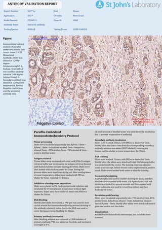
  • 1. Figure: Immunohistochemical analysis of paraffin embedded Human liver cancer tissue. 1: CD5 Monoclonal Antibody(10G8) was diluted at 1:200 (4 degree Celsius,overnight). 2: Sodium citrate pH 6.0 was used for antibody retrieval (>98 degree Celsius,20min). 3: Secondary antibody was diluted at 1:200 (room temperature, 30min). Negative control was used by secondary antibody only. Report Number 96973-a Host Mouse Application IHC-P Clonality Monoclonal Model Number STJ96973 Clone ID 10G8 Antibody Name Anti-CD5 antibody Testing Species HUMAN Testing Tissue LIVER CANCER ANTIBODY VALIDATION REPORT a. (A small amount of distilled water was added into the incubation box to prevent evaporation of antibody). 51. Secondary antibody incubation a. Slides were washed 3 times, with PBS on a shaker for 5min. Shortly after the slides were dried the corresponding secondary antibody solution was added (HRP labelled), covering the tissues, and incubated at room temperature for 30min. b. 52. DAB staining a. Slides were washed 3 times, with PBS on a shaker for 5min. b. Shortly after, the slides were dried and fresh DAB staining buffer was added inside the circles. The staining time was adjusted under a microscope. Yellow-brown colour represented a positive result. Slides were washed with water to stop the staining. c. 53. Haematoxylin staining a. Haematoxylin was used to counter-staining for 1min, and then the slides were washed with water. 1% Hydrochloric acid and alcohol was added for several seconds and then washed with water. Ammonia was used to reveal blue colour, and then flushed with water. b. 54. Desolation and Clearing i. Slides were incubated sequentially into: 75% alcohol 5min, 85% alcohol 5min, Anhydrous ethanol - 5min, Anhydrous ethanol - 5min & Xylene - 5min. Shortly after slides were dried and neutral gum was used to seal the slides. ii. 55. Visualization a. Results were validated with microscope, and the slides were scanned. Paraffin-Embedded Immunohistochemistry Protocol 45. 46. Tissue processing a. Slides were incubated sequentially into Xylene; 15min – Xylene, 15min - Anhydrous ethanol, 5min - Anhydrous ethanol, 5min - 85% alcohol, 5min - 75% alcohol & 5min – wash in distilled water. b. 47. Antigen retrieval a. Tissue slides were incubated with citric acid (PH6.0) antigen retrieval buffer and microwaved for antigen retrieval (heated until boiled and then stopped heating) for 8min. Slides were then heated with medium power for 7min. During this process slides were kept from drying out. After cooling down at room temperature, slides were washed with PBS on shaker for 5min, repeated for 3 times. b. 48. Inhibition of endogenous peroxidase a. Slides were placed in 3% Hydrogen peroxide solution, and incubated for 10 min at room temperature without light exposure. Slides were then washed 3 times with PBS on a shaker for 5mins. b. 49. BSA Blocking a. Shortly after slides were dried, a PAP pen was used to draw circles around the tissue sections (and to prevent draining of the antibody solution). Inside the circles, BSA was used to cover the tissue evenly, blocking for 30min. b. 50. Primary antibody incubation After blocking solution was removed a 1:200 solution of primary antibody/PBS was added on the slide, and incubated overnight at 4°C. St John's Laboratory Ltd. www.stjohnslabs.com
  • 2. Figure: Immunohistochemical analysis of paraffin embedded Human stomach tissue. 1: CD5 Monoclonal Antibody(10G8) was diluted at 1:200 (4 degree Celsius,overnight). 2: Sodium citrate pH 6.0 was used for antibody retrieval (>98 degree Celsius,20min). 3: Secondary antibody was diluted at 1:200 (room temperature, 30min). Negative control was used by secondary antibody only. Report Number 96973-b Host Mouse Application IHC-P Clonality Monoclonal Model Number STJ96973 Clone ID 10G8 Antibody Name Anti-CD5 antibody Testing Species HUMAN Testing Tissue STOMACH ANTIBODY VALIDATION REPORT a. (A small amount of distilled water was added into the incubation box to prevent evaporation of antibody). 40. Secondary antibody incubation a. Slides were washed 3 times, with PBS on a shaker for 5min. Shortly after the slides were dried the corresponding secondary antibody solution was added (HRP labelled), covering the tissues, and incubated at room temperature for 30min. b. 41. DAB staining a. Slides were washed 3 times, with PBS on a shaker for 5min. b. Shortly after, the slides were dried and fresh DAB staining buffer was added inside the circles. The staining time was adjusted under a microscope. Yellow-brown colour represented a positive result. Slides were washed with water to stop the staining. c. 42. Haematoxylin staining a. Haematoxylin was used to counter-staining for 1min, and then the slides were washed with water. 1% Hydrochloric acid and alcohol was added for several seconds and then washed with water. Ammonia was used to reveal blue colour, and then flushed with water. b. 43. Desolation and Clearing i. Slides were incubated sequentially into: 75% alcohol 5min, 85% alcohol 5min, Anhydrous ethanol - 5min, Anhydrous ethanol - 5min & Xylene - 5min. Shortly after slides were dried and neutral gum was used to seal the slides. ii. 44. Visualization a. Results were validated with microscope, and the slides were scanned. Paraffin-Embedded Immunohistochemistry Protocol 34. 35. Tissue processing a. Slides were incubated sequentially into Xylene; 15min – Xylene, 15min - Anhydrous ethanol, 5min - Anhydrous ethanol, 5min - 85% alcohol, 5min - 75% alcohol & 5min – wash in distilled water. b. 36. Antigen retrieval a. Tissue slides were incubated with citric acid (PH6.0) antigen retrieval buffer and microwaved for antigen retrieval (heated until boiled and then stopped heating) for 8min. Slides were then heated with medium power for 7min. During this process slides were kept from drying out. After cooling down at room temperature, slides were washed with PBS on shaker for 5min, repeated for 3 times. b. 37. Inhibition of endogenous peroxidase a. Slides were placed in 3% Hydrogen peroxide solution, and incubated for 10 min at room temperature without light exposure. Slides were then washed 3 times with PBS on a shaker for 5mins. b. 38. BSA Blocking a. Shortly after slides were dried, a PAP pen was used to draw circles around the tissue sections (and to prevent draining of the antibody solution). Inside the circles, BSA was used to cover the tissue evenly, blocking for 30min. b. 39. Primary antibody incubation After blocking solution was removed a 1:200 solution of primary antibody/PBS was added on the slide, and incubated overnight at 4°C. St John's Laboratory Ltd. www.stjohnslabs.com
  • 3. Figure: Immunohistochemical analysis of paraffin embedded Human stomach cancer tissue. 1: CD5 Monoclonal Antibody(10G8) was diluted at 1:200 (4 degree Celsius,overnight). 2: Sodium citrate pH 6.0 was used for antibody retrieval (>98 degree Celsius,20min). 3: Secondary antibody was diluted at 1:200 (room temperature, 30min). Negative control was used by secondary antibody only. Report Number 96973-c Host Mouse Application IHC-P Clonality Monoclonal Model Number STJ96973 Clone ID 10G8 Antibody Name Anti-CD5 antibody Testing Species HUMAN Testing Tissue STOMACH CANCER ANTIBODY VALIDATION REPORT a. (A small amount of distilled water was added into the incubation box to prevent evaporation of antibody). 29. Secondary antibody incubation a. Slides were washed 3 times, with PBS on a shaker for 5min. Shortly after the slides were dried the corresponding secondary antibody solution was added (HRP labelled), covering the tissues, and incubated at room temperature for 30min. b. 30. DAB staining a. Slides were washed 3 times, with PBS on a shaker for 5min. b. Shortly after, the slides were dried and fresh DAB staining buffer was added inside the circles. The staining time was adjusted under a microscope. Yellow-brown colour represented a positive result. Slides were washed with water to stop the staining. c. 31. Haematoxylin staining a. Haematoxylin was used to counter-staining for 1min, and then the slides were washed with water. 1% Hydrochloric acid and alcohol was added for several seconds and then washed with water. Ammonia was used to reveal blue colour, and then flushed with water. b. 32. Desolation and Clearing i. Slides were incubated sequentially into: 75% alcohol 5min, 85% alcohol 5min, Anhydrous ethanol - 5min, Anhydrous ethanol - 5min & Xylene - 5min. Shortly after slides were dried and neutral gum was used to seal the slides. ii. 33. Visualization a. Results were validated with microscope, and the slides were scanned. Paraffin-Embedded Immunohistochemistry Protocol 23. 24. Tissue processing a. Slides were incubated sequentially into Xylene; 15min – Xylene, 15min - Anhydrous ethanol, 5min - Anhydrous ethanol, 5min - 85% alcohol, 5min - 75% alcohol & 5min – wash in distilled water. b. 25. Antigen retrieval a. Tissue slides were incubated with citric acid (PH6.0) antigen retrieval buffer and microwaved for antigen retrieval (heated until boiled and then stopped heating) for 8min. Slides were then heated with medium power for 7min. During this process slides were kept from drying out. After cooling down at room temperature, slides were washed with PBS on shaker for 5min, repeated for 3 times. b. 26. Inhibition of endogenous peroxidase a. Slides were placed in 3% Hydrogen peroxide solution, and incubated for 10 min at room temperature without light exposure. Slides were then washed 3 times with PBS on a shaker for 5mins. b. 27. BSA Blocking a. Shortly after slides were dried, a PAP pen was used to draw circles around the tissue sections (and to prevent draining of the antibody solution). Inside the circles, BSA was used to cover the tissue evenly, blocking for 30min. b. 28. Primary antibody incubation After blocking solution was removed a 1:200 solution of primary antibody/PBS was added on the slide, and incubated overnight at 4°C. St John's Laboratory Ltd. www.stjohnslabs.com
  • 4. Figure: Immunohistochemical analysis of paraffin embedded Rat kidney tissue. 1: CD5 Monoclonal Antibody(10G8) was diluted at 1:200 (4 degree Celsius,overnight). 2: Sodium citrate pH 6.0 was used for antibody retrieval (>98 degree Celsius,20min). 3: Secondary antibody was diluted at 1:200 (room temperature, 30min). Negative control was used by secondary antibody only. Report Number 96973-d Host Mouse Application IHC-P Clonality Monoclonal Model Number STJ96973 Clone ID 10G8 Antibody Name Anti-CD5 antibody Testing Species RAT Testing Tissue KIDNEY ANTIBODY VALIDATION REPORT a. (A small amount of distilled water was added into the incubation box to prevent evaporation of antibody). 18. Secondary antibody incubation a. Slides were washed 3 times, with PBS on a shaker for 5min. Shortly after the slides were dried the corresponding secondary antibody solution was added (HRP labelled), covering the tissues, and incubated at room temperature for 30min. b. 19. DAB staining a. Slides were washed 3 times, with PBS on a shaker for 5min. b. Shortly after, the slides were dried and fresh DAB staining buffer was added inside the circles. The staining time was adjusted under a microscope. Yellow-brown colour represented a positive result. Slides were washed with water to stop the staining. c. 20. Haematoxylin staining a. Haematoxylin was used to counter-staining for 1min, and then the slides were washed with water. 1% Hydrochloric acid and alcohol was added for several seconds and then washed with water. Ammonia was used to reveal blue colour, and then flushed with water. b. 21. Desolation and Clearing i. Slides were incubated sequentially into: 75% alcohol 5min, 85% alcohol 5min, Anhydrous ethanol - 5min, Anhydrous ethanol - 5min & Xylene - 5min. Shortly after slides were dried and neutral gum was used to seal the slides. ii. 22. Visualization a. Results were validated with microscope, and the slides were scanned. Paraffin-Embedded Immunohistochemistry Protocol 12. 13. Tissue processing a. Slides were incubated sequentially into Xylene; 15min – Xylene, 15min - Anhydrous ethanol, 5min - Anhydrous ethanol, 5min - 85% alcohol, 5min - 75% alcohol & 5min – wash in distilled water. b. 14. Antigen retrieval a. Tissue slides were incubated with citric acid (PH6.0) antigen retrieval buffer and microwaved for antigen retrieval (heated until boiled and then stopped heating) for 8min. Slides were then heated with medium power for 7min. During this process slides were kept from drying out. After cooling down at room temperature, slides were washed with PBS on shaker for 5min, repeated for 3 times. b. 15. Inhibition of endogenous peroxidase a. Slides were placed in 3% Hydrogen peroxide solution, and incubated for 10 min at room temperature without light exposure. Slides were then washed 3 times with PBS on a shaker for 5mins. b. 16. BSA Blocking a. Shortly after slides were dried, a PAP pen was used to draw circles around the tissue sections (and to prevent draining of the antibody solution). Inside the circles, BSA was used to cover the tissue evenly, blocking for 30min. b. 17. Primary antibody incubation After blocking solution was removed a 1:200 solution of primary antibody/PBS was added on the slide, and incubated overnight at 4°C. St John's Laboratory Ltd. www.stjohnslabs.com
  • 5. Figure: Immunohistochemical analysis of paraffin embedded Rat spinal cord tissue. 1: CD5 Monoclonal Antibody(10G8) was diluted at 1:200 (4 degree Celsius,overnight). 2: Sodium citrate pH 6.0 was used for antibody retrieval (>98 degree Celsius,20min). 3: Secondary antibody was diluted at 1:200 (room temperature, 30min). Negative control was used by secondary antibody only. Report Number 96973-e Host Mouse Application IHC-P Clonality Monoclonal Model Number STJ96973 Clone ID 10G8 Antibody Name Anti-CD5 antibody Testing Species RAT Testing Tissue SPINAL ANTIBODY VALIDATION REPORT a. (A small amount of distilled water was added into the incubation box to prevent evaporation of antibody). 7. Secondary antibody incubation a. Slides were washed 3 times, with PBS on a shaker for 5min. Shortly after the slides were dried the corresponding secondary antibody solution was added (HRP labelled), covering the tissues, and incubated at room temperature for 30min. b. 8. DAB staining a. Slides were washed 3 times, with PBS on a shaker for 5min. b. Shortly after, the slides were dried and fresh DAB staining buffer was added inside the circles. The staining time was adjusted under a microscope. Yellow-brown colour represented a positive result. Slides were washed with water to stop the staining. c. 9. Haematoxylin staining a. Haematoxylin was used to counter-staining for 1min, and then the slides were washed with water. 1% Hydrochloric acid and alcohol was added for several seconds and then washed with water. Ammonia was used to reveal blue colour, and then flushed with water. b. 10. Desolation and Clearing i. Slides were incubated sequentially into: 75% alcohol 5min, 85% alcohol 5min, Anhydrous ethanol - 5min, Anhydrous ethanol - 5min & Xylene - 5min. Shortly after slides were dried and neutral gum was used to seal the slides. ii. 11. Visualization a. Results were validated with microscope, and the slides were scanned. Paraffin-Embedded Immunohistochemistry Protocol 1. 2. Tissue processing a. Slides were incubated sequentially into Xylene; 15min – Xylene, 15min - Anhydrous ethanol, 5min - Anhydrous ethanol, 5min - 85% alcohol, 5min - 75% alcohol & 5min – wash in distilled water. b. 3. Antigen retrieval a. Tissue slides were incubated with citric acid (PH6.0) antigen retrieval buffer and microwaved for antigen retrieval (heated until boiled and then stopped heating) for 8min. Slides were then heated with medium power for 7min. During this process slides were kept from drying out. After cooling down at room temperature, slides were washed with PBS on shaker for 5min, repeated for 3 times. b. 4. Inhibition of endogenous peroxidase a. Slides were placed in 3% Hydrogen peroxide solution, and incubated for 10 min at room temperature without light exposure. Slides were then washed 3 times with PBS on a shaker for 5mins. b. 5. BSA Blocking a. Shortly after slides were dried, a PAP pen was used to draw circles around the tissue sections (and to prevent draining of the antibody solution). Inside the circles, BSA was used to cover the tissue evenly, blocking for 30min. b. 6. Primary antibody incubation After blocking solution was removed a 1:200 solution of primary antibody/PBS was added on the slide, and incubated overnight at 4°C. St John's Laboratory Ltd. www.stjohnslabs.com
  • 6. Figure: Immunohistochemical analysis of paraffin embedded Rat brain tissue. 1: CD5 Monoclonal Antibody(10G8) was diluted at 1:200 (4 degree Celsius,overnight). 2: Sodium citrate pH 6.0 was used for antibody retrieval (>98 degree Celsius,20min). 3: Secondary antibody was diluted at 1:200 (room temperature, 30min). Negative control was used by secondary antibody only. Report Number 96973-f Host Mouse Application IHC-P Clonality Monoclonal Model Number STJ96973 Clone ID 10G8 Antibody Name Anti-CD5 antibody Testing Species RAT Testing Tissue BRAIN ANTIBODY VALIDATION REPORT c. (A small amount of distilled water was added into the incubation box to prevent evaporation of antibody). 56. Secondary antibody incubation a. Slides were washed 3 times, with PBS on a shaker for 5min. Shortly after the slides were dried the corresponding secondary antibody solution was added (HRP labelled), covering the tissues, and incubated at room temperature for 30min. b. 57. DAB staining a. Slides were washed 3 times, with PBS on a shaker for 5min. b. Shortly after, the slides were dried and fresh DAB staining buffer was added inside the circles. The staining time was adjusted under a microscope. Yellow-brown colour represented a positive result. Slides were washed with water to stop the staining. c. 58. Haematoxylin staining a. Haematoxylin was used to counter-staining for 1min, and then the slides were washed with water. 1% Hydrochloric acid and alcohol was added for several seconds and then washed with water. Ammonia was used to reveal blue colour, and then flushed with water. b. 59. Desolation and Clearing i. Slides were incubated sequentially into: 75% alcohol 5min, 85% alcohol 5min, Anhydrous ethanol - 5min, Anhydrous ethanol - 5min & Xylene - 5min. Shortly after slides were dried and neutral gum was used to seal the slides. ii. 60. Visualization a. Results were validated with microscope, and the slides were scanned. Paraffin-Embedded Immunohistochemistry Protocol 61. 62. Tissue processing a. Slides were incubated sequentially into Xylene; 15min – Xylene, 15min - Anhydrous ethanol, 5min - Anhydrous ethanol, 5min - 85% alcohol, 5min - 75% alcohol & 5min – wash in distilled water. b. 63. Antigen retrieval a. Tissue slides were incubated with citric acid (PH6.0) antigen retrieval buffer and microwaved for antigen retrieval (heated until boiled and then stopped heating) for 8min. Slides were then heated with medium power for 7min. During this process slides were kept from drying out. After cooling down at room temperature, slides were washed with PBS on shaker for 5min, repeated for 3 times. b. 64. Inhibition of endogenous peroxidase a. Slides were placed in 3% Hydrogen peroxide solution, and incubated for 10 min at room temperature without light exposure. Slides were then washed 3 times with PBS on a shaker for 5mins. b. 65. BSA Blocking a. Shortly after slides were dried, a PAP pen was used to draw circles around the tissue sections (and to prevent draining of the antibody solution). Inside the circles, BSA was used to cover the tissue evenly, blocking for 30min. b. 66. Primary antibody incubation After blocking solution was removed a 1:200 solution of primary antibody/PBS was added on the slide, and incubated overnight at 4°C. St John's Laboratory Ltd. www.stjohnslabs.com
  • 7. Figure: Immunohistochemical analysis of paraffin embedded Rat spleen tissue. 1: CD5 Monoclonal Antibody(10G8) was diluted at 1:200 (4 degree Celsius,overnight). 2: Sodium citrate pH 6.0 was used for antibody retrieval (>98 degree Celsius,20min). 3: Secondary antibody was diluted at 1:200 (room temperature, 30min). Negative control was used by secondary antibody only. Report Number 96973-g Host Mouse Application IHC-P Clonality Monoclonal Model Number STJ96973 Clone ID 10G8 Antibody Name Anti-CD5 antibody Testing Species RAT Testing Tissue SPLEEN ANTIBODY VALIDATION REPORT b. (A small amount of distilled water was added into the incubation box to prevent evaporation of antibody). 67. Secondary antibody incubation a. Slides were washed 3 times, with PBS on a shaker for 5min. Shortly after the slides were dried the corresponding secondary antibody solution was added (HRP labelled), covering the tissues, and incubated at room temperature for 30min. b. 68. DAB staining a. Slides were washed 3 times, with PBS on a shaker for 5min. b. Shortly after, the slides were dried and fresh DAB staining buffer was added inside the circles. The staining time was adjusted under a microscope. Yellow-brown colour represented a positive result. Slides were washed with water to stop the staining. c. 69. Haematoxylin staining a. Haematoxylin was used to counter-staining for 1min, and then the slides were washed with water. 1% Hydrochloric acid and alcohol was added for several seconds and then washed with water. Ammonia was used to reveal blue colour, and then flushed with water. b. 70. Desolation and Clearing i. Slides were incubated sequentially into: 75% alcohol 5min, 85% alcohol 5min, Anhydrous ethanol - 5min, Anhydrous ethanol - 5min & Xylene - 5min. Shortly after slides were dried and neutral gum was used to seal the slides. ii. 71. Visualization a. Results were validated with microscope, and the slides were scanned. Paraffin-Embedded Immunohistochemistry Protocol 72. 73. Tissue processing a. Slides were incubated sequentially into Xylene; 15min – Xylene, 15min - Anhydrous ethanol, 5min - Anhydrous ethanol, 5min - 85% alcohol, 5min - 75% alcohol & 5min – wash in distilled water. b. 74. Antigen retrieval a. Tissue slides were incubated with citric acid (PH6.0) antigen retrieval buffer and microwaved for antigen retrieval (heated until boiled and then stopped heating) for 8min. Slides were then heated with medium power for 7min. During this process slides were kept from drying out. After cooling down at room temperature, slides were washed with PBS on shaker for 5min, repeated for 3 times. b. 75. Inhibition of endogenous peroxidase a. Slides were placed in 3% Hydrogen peroxide solution, and incubated for 10 min at room temperature without light exposure. Slides were then washed 3 times with PBS on a shaker for 5mins. b. 76. BSA Blocking a. Shortly after slides were dried, a PAP pen was used to draw circles around the tissue sections (and to prevent draining of the antibody solution). Inside the circles, BSA was used to cover the tissue evenly, blocking for 30min. b. 77. Primary antibody incubation After blocking solution was removed a 1:200 solution of primary antibody/PBS was added on the slide, and incubated overnight at 4°C. St John's Laboratory Ltd. www.stjohnslabs.com
  • 8. Figure: Immunohistochemical analysis of paraffin embedded Mouse kidney tissue. 1: CD5 Monoclonal Antibody(10G8) was diluted at 1:200 (4 degree Celsius,overnight). 2: Sodium citrate pH 6.0 was used for antibody retrieval (>98 degree Celsius,20min). 3: Secondary antibody was diluted at 1:200 (room temperature, 30min). Negative control was used by secondary antibody only. Report Number 96973-h Host Mouse Application IHC-P Clonality Monoclonal Model Number STJ96973 Clone ID 10G8 Antibody Name Anti-CD5 antibody Testing Species MOUSE Testing Tissue KIDNEY ANTIBODY VALIDATION REPORT b. (A small amount of distilled water was added into the incubation box to prevent evaporation of antibody). 78. Secondary antibody incubation a. Slides were washed 3 times, with PBS on a shaker for 5min. Shortly after the slides were dried the corresponding secondary antibody solution was added (HRP labelled), covering the tissues, and incubated at room temperature for 30min. b. 79. DAB staining a. Slides were washed 3 times, with PBS on a shaker for 5min. b. Shortly after, the slides were dried and fresh DAB staining buffer was added inside the circles. The staining time was adjusted under a microscope. Yellow-brown colour represented a positive result. Slides were washed with water to stop the staining. c. 80. Haematoxylin staining a. Haematoxylin was used to counter-staining for 1min, and then the slides were washed with water. 1% Hydrochloric acid and alcohol was added for several seconds and then washed with water. Ammonia was used to reveal blue colour, and then flushed with water. b. 81. Desolation and Clearing i. Slides were incubated sequentially into: 75% alcohol 5min, 85% alcohol 5min, Anhydrous ethanol - 5min, Anhydrous ethanol - 5min & Xylene - 5min. Shortly after slides were dried and neutral gum was used to seal the slides. ii. 82. Visualization a. Results were validated with microscope, and the slides were scanned. Paraffin-Embedded Immunohistochemistry Protocol 83. 84. Tissue processing a. Slides were incubated sequentially into Xylene; 15min – Xylene, 15min - Anhydrous ethanol, 5min - Anhydrous ethanol, 5min - 85% alcohol, 5min - 75% alcohol & 5min – wash in distilled water. b. 85. Antigen retrieval a. Tissue slides were incubated with citric acid (PH6.0) antigen retrieval buffer and microwaved for antigen retrieval (heated until boiled and then stopped heating) for 8min. Slides were then heated with medium power for 7min. During this process slides were kept from drying out. After cooling down at room temperature, slides were washed with PBS on shaker for 5min, repeated for 3 times. b. 86. Inhibition of endogenous peroxidase a. Slides were placed in 3% Hydrogen peroxide solution, and incubated for 10 min at room temperature without light exposure. Slides were then washed 3 times with PBS on a shaker for 5mins. b. 87. BSA Blocking a. Shortly after slides were dried, a PAP pen was used to draw circles around the tissue sections (and to prevent draining of the antibody solution). Inside the circles, BSA was used to cover the tissue evenly, blocking for 30min. b. 88. Primary antibody incubation After blocking solution was removed a 1:200 solution of primary antibody/PBS was added on the slide, and incubated overnight at 4°C. St John's Laboratory Ltd. www.stjohnslabs.com
  • 9. Figure: Immunohistochemical analysis of paraffin embedded Mouse brain tissue. 1: CD5 Monoclonal Antibody(10G8) was diluted at 1:200 (4 degree Celsius,overnight). 2: Sodium citrate pH 6.0 was used for antibody retrieval (>98 degree Celsius,20min). 3: Secondary antibody was diluted at 1:200 (room temperature, 30min). Negative control was used by secondary antibody only. Report Number 96973-i Host Mouse Application IHC-P Clonality Monoclonal Model Number STJ96973 Clone ID 10G8 Antibody Name Anti-CD5 antibody Testing Species MOUSE Testing Tissue BRAIN ANTIBODY VALIDATION REPORT b. (A small amount of distilled water was added into the incubation box to prevent evaporation of antibody). 89. Secondary antibody incubation a. Slides were washed 3 times, with PBS on a shaker for 5min. Shortly after the slides were dried the corresponding secondary antibody solution was added (HRP labelled), covering the tissues, and incubated at room temperature for 30min. b. 90. DAB staining a. Slides were washed 3 times, with PBS on a shaker for 5min. b. Shortly after, the slides were dried and fresh DAB staining buffer was added inside the circles. The staining time was adjusted under a microscope. Yellow-brown colour represented a positive result. Slides were washed with water to stop the staining. c. 91. Haematoxylin staining a. Haematoxylin was used to counter-staining for 1min, and then the slides were washed with water. 1% Hydrochloric acid and alcohol was added for several seconds and then washed with water. Ammonia was used to reveal blue colour, and then flushed with water. b. 92. Desolation and Clearing i. Slides were incubated sequentially into: 75% alcohol 5min, 85% alcohol 5min, Anhydrous ethanol - 5min, Anhydrous ethanol - 5min & Xylene - 5min. Shortly after slides were dried and neutral gum was used to seal the slides. ii. 93. Visualization a. Results were validated with microscope, and the slides were scanned. Paraffin-Embedded Immunohistochemistry Protocol 94. 95. Tissue processing a. Slides were incubated sequentially into Xylene; 15min – Xylene, 15min - Anhydrous ethanol, 5min - Anhydrous ethanol, 5min - 85% alcohol, 5min - 75% alcohol & 5min – wash in distilled water. b. 96. Antigen retrieval a. Tissue slides were incubated with citric acid (PH6.0) antigen retrieval buffer and microwaved for antigen retrieval (heated until boiled and then stopped heating) for 8min. Slides were then heated with medium power for 7min. During this process slides were kept from drying out. After cooling down at room temperature, slides were washed with PBS on shaker for 5min, repeated for 3 times. b. 97. Inhibition of endogenous peroxidase a. Slides were placed in 3% Hydrogen peroxide solution, and incubated for 10 min at room temperature without light exposure. Slides were then washed 3 times with PBS on a shaker for 5mins. b. 98. BSA Blocking a. Shortly after slides were dried, a PAP pen was used to draw circles around the tissue sections (and to prevent draining of the antibody solution). Inside the circles, BSA was used to cover the tissue evenly, blocking for 30min. b. 99. Primary antibody incubation After blocking solution was removed a 1:200 solution of primary antibody/PBS was added on the slide, and incubated overnight at 4°C. St John's Laboratory Ltd. www.stjohnslabs.com
  • 10. Figure: Immunohistochemical analysis of paraffin embedded Mouse spleen tissue. 1: CD5 Monoclonal Antibody(10G8) was diluted at 1:200 (4 degree Celsius,overnight). 2: Sodium citrate pH 6.0 was used for antibody retrieval (>98 degree Celsius,20min). 3: Secondary antibody was diluted at 1:200 (room temperature, 30min). Negative control was used by secondary antibody only. Report Number 96973-j Host Mouse Application IHC-P Clonality Monoclonal Model Number STJ96973 Clone ID 10G8 Antibody Name Anti-CD5 antibody Testing Species MOUSE Testing Tissue SPLEEN ANTIBODY VALIDATION REPORT b. (A small amount of distilled water was added into the incubation box to prevent evaporation of antibody). 100. Secondary antibody incubation a. Slides were washed 3 times, with PBS on a shaker for 5min. Shortly after the slides were dried the corresponding secondary antibody solution was added (HRP labelled), covering the tissues, and incubated at room temperature for 30min. b. 101. DAB staining a. Slides were washed 3 times, with PBS on a shaker for 5min. b. Shortly after, the slides were dried and fresh DAB staining buffer was added inside the circles. The staining time was adjusted under a microscope. Yellow-brown colour represented a positive result. Slides were washed with water to stop the staining. c. 102. Haematoxylin staining a. Haematoxylin was used to counter-staining for 1min, and then the slides were washed with water. 1% Hydrochloric acid and alcohol was added for several seconds and then washed with water. Ammonia was used to reveal blue colour, and then flushed with water. b. 103. Desolation and Clearing i. Slides were incubated sequentially into: 75% alcohol 5min, 85% alcohol 5min, Anhydrous ethanol - 5min, Anhydrous ethanol - 5min & Xylene - 5min. Shortly after slides were dried and neutral gum was used to seal the slides. ii. 104. Visualization a. Results were validated with microscope, and the slides were scanned. Paraffin-Embedded Immunohistochemistry Protocol 105. 106. Tissue processing a. Slides were incubated sequentially into Xylene; 15min – Xylene, 15min - Anhydrous ethanol, 5min - Anhydrous ethanol, 5min - 85% alcohol, 5min - 75% alcohol & 5min – wash in distilled water. b. 107. Antigen retrieval a. Tissue slides were incubated with citric acid (PH6.0) antigen retrieval buffer and microwaved for antigen retrieval (heated until boiled and then stopped heating) for 8min. Slides were then heated with medium power for 7min. During this process slides were kept from drying out. After cooling down at room temperature, slides were washed with PBS on shaker for 5min, repeated for 3 times. b. 108. Inhibition of endogenous peroxidase a. Slides were placed in 3% Hydrogen peroxide solution, and incubated for 10 min at room temperature without light exposure. Slides were then washed 3 times with PBS on a shaker for 5mins. b. 109. BSA Blocking a. Shortly after slides were dried, a PAP pen was used to draw circles around the tissue sections (and to prevent draining of the antibody solution). Inside the circles, BSA was used to cover the tissue evenly, blocking for 30min. b. 110. Primary antibody incubation After blocking solution was removed a 1:200 solution of primary antibody/PBS was added on the slide, and incubated overnight at 4°C. St John's Laboratory Ltd. www.stjohnslabs.com