SlideShare a Scribd company logo
1 of 6
Download to read offline
ANTIBODY VALIDATION REPORT
Report Number 90750-a
Application Immunofluorescence
Model Number STJ90750
Antibody Name Anti-Phospho-Akt (T308) antibody
Host Rabbit
Clonality Polyclonal
Clone ID NA
Species HUMAN Tissue LUNG
Image
Description
Immunofluorescence analysis of Human
lung tissue. 1: Akt (phospho Thr308)
Polyclonal Antibody(red) was diluted at
1:200 (4 degree Celsius,overnight). 2:
Cy3 labled Secondary antibody was
diluted at 1:300 (room temperature,
50min).3: Picture B: DAPI(blue) 10min.
Picture A:Target. Picture B: DAPI.
Picture C: merge of A+B.
Primary Antibody Incubation
After blocking solution was removed a 1:200 primary antibody/PBS
solution was added on the slide, and incubated overnight at 4°C (a
small amount of distilled water was added into the incubation box to
prevent evaporation of antibody).
Secondary Antibody Incubation
slides were washed with PBS on a shaker for 5min, and repeated 3
times. Shortly after the slides were dried and corresponding
secondary antibody solution was added (HRP labelled), covering the
tissues, and incubated at room temperature for 50min.
DAPI Counter-Staining
slides were washed with PBS on a shaker for 5min, repeated 3 times
and then dried. DAPI staining solution was added inside the PAP
circles and incubated for 10 min at room temperature without light
exposure.
Mounting
Slides were washed with PBS on a shaker for 5min, and repeated 3
times. Shortly after slides were dried, anti-quench mountings were
used to mount slides.
Visualization
The slides were observed and placed under a NIKON inverted
fluorescence microscope (Ultra violet excitation 330-380nm,
emission 420nm; FITC green excitation 465-495nm, emission 515-
555 nm; CY3 red excitation 510-560nm, emission 590nm)
Immunofluorescence Protocol
Tissue Processing
Slides were incubated sequentially into: Xylene - 15min, Anhydrous
ethanol – 15 min, Anhydrous ethanol – 5 min, 85% alcohol – 5 min,
75% alcohol – 5 min & washed with distilled water – 5 min.
Antigen Retrieval
Tissue slides were incubated with citric acid (PH6.0) antigen
retrieval buffer, and microwaved for antigen retrieval (heated until
boiled and then stop heating) for 8min. Slides were then heated with
medium power for 7min. During this process slides are kept from
drying out. After cooling down at room temperature, slides were
washed with PBS on a shaker for 5min, and repeated 3 times.
Anti-Quench
shortly after slides were dried, a PAP pen was used to draw circles
around the tissues (to prevent draining of the antibody). Inside the
circles, anti-quench mountings were added and incubated for 5 min,
and then flushed with water for 10min.
BSA Blocking
Inside the circles, BSA was used to cover the tissue evenly, blocking
for 30min.
ANTIBODY VALIDATION REPORT
Report Number 90750-b
Application Immunofluorescence
Model Number STJ90750
Antibody Name Anti-Phospho-Akt (T308) antibody
Host Rabbit
Clonality Polyclonal
Clone ID NA
Species HUMAN Tissue LUNG
Image
Description
Immunofluorescence analysis of Human
lung tissue. 1: Akt (phospho Thr308)
Polyclonal Antibody(red) was diluted at
1:200 (4 degree Celsius,overnight). 2:
Cy3 labled Secondary antibody was
diluted at 1:300 (room temperature,
50min).3: Picture B: DAPI(blue) 10min.
Picture A:Target. Picture B: DAPI.
Picture C: merge of A+B.
Primary Antibody Incubation
After blocking solution was removed a 1:200 primary antibody/PBS
solution was added on the slide, and incubated overnight at 4°C (a
small amount of distilled water was added into the incubation box to
prevent evaporation of antibody).
Secondary Antibody Incubation
slides were washed with PBS on a shaker for 5min, and repeated 3
times. Shortly after the slides were dried and corresponding
secondary antibody solution was added (HRP labelled), covering the
tissues, and incubated at room temperature for 50min.
DAPI Counter-Staining
slides were washed with PBS on a shaker for 5min, repeated 3 times
and then dried. DAPI staining solution was added inside the PAP
circles and incubated for 10 min at room temperature without light
exposure.
Mounting
Slides were washed with PBS on a shaker for 5min, and repeated 3
times. Shortly after slides were dried, anti-quench mountings were
used to mount slides.
Visualization
The slides were observed and placed under a NIKON inverted
fluorescence microscope (Ultra violet excitation 330-380nm,
emission 420nm; FITC green excitation 465-495nm, emission 515-
555 nm; CY3 red excitation 510-560nm, emission 590nm)
Immunofluorescence Protocol
Tissue Processing
Slides were incubated sequentially into: Xylene - 15min, Anhydrous
ethanol – 15 min, Anhydrous ethanol – 5 min, 85% alcohol – 5 min,
75% alcohol – 5 min & washed with distilled water – 5 min.
Antigen Retrieval
Tissue slides were incubated with citric acid (PH6.0) antigen
retrieval buffer, and microwaved for antigen retrieval (heated until
boiled and then stop heating) for 8min. Slides were then heated with
medium power for 7min. During this process slides are kept from
drying out. After cooling down at room temperature, slides were
washed with PBS on a shaker for 5min, and repeated 3 times.
Anti-Quench
shortly after slides were dried, a PAP pen was used to draw circles
around the tissues (to prevent draining of the antibody). Inside the
circles, anti-quench mountings were added and incubated for 5 min,
and then flushed with water for 10min.
BSA Blocking
Inside the circles, BSA was used to cover the tissue evenly, blocking
for 30min.
ANTIBODY VALIDATION REPORT
Report Number 90750-c
Application Immunofluorescence
Model Number STJ90750
Antibody Name Anti-Phospho-Akt (T308) antibody
Host Rabbit
Clonality Polyclonal
Clone ID NA
Species HUMAN Tissue STOMACH
Image
Description
Immunofluorescence analysis of Human
stomach tissue. 1: Akt (phospho
Thr308) Polyclonal Antibody(red) was
diluted at 1:200 (4 degree
Celsius,overnight). 2: Cy3 labled
Secondary antibody was diluted at
1:300 (room temperature, 50min).3:
Picture B: DAPI(blue) 10min. Picture
A:Target. Picture B: DAPI. Picture C:
merge of A+B.
Primary Antibody Incubation
After blocking solution was removed a 1:200 primary antibody/PBS
solution was added on the slide, and incubated overnight at 4°C (a
small amount of distilled water was added into the incubation box to
prevent evaporation of antibody).
Secondary Antibody Incubation
slides were washed with PBS on a shaker for 5min, and repeated 3
times. Shortly after the slides were dried and corresponding
secondary antibody solution was added (HRP labelled), covering the
tissues, and incubated at room temperature for 50min.
DAPI Counter-Staining
slides were washed with PBS on a shaker for 5min, repeated 3 times
and then dried. DAPI staining solution was added inside the PAP
circles and incubated for 10 min at room temperature without light
exposure.
Mounting
Slides were washed with PBS on a shaker for 5min, and repeated 3
times. Shortly after slides were dried, anti-quench mountings were
used to mount slides.
Visualization
The slides were observed and placed under a NIKON inverted
fluorescence microscope (Ultra violet excitation 330-380nm,
emission 420nm; FITC green excitation 465-495nm, emission 515-
555 nm; CY3 red excitation 510-560nm, emission 590nm)
Immunofluorescence Protocol
Tissue Processing
Slides were incubated sequentially into: Xylene - 15min, Anhydrous
ethanol – 15 min, Anhydrous ethanol – 5 min, 85% alcohol – 5 min,
75% alcohol – 5 min & washed with distilled water – 5 min.
Antigen Retrieval
Tissue slides were incubated with citric acid (PH6.0) antigen
retrieval buffer, and microwaved for antigen retrieval (heated until
boiled and then stop heating) for 8min. Slides were then heated with
medium power for 7min. During this process slides are kept from
drying out. After cooling down at room temperature, slides were
washed with PBS on a shaker for 5min, and repeated 3 times.
Anti-Quench
shortly after slides were dried, a PAP pen was used to draw circles
around the tissues (to prevent draining of the antibody). Inside the
circles, anti-quench mountings were added and incubated for 5 min,
and then flushed with water for 10min.
BSA Blocking
Inside the circles, BSA was used to cover the tissue evenly, blocking
for 30min.
ANTIBODY VALIDATION REPORT
Report Number 90750-d
Application Immunofluorescence
Model Number STJ90750
Antibody Name Anti-Phospho-Akt (T308) antibody
Host Rabbit
Clonality Polyclonal
Clone ID NA
Species HUMAN Tissue STOMACH
Image
Description
Immunofluorescence analysis of Human
stomach tissue. 1: Akt (phospho
Thr308) Polyclonal Antibody(red) was
diluted at 1:200 (4 degree
Celsius,overnight). 2: Cy3 labled
Secondary antibody was diluted at
1:300 (room temperature, 50min).3:
Picture B: DAPI(blue) 10min. Picture
A:Target. Picture B: DAPI. Picture C:
merge of A+B.
Primary Antibody Incubation
After blocking solution was removed a 1:200 primary antibody/PBS
solution was added on the slide, and incubated overnight at 4°C (a
small amount of distilled water was added into the incubation box to
prevent evaporation of antibody).
Secondary Antibody Incubation
slides were washed with PBS on a shaker for 5min, and repeated 3
times. Shortly after the slides were dried and corresponding
secondary antibody solution was added (HRP labelled), covering the
tissues, and incubated at room temperature for 50min.
DAPI Counter-Staining
slides were washed with PBS on a shaker for 5min, repeated 3 times
and then dried. DAPI staining solution was added inside the PAP
circles and incubated for 10 min at room temperature without light
exposure.
Mounting
Slides were washed with PBS on a shaker for 5min, and repeated 3
times. Shortly after slides were dried, anti-quench mountings were
used to mount slides.
Visualization
The slides were observed and placed under a NIKON inverted
fluorescence microscope (Ultra violet excitation 330-380nm,
emission 420nm; FITC green excitation 465-495nm, emission 515-
555 nm; CY3 red excitation 510-560nm, emission 590nm)
Immunofluorescence Protocol
Tissue Processing
Slides were incubated sequentially into: Xylene - 15min, Anhydrous
ethanol – 15 min, Anhydrous ethanol – 5 min, 85% alcohol – 5 min,
75% alcohol – 5 min & washed with distilled water – 5 min.
Antigen Retrieval
Tissue slides were incubated with citric acid (PH6.0) antigen
retrieval buffer, and microwaved for antigen retrieval (heated until
boiled and then stop heating) for 8min. Slides were then heated with
medium power for 7min. During this process slides are kept from
drying out. After cooling down at room temperature, slides were
washed with PBS on a shaker for 5min, and repeated 3 times.
Anti-Quench
shortly after slides were dried, a PAP pen was used to draw circles
around the tissues (to prevent draining of the antibody). Inside the
circles, anti-quench mountings were added and incubated for 5 min,
and then flushed with water for 10min.
BSA Blocking
Inside the circles, BSA was used to cover the tissue evenly, blocking
for 30min.
ANTIBODY VALIDATION REPORT
Report Number 90750-e
Application Immunofluorescence
Model Number STJ90750
Antibody Name Anti-Phospho-Akt (T308) antibody
Host Rabbit
Clonality Polyclonal
Clone ID NA
Species RAT Tissue KIDNEY
Image
Description
Immunofluorescence analysis of Rat
kidney tissue. 1: Akt (phospho Thr308)
Polyclonal Antibody(red) was diluted at
1:200 (4 degree Celsius,overnight). 2:
Cy3 labled Secondary antibody was
diluted at 1:300 (room temperature,
50min).3: Picture B: DAPI(blue) 10min.
Picture A:Target. Picture B: DAPI.
Picture C: merge of A+B.
Primary Antibody Incubation
After blocking solution was removed a 1:200 primary antibody/PBS
solution was added on the slide, and incubated overnight at 4°C (a
small amount of distilled water was added into the incubation box to
prevent evaporation of antibody).
Secondary Antibody Incubation
slides were washed with PBS on a shaker for 5min, and repeated 3
times. Shortly after the slides were dried and corresponding
secondary antibody solution was added (HRP labelled), covering the
tissues, and incubated at room temperature for 50min.
DAPI Counter-Staining
slides were washed with PBS on a shaker for 5min, repeated 3 times
and then dried. DAPI staining solution was added inside the PAP
circles and incubated for 10 min at room temperature without light
exposure.
Mounting
Slides were washed with PBS on a shaker for 5min, and repeated 3
times. Shortly after slides were dried, anti-quench mountings were
used to mount slides.
Visualization
The slides were observed and placed under a NIKON inverted
fluorescence microscope (Ultra violet excitation 330-380nm,
emission 420nm; FITC green excitation 465-495nm, emission 515-
555 nm; CY3 red excitation 510-560nm, emission 590nm)
Immunofluorescence Protocol
Tissue Processing
Slides were incubated sequentially into: Xylene - 15min, Anhydrous
ethanol – 15 min, Anhydrous ethanol – 5 min, 85% alcohol – 5 min,
75% alcohol – 5 min & washed with distilled water – 5 min.
Antigen Retrieval
Tissue slides were incubated with citric acid (PH6.0) antigen
retrieval buffer, and microwaved for antigen retrieval (heated until
boiled and then stop heating) for 8min. Slides were then heated with
medium power for 7min. During this process slides are kept from
drying out. After cooling down at room temperature, slides were
washed with PBS on a shaker for 5min, and repeated 3 times.
Anti-Quench
shortly after slides were dried, a PAP pen was used to draw circles
around the tissues (to prevent draining of the antibody). Inside the
circles, anti-quench mountings were added and incubated for 5 min,
and then flushed with water for 10min.
BSA Blocking
Inside the circles, BSA was used to cover the tissue evenly, blocking
for 30min.
ANTIBODY VALIDATION REPORT
Report Number 90750-f
Application Immunofluorescence
Model Number STJ90750
Antibody Name Anti-Phospho-Akt (T308) antibody
Host Rabbit
Clonality Polyclonal
Clone ID NA
Species RAT Tissue KIDNEY
Image
Description
Immunofluorescence analysis of Rat
kidney tissue. 1: Akt (phospho Thr308)
Polyclonal Antibody(red) was diluted at
1:200 (4 degree Celsius,overnight). 2:
Cy3 labled Secondary antibody was
diluted at 1:300 (room temperature,
50min).3: Picture B: DAPI(blue) 10min.
Picture A:Target. Picture B: DAPI.
Picture C: merge of A+B.
Primary Antibody Incubation
After blocking solution was removed a 1:200 primary antibody/PBS
solution was added on the slide, and incubated overnight at 4°C (a
small amount of distilled water was added into the incubation box to
prevent evaporation of antibody).
Secondary Antibody Incubation
slides were washed with PBS on a shaker for 5min, and repeated 3
times. Shortly after the slides were dried and corresponding
secondary antibody solution was added (HRP labelled), covering the
tissues, and incubated at room temperature for 50min.
DAPI Counter-Staining
slides were washed with PBS on a shaker for 5min, repeated 3 times
and then dried. DAPI staining solution was added inside the PAP
circles and incubated for 10 min at room temperature without light
exposure.
Mounting
Slides were washed with PBS on a shaker for 5min, and repeated 3
times. Shortly after slides were dried, anti-quench mountings were
used to mount slides.
Visualization
The slides were observed and placed under a NIKON inverted
fluorescence microscope (Ultra violet excitation 330-380nm,
emission 420nm; FITC green excitation 465-495nm, emission 515-
555 nm; CY3 red excitation 510-560nm, emission 590nm)
Immunofluorescence Protocol
Tissue Processing
Slides were incubated sequentially into: Xylene - 15min, Anhydrous
ethanol – 15 min, Anhydrous ethanol – 5 min, 85% alcohol – 5 min,
75% alcohol – 5 min & washed with distilled water – 5 min.
Antigen Retrieval
Tissue slides were incubated with citric acid (PH6.0) antigen
retrieval buffer, and microwaved for antigen retrieval (heated until
boiled and then stop heating) for 8min. Slides were then heated with
medium power for 7min. During this process slides are kept from
drying out. After cooling down at room temperature, slides were
washed with PBS on a shaker for 5min, and repeated 3 times.
Anti-Quench
shortly after slides were dried, a PAP pen was used to draw circles
around the tissues (to prevent draining of the antibody). Inside the
circles, anti-quench mountings were added and incubated for 5 min,
and then flushed with water for 10min.
BSA Blocking
Inside the circles, BSA was used to cover the tissue evenly, blocking
for 30min.

More Related Content

What's hot

What's hot (20)

Immunofluorescence Antibody Validation Report for Anti-Collagen IV Antibody (...
Immunofluorescence Antibody Validation Report for Anti-Collagen IV Antibody (...Immunofluorescence Antibody Validation Report for Anti-Collagen IV Antibody (...
Immunofluorescence Antibody Validation Report for Anti-Collagen IV Antibody (...
 
Immunofluorescence Antibody Validation Report for Anti-Survivin Antibody (STJ...
Immunofluorescence Antibody Validation Report for Anti-Survivin Antibody (STJ...Immunofluorescence Antibody Validation Report for Anti-Survivin Antibody (STJ...
Immunofluorescence Antibody Validation Report for Anti-Survivin Antibody (STJ...
 
Immunofluorescence Antibody Validation Report for Anti-HSP90β Antibody (STJ97...
Immunofluorescence Antibody Validation Report for Anti-HSP90β Antibody (STJ97...Immunofluorescence Antibody Validation Report for Anti-HSP90β Antibody (STJ97...
Immunofluorescence Antibody Validation Report for Anti-HSP90β Antibody (STJ97...
 
Immunofluorescence Antibody Validation Report for Anti-PPAR Delta Antibody (S...
Immunofluorescence Antibody Validation Report for Anti-PPAR Delta Antibody (S...Immunofluorescence Antibody Validation Report for Anti-PPAR Delta Antibody (S...
Immunofluorescence Antibody Validation Report for Anti-PPAR Delta Antibody (S...
 
Immunofluorescence Antibody Validation Report for Anti-HP-1α Antibody (STJ98895)
Immunofluorescence Antibody Validation Report for Anti-HP-1α Antibody (STJ98895)Immunofluorescence Antibody Validation Report for Anti-HP-1α Antibody (STJ98895)
Immunofluorescence Antibody Validation Report for Anti-HP-1α Antibody (STJ98895)
 
Immunofluorescence Antibody Validation Report for Anti-Peroxiredoxin 1 Antibo...
Immunofluorescence Antibody Validation Report for Anti-Peroxiredoxin 1 Antibo...Immunofluorescence Antibody Validation Report for Anti-Peroxiredoxin 1 Antibo...
Immunofluorescence Antibody Validation Report for Anti-Peroxiredoxin 1 Antibo...
 
Immunofluorescence Antibody Validation Report for Anti-PTEN Antibody (STJ98583)
Immunofluorescence Antibody Validation Report for Anti-PTEN Antibody (STJ98583)Immunofluorescence Antibody Validation Report for Anti-PTEN Antibody (STJ98583)
Immunofluorescence Antibody Validation Report for Anti-PTEN Antibody (STJ98583)
 
Immunofluorescence Antibody Validation Report for Anti-eIF4A1 Antibody (STJ97...
Immunofluorescence Antibody Validation Report for Anti-eIF4A1 Antibody (STJ97...Immunofluorescence Antibody Validation Report for Anti-eIF4A1 Antibody (STJ97...
Immunofluorescence Antibody Validation Report for Anti-eIF4A1 Antibody (STJ97...
 
Immunofluorescence Antibody Validation Report for Anti-Ub Antibody (STJ97236)
Immunofluorescence Antibody Validation Report for Anti-Ub Antibody (STJ97236)Immunofluorescence Antibody Validation Report for Anti-Ub Antibody (STJ97236)
Immunofluorescence Antibody Validation Report for Anti-Ub Antibody (STJ97236)
 
Immunofluorescence Antibody Validation Report for Anti-p38 Antibody (STJ94878)
Immunofluorescence Antibody Validation Report for Anti-p38 Antibody (STJ94878)Immunofluorescence Antibody Validation Report for Anti-p38 Antibody (STJ94878)
Immunofluorescence Antibody Validation Report for Anti-p38 Antibody (STJ94878)
 
Immunofluorescence Antibody Validation Report for Anti-EPAS-1 Antibody (STJ96...
Immunofluorescence Antibody Validation Report for Anti-EPAS-1 Antibody (STJ96...Immunofluorescence Antibody Validation Report for Anti-EPAS-1 Antibody (STJ96...
Immunofluorescence Antibody Validation Report for Anti-EPAS-1 Antibody (STJ96...
 
Immunofluorescence Antibody Validation Report for Anti-Phospho-Smad3 (S425) A...
Immunofluorescence Antibody Validation Report for Anti-Phospho-Smad3 (S425) A...Immunofluorescence Antibody Validation Report for Anti-Phospho-Smad3 (S425) A...
Immunofluorescence Antibody Validation Report for Anti-Phospho-Smad3 (S425) A...
 
Immunofluorescence Antibody Validation Report for Anti-Epsilon Tubulin Antibo...
Immunofluorescence Antibody Validation Report for Anti-Epsilon Tubulin Antibo...Immunofluorescence Antibody Validation Report for Anti-Epsilon Tubulin Antibo...
Immunofluorescence Antibody Validation Report for Anti-Epsilon Tubulin Antibo...
 
Immunofluorescence Antibody Validation Report for Anti-Phospho-NFκB-p65 (S529...
Immunofluorescence Antibody Validation Report for Anti-Phospho-NFκB-p65 (S529...Immunofluorescence Antibody Validation Report for Anti-Phospho-NFκB-p65 (S529...
Immunofluorescence Antibody Validation Report for Anti-Phospho-NFκB-p65 (S529...
 
Immunofluorescence Antibody Validation Report for Anti-Histone H4 (Acetyl Lys...
Immunofluorescence Antibody Validation Report for Anti-Histone H4 (Acetyl Lys...Immunofluorescence Antibody Validation Report for Anti-Histone H4 (Acetyl Lys...
Immunofluorescence Antibody Validation Report for Anti-Histone H4 (Acetyl Lys...
 
Immunofluorescence Antibody Validation Report for Anti-β-tubulin (HRP)Antibo...
Immunofluorescence Antibody Validation Report for Anti-β-tubulin  (HRP)Antibo...Immunofluorescence Antibody Validation Report for Anti-β-tubulin  (HRP)Antibo...
Immunofluorescence Antibody Validation Report for Anti-β-tubulin (HRP)Antibo...
 
Immunofluorescence Antibody Validation Report for Anti-EFHD1 Antibody (STJ96987)
Immunofluorescence Antibody Validation Report for Anti-EFHD1 Antibody (STJ96987)Immunofluorescence Antibody Validation Report for Anti-EFHD1 Antibody (STJ96987)
Immunofluorescence Antibody Validation Report for Anti-EFHD1 Antibody (STJ96987)
 
Immunofluorescence Antibody Validation Report for Anti-Luciferase Antibody (S...
Immunofluorescence Antibody Validation Report for Anti-Luciferase Antibody (S...Immunofluorescence Antibody Validation Report for Anti-Luciferase Antibody (S...
Immunofluorescence Antibody Validation Report for Anti-Luciferase Antibody (S...
 
Immunofluorescence Antibody Validation Report for Anti-CD1 Antibody (STJ96970)
Immunofluorescence Antibody Validation Report for Anti-CD1 Antibody (STJ96970)Immunofluorescence Antibody Validation Report for Anti-CD1 Antibody (STJ96970)
Immunofluorescence Antibody Validation Report for Anti-CD1 Antibody (STJ96970)
 
Immunofluorescence Antibody Validation Report for Anti-CYCS Antibody (STJ98953)
Immunofluorescence Antibody Validation Report for Anti-CYCS Antibody (STJ98953)Immunofluorescence Antibody Validation Report for Anti-CYCS Antibody (STJ98953)
Immunofluorescence Antibody Validation Report for Anti-CYCS Antibody (STJ98953)
 

Viewers also liked

Viewers also liked (11)

Immunohistochemistry Antibody Validation Report for Anti-Phospho-Akt (T308) A...
Immunohistochemistry Antibody Validation Report for Anti-Phospho-Akt (T308) A...Immunohistochemistry Antibody Validation Report for Anti-Phospho-Akt (T308) A...
Immunohistochemistry Antibody Validation Report for Anti-Phospho-Akt (T308) A...
 
Yang{JMCC_2013]
Yang{JMCC_2013]Yang{JMCC_2013]
Yang{JMCC_2013]
 
npy ppt
npy pptnpy ppt
npy ppt
 
Immunofluorescence Antibody Validation Report for Anti-Phospho-p38 (T180/Y182...
Immunofluorescence Antibody Validation Report for Anti-Phospho-p38 (T180/Y182...Immunofluorescence Antibody Validation Report for Anti-Phospho-p38 (T180/Y182...
Immunofluorescence Antibody Validation Report for Anti-Phospho-p38 (T180/Y182...
 
Immunofluorescence Antibody Validation Report for Anti-AP-1 Antibody (STJ91616)
Immunofluorescence Antibody Validation Report for Anti-AP-1 Antibody (STJ91616)Immunofluorescence Antibody Validation Report for Anti-AP-1 Antibody (STJ91616)
Immunofluorescence Antibody Validation Report for Anti-AP-1 Antibody (STJ91616)
 
Immunofluorescence Antibody Validation Report for Anti-Phospho-p38 (T180) Ant...
Immunofluorescence Antibody Validation Report for Anti-Phospho-p38 (T180) Ant...Immunofluorescence Antibody Validation Report for Anti-Phospho-p38 (T180) Ant...
Immunofluorescence Antibody Validation Report for Anti-Phospho-p38 (T180) Ant...
 
Immunofluorescence Antibody Validation Report for Anti-Bad Antibody (STJ91800)
Immunofluorescence Antibody Validation Report for Anti-Bad Antibody (STJ91800)Immunofluorescence Antibody Validation Report for Anti-Bad Antibody (STJ91800)
Immunofluorescence Antibody Validation Report for Anti-Bad Antibody (STJ91800)
 
Antibody Customer Review For ASC Polyclonal Antibody (STJ91730)
Antibody Customer Review For ASC Polyclonal Antibody (STJ91730)Antibody Customer Review For ASC Polyclonal Antibody (STJ91730)
Antibody Customer Review For ASC Polyclonal Antibody (STJ91730)
 
Akt (protein kinase)
Akt (protein kinase)Akt (protein kinase)
Akt (protein kinase)
 
Immunofluorescence Antibody Validation Report for Anti-Phospho-PI 3-kinase p8...
Immunofluorescence Antibody Validation Report for Anti-Phospho-PI 3-kinase p8...Immunofluorescence Antibody Validation Report for Anti-Phospho-PI 3-kinase p8...
Immunofluorescence Antibody Validation Report for Anti-Phospho-PI 3-kinase p8...
 
Immunofluorescence Antibody Validation Report for Anti-Catalase Antibody (STJ...
Immunofluorescence Antibody Validation Report for Anti-Catalase Antibody (STJ...Immunofluorescence Antibody Validation Report for Anti-Catalase Antibody (STJ...
Immunofluorescence Antibody Validation Report for Anti-Catalase Antibody (STJ...
 

Similar to Immunofluorescence Antibody Validation Report for Anti-Phospho-Akt (T308) Antibody (STJ90750)

Similar to Immunofluorescence Antibody Validation Report for Anti-Phospho-Akt (T308) Antibody (STJ90750) (20)

Immunofluorescence Antibody Validation Report for Anti-SIRT1 Antibody (STJ95667)
Immunofluorescence Antibody Validation Report for Anti-SIRT1 Antibody (STJ95667)Immunofluorescence Antibody Validation Report for Anti-SIRT1 Antibody (STJ95667)
Immunofluorescence Antibody Validation Report for Anti-SIRT1 Antibody (STJ95667)
 
Immunofluorescence Antibody Validation Report for Anti-TIMP-1 Antibody (STJ96...
Immunofluorescence Antibody Validation Report for Anti-TIMP-1 Antibody (STJ96...Immunofluorescence Antibody Validation Report for Anti-TIMP-1 Antibody (STJ96...
Immunofluorescence Antibody Validation Report for Anti-TIMP-1 Antibody (STJ96...
 
Immunofluorescence Antibody Validation Report for Anti-Phospho-NFκB-p65 (S536...
Immunofluorescence Antibody Validation Report for Anti-Phospho-NFκB-p65 (S536...Immunofluorescence Antibody Validation Report for Anti-Phospho-NFκB-p65 (S536...
Immunofluorescence Antibody Validation Report for Anti-Phospho-NFκB-p65 (S536...
 
Immunofluorescence Antibody Validation Report for Anti-PERK Antibody (STJ95031)
Immunofluorescence Antibody Validation Report for Anti-PERK Antibody (STJ95031)Immunofluorescence Antibody Validation Report for Anti-PERK Antibody (STJ95031)
Immunofluorescence Antibody Validation Report for Anti-PERK Antibody (STJ95031)
 
Immunofluorescence Antibody Validation Report for Anti-Tak1 Antibody (STJ95901)
Immunofluorescence Antibody Validation Report for Anti-Tak1 Antibody (STJ95901)Immunofluorescence Antibody Validation Report for Anti-Tak1 Antibody (STJ95901)
Immunofluorescence Antibody Validation Report for Anti-Tak1 Antibody (STJ95901)
 
Immunofluorescence Antibody Validation Report for Anti-Flk-1 Antibody (STJ93088)
Immunofluorescence Antibody Validation Report for Anti-Flk-1 Antibody (STJ93088)Immunofluorescence Antibody Validation Report for Anti-Flk-1 Antibody (STJ93088)
Immunofluorescence Antibody Validation Report for Anti-Flk-1 Antibody (STJ93088)
 
Immunofluorescence Antibody Validation Report for Anti-ATG5 Antibody (STJ98903)
Immunofluorescence Antibody Validation Report for Anti-ATG5 Antibody (STJ98903)Immunofluorescence Antibody Validation Report for Anti-ATG5 Antibody (STJ98903)
Immunofluorescence Antibody Validation Report for Anti-ATG5 Antibody (STJ98903)
 
Immunofluorescence Antibody Validation Report for Anti-Phospho-p38 (Y323) Ant...
Immunofluorescence Antibody Validation Report for Anti-Phospho-p38 (Y323) Ant...Immunofluorescence Antibody Validation Report for Anti-Phospho-p38 (Y323) Ant...
Immunofluorescence Antibody Validation Report for Anti-Phospho-p38 (Y323) Ant...
 
Immunofluorescence Antibody Validation Report for Anti-Tubulin α Antibody (ST...
Immunofluorescence Antibody Validation Report for Anti-Tubulin α Antibody (ST...Immunofluorescence Antibody Validation Report for Anti-Tubulin α Antibody (ST...
Immunofluorescence Antibody Validation Report for Anti-Tubulin α Antibody (ST...
 
Immunofluorescence Antibody Validation Report for Anti-Histone H3 (Acetyl K9)...
Immunofluorescence Antibody Validation Report for Anti-Histone H3 (Acetyl K9)...Immunofluorescence Antibody Validation Report for Anti-Histone H3 (Acetyl K9)...
Immunofluorescence Antibody Validation Report for Anti-Histone H3 (Acetyl K9)...
 
Immunofluorescence Antibody Validation Report for Anti-Phospho-Stat3 (Y705) A...
Immunofluorescence Antibody Validation Report for Anti-Phospho-Stat3 (Y705) A...Immunofluorescence Antibody Validation Report for Anti-Phospho-Stat3 (Y705) A...
Immunofluorescence Antibody Validation Report for Anti-Phospho-Stat3 (Y705) A...
 
Immunofluorescence Antibody Validation Report for Anti-Amyloid-β Antibody (ST...
Immunofluorescence Antibody Validation Report for Anti-Amyloid-β Antibody (ST...Immunofluorescence Antibody Validation Report for Anti-Amyloid-β Antibody (ST...
Immunofluorescence Antibody Validation Report for Anti-Amyloid-β Antibody (ST...
 
Immunofluorescence Antibody Validation Report for Anti-PPAR-γ Antibody (STJ95...
Immunofluorescence Antibody Validation Report for Anti-PPAR-γ Antibody (STJ95...Immunofluorescence Antibody Validation Report for Anti-PPAR-γ Antibody (STJ95...
Immunofluorescence Antibody Validation Report for Anti-PPAR-γ Antibody (STJ95...
 
Immunofluorescence Antibody Validation Report for Anti-Cleaved-Notch 2 (D1733...
Immunofluorescence Antibody Validation Report for Anti-Cleaved-Notch 2 (D1733...Immunofluorescence Antibody Validation Report for Anti-Cleaved-Notch 2 (D1733...
Immunofluorescence Antibody Validation Report for Anti-Cleaved-Notch 2 (D1733...
 
Immunofluorescence Antibody Validation Report for Anti-Histone H3 Antibody (S...
Immunofluorescence Antibody Validation Report for Anti-Histone H3 Antibody (S...Immunofluorescence Antibody Validation Report for Anti-Histone H3 Antibody (S...
Immunofluorescence Antibody Validation Report for Anti-Histone H3 Antibody (S...
 
Immunofluorescence Antibody Validation Report for Anti-PPAR-α Antibody (STJ95...
Immunofluorescence Antibody Validation Report for Anti-PPAR-α Antibody (STJ95...Immunofluorescence Antibody Validation Report for Anti-PPAR-α Antibody (STJ95...
Immunofluorescence Antibody Validation Report for Anti-PPAR-α Antibody (STJ95...
 
Immunofluorescence Antibody Validation Report for Anti-FAS Antibody (STJ93041)
Immunofluorescence Antibody Validation Report for Anti-FAS Antibody (STJ93041)Immunofluorescence Antibody Validation Report for Anti-FAS Antibody (STJ93041)
Immunofluorescence Antibody Validation Report for Anti-FAS Antibody (STJ93041)
 
Immunofluorescence Antibody Validation Report for Anti-Phospho-Stat3 (S727) A...
Immunofluorescence Antibody Validation Report for Anti-Phospho-Stat3 (S727) A...Immunofluorescence Antibody Validation Report for Anti-Phospho-Stat3 (S727) A...
Immunofluorescence Antibody Validation Report for Anti-Phospho-Stat3 (S727) A...
 
Immunofluorescence Antibody Validation Report for Anti-VE-Cadherin Antibody (...
Immunofluorescence Antibody Validation Report for Anti-VE-Cadherin Antibody (...Immunofluorescence Antibody Validation Report for Anti-VE-Cadherin Antibody (...
Immunofluorescence Antibody Validation Report for Anti-VE-Cadherin Antibody (...
 
Immunofluorescence Antibody Validation Report for Anti-Phospho-ERK 1/2 (Y222/...
Immunofluorescence Antibody Validation Report for Anti-Phospho-ERK 1/2 (Y222/...Immunofluorescence Antibody Validation Report for Anti-Phospho-ERK 1/2 (Y222/...
Immunofluorescence Antibody Validation Report for Anti-Phospho-ERK 1/2 (Y222/...
 

More from St John's Laboratory Ltd

Western Blot Customer Review Anti Glucocorticoid Receptor antibody (STJ97101)
Western Blot Customer Review Anti Glucocorticoid Receptor antibody (STJ97101)Western Blot Customer Review Anti Glucocorticoid Receptor antibody (STJ97101)
Western Blot Customer Review Anti Glucocorticoid Receptor antibody (STJ97101)
St John's Laboratory Ltd
 
Immunohistochemistry Antibody Validation Report for Anti-Caspase-8 Antibody (...
Immunohistochemistry Antibody Validation Report for Anti-Caspase-8 Antibody (...Immunohistochemistry Antibody Validation Report for Anti-Caspase-8 Antibody (...
Immunohistochemistry Antibody Validation Report for Anti-Caspase-8 Antibody (...
St John's Laboratory Ltd
 

More from St John's Laboratory Ltd (20)

Immunohistochemistry Customer Review Anti-Smo antibody (STJ95710)
Immunohistochemistry Customer Review Anti-Smo antibody (STJ95710)Immunohistochemistry Customer Review Anti-Smo antibody (STJ95710)
Immunohistochemistry Customer Review Anti-Smo antibody (STJ95710)
 
Western Blot Customer Review Anti Glucocorticoid Receptor antibody (STJ97101)
Western Blot Customer Review Anti Glucocorticoid Receptor antibody (STJ97101)Western Blot Customer Review Anti Glucocorticoid Receptor antibody (STJ97101)
Western Blot Customer Review Anti Glucocorticoid Receptor antibody (STJ97101)
 
Western Blot Customer Review Anti-Phospho-Cofilin (S3) Antibody (STJ90230)
Western Blot Customer Review Anti-Phospho-Cofilin (S3) Antibody (STJ90230)Western Blot Customer Review Anti-Phospho-Cofilin (S3) Antibody (STJ90230)
Western Blot Customer Review Anti-Phospho-Cofilin (S3) Antibody (STJ90230)
 
Scientist of the Month - May Byron Baron
Scientist of the Month - May Byron BaronScientist of the Month - May Byron Baron
Scientist of the Month - May Byron Baron
 
Western Blot Customer Review Anti-WAVE2 Antibody (STJ96263)
Western Blot Customer Review Anti-WAVE2 Antibody (STJ96263)Western Blot Customer Review Anti-WAVE2 Antibody (STJ96263)
Western Blot Customer Review Anti-WAVE2 Antibody (STJ96263)
 
Western Blot Customer Review Anti-Endophilin I Antibody (STJ92924)
Western Blot Customer Review Anti-Endophilin I Antibody (STJ92924)Western Blot Customer Review Anti-Endophilin I Antibody (STJ92924)
Western Blot Customer Review Anti-Endophilin I Antibody (STJ92924)
 
Western Blot Customer Review Anti-β-tubulin Antibody (STJ97037)
Western Blot Customer Review Anti-β-tubulin Antibody (STJ97037)Western Blot Customer Review Anti-β-tubulin Antibody (STJ97037)
Western Blot Customer Review Anti-β-tubulin Antibody (STJ97037)
 
Immunohistochemistry Antibody Validation Report for Anti-Caspase-8 Antibody (...
Immunohistochemistry Antibody Validation Report for Anti-Caspase-8 Antibody (...Immunohistochemistry Antibody Validation Report for Anti-Caspase-8 Antibody (...
Immunohistochemistry Antibody Validation Report for Anti-Caspase-8 Antibody (...
 
Immunohistochemistry Antibody Validation Report for Anti-Gamma Tubulin Antibo...
Immunohistochemistry Antibody Validation Report for Anti-Gamma Tubulin Antibo...Immunohistochemistry Antibody Validation Report for Anti-Gamma Tubulin Antibo...
Immunohistochemistry Antibody Validation Report for Anti-Gamma Tubulin Antibo...
 
Immunohistochemistry Antibody Validation Report for Anti-Epsilon Tubulin Anti...
Immunohistochemistry Antibody Validation Report for Anti-Epsilon Tubulin Anti...Immunohistochemistry Antibody Validation Report for Anti-Epsilon Tubulin Anti...
Immunohistochemistry Antibody Validation Report for Anti-Epsilon Tubulin Anti...
 
Immunohistochemistry Antibody Validation Report for Anti-Luciferase Antibody ...
Immunohistochemistry Antibody Validation Report for Anti-Luciferase Antibody ...Immunohistochemistry Antibody Validation Report for Anti-Luciferase Antibody ...
Immunohistochemistry Antibody Validation Report for Anti-Luciferase Antibody ...
 
Immunohistochemistry Antibody Validation Report for Anti-LC3A Antibody (STJ97...
Immunohistochemistry Antibody Validation Report for Anti-LC3A Antibody (STJ97...Immunohistochemistry Antibody Validation Report for Anti-LC3A Antibody (STJ97...
Immunohistochemistry Antibody Validation Report for Anti-LC3A Antibody (STJ97...
 
Immunohistochemistry Antibody Validation Report for Anti-CHOP Antibody (STJ97...
Immunohistochemistry Antibody Validation Report for Anti-CHOP Antibody (STJ97...Immunohistochemistry Antibody Validation Report for Anti-CHOP Antibody (STJ97...
Immunohistochemistry Antibody Validation Report for Anti-CHOP Antibody (STJ97...
 
Immunohistochemistry Antibody Validation Report for Anti-phospho-MLKL (S358) ...
Immunohistochemistry Antibody Validation Report for Anti-phospho-MLKL (S358) ...Immunohistochemistry Antibody Validation Report for Anti-phospho-MLKL (S358) ...
Immunohistochemistry Antibody Validation Report for Anti-phospho-MLKL (S358) ...
 
Immunohistochemistry Antibody Validation Report for Anti-ERK1 Antibody (STJ97...
Immunohistochemistry Antibody Validation Report for Anti-ERK1 Antibody (STJ97...Immunohistochemistry Antibody Validation Report for Anti-ERK1 Antibody (STJ97...
Immunohistochemistry Antibody Validation Report for Anti-ERK1 Antibody (STJ97...
 
Immunohistochemistry Antibody Validation Report for Anti-PDGFRα Antibody (STJ...
Immunohistochemistry Antibody Validation Report for Anti-PDGFRα Antibody (STJ...Immunohistochemistry Antibody Validation Report for Anti-PDGFRα Antibody (STJ...
Immunohistochemistry Antibody Validation Report for Anti-PDGFRα Antibody (STJ...
 
Immunohistochemistry Antibody Validation Report for Anti-Caspase-1 Antibody (...
Immunohistochemistry Antibody Validation Report for Anti-Caspase-1 Antibody (...Immunohistochemistry Antibody Validation Report for Anti-Caspase-1 Antibody (...
Immunohistochemistry Antibody Validation Report for Anti-Caspase-1 Antibody (...
 
Immunohistochemistry Antibody Validation Report for Anti-Amyloid-β Antibody (...
Immunohistochemistry Antibody Validation Report for Anti-Amyloid-β Antibody (...Immunohistochemistry Antibody Validation Report for Anti-Amyloid-β Antibody (...
Immunohistochemistry Antibody Validation Report for Anti-Amyloid-β Antibody (...
 
Immunohistochemistry Antibody Validation Report for Anti-Bcl-2 Antibody (STJ9...
Immunohistochemistry Antibody Validation Report for Anti-Bcl-2 Antibody (STJ9...Immunohistochemistry Antibody Validation Report for Anti-Bcl-2 Antibody (STJ9...
Immunohistochemistry Antibody Validation Report for Anti-Bcl-2 Antibody (STJ9...
 
Immunohistochemistry Antibody Validation Report for Anti-α-SMA Antibody (STJ9...
Immunohistochemistry Antibody Validation Report for Anti-α-SMA Antibody (STJ9...Immunohistochemistry Antibody Validation Report for Anti-α-SMA Antibody (STJ9...
Immunohistochemistry Antibody Validation Report for Anti-α-SMA Antibody (STJ9...
 

Recently uploaded

The Mariana Trench remarkable geological features on Earth.pptx
The Mariana Trench remarkable geological features on Earth.pptxThe Mariana Trench remarkable geological features on Earth.pptx
The Mariana Trench remarkable geological features on Earth.pptx
seri bangash
 
Cyathodium bryophyte: morphology, anatomy, reproduction etc.
Cyathodium bryophyte: morphology, anatomy, reproduction etc.Cyathodium bryophyte: morphology, anatomy, reproduction etc.
Cyathodium bryophyte: morphology, anatomy, reproduction etc.
Cherry
 
development of diagnostic enzyme assay to detect leuser virus
development of diagnostic enzyme assay to detect leuser virusdevelopment of diagnostic enzyme assay to detect leuser virus
development of diagnostic enzyme assay to detect leuser virus
NazaninKarimi6
 
CYTOGENETIC MAP................ ppt.pptx
CYTOGENETIC MAP................ ppt.pptxCYTOGENETIC MAP................ ppt.pptx
CYTOGENETIC MAP................ ppt.pptx
Cherry
 
Human genetics..........................pptx
Human genetics..........................pptxHuman genetics..........................pptx
Human genetics..........................pptx
Cherry
 
POGONATUM : morphology, anatomy, reproduction etc.
POGONATUM : morphology, anatomy, reproduction etc.POGONATUM : morphology, anatomy, reproduction etc.
POGONATUM : morphology, anatomy, reproduction etc.
Cherry
 
Reboulia: features, anatomy, morphology etc.
Reboulia: features, anatomy, morphology etc.Reboulia: features, anatomy, morphology etc.
Reboulia: features, anatomy, morphology etc.
Cherry
 

Recently uploaded (20)

Thyroid Physiology_Dr.E. Muralinath_ Associate Professor
Thyroid Physiology_Dr.E. Muralinath_ Associate ProfessorThyroid Physiology_Dr.E. Muralinath_ Associate Professor
Thyroid Physiology_Dr.E. Muralinath_ Associate Professor
 
Gwalior ❤CALL GIRL 84099*07087 ❤CALL GIRLS IN Gwalior ESCORT SERVICE❤CALL GIRL
Gwalior ❤CALL GIRL 84099*07087 ❤CALL GIRLS IN Gwalior ESCORT SERVICE❤CALL GIRLGwalior ❤CALL GIRL 84099*07087 ❤CALL GIRLS IN Gwalior ESCORT SERVICE❤CALL GIRL
Gwalior ❤CALL GIRL 84099*07087 ❤CALL GIRLS IN Gwalior ESCORT SERVICE❤CALL GIRL
 
The Mariana Trench remarkable geological features on Earth.pptx
The Mariana Trench remarkable geological features on Earth.pptxThe Mariana Trench remarkable geological features on Earth.pptx
The Mariana Trench remarkable geological features on Earth.pptx
 
Selaginella: features, morphology ,anatomy and reproduction.
Selaginella: features, morphology ,anatomy and reproduction.Selaginella: features, morphology ,anatomy and reproduction.
Selaginella: features, morphology ,anatomy and reproduction.
 
Role of AI in seed science Predictive modelling and Beyond.pptx
Role of AI in seed science  Predictive modelling and  Beyond.pptxRole of AI in seed science  Predictive modelling and  Beyond.pptx
Role of AI in seed science Predictive modelling and Beyond.pptx
 
Cot curve, melting temperature, unique and repetitive DNA
Cot curve, melting temperature, unique and repetitive DNACot curve, melting temperature, unique and repetitive DNA
Cot curve, melting temperature, unique and repetitive DNA
 
Plasmid: types, structure and functions.
Plasmid: types, structure and functions.Plasmid: types, structure and functions.
Plasmid: types, structure and functions.
 
TransientOffsetin14CAftertheCarringtonEventRecordedbyPolarTreeRings
TransientOffsetin14CAftertheCarringtonEventRecordedbyPolarTreeRingsTransientOffsetin14CAftertheCarringtonEventRecordedbyPolarTreeRings
TransientOffsetin14CAftertheCarringtonEventRecordedbyPolarTreeRings
 
Cyathodium bryophyte: morphology, anatomy, reproduction etc.
Cyathodium bryophyte: morphology, anatomy, reproduction etc.Cyathodium bryophyte: morphology, anatomy, reproduction etc.
Cyathodium bryophyte: morphology, anatomy, reproduction etc.
 
PATNA CALL GIRLS 8617370543 LOW PRICE ESCORT SERVICE
PATNA CALL GIRLS 8617370543 LOW PRICE ESCORT SERVICEPATNA CALL GIRLS 8617370543 LOW PRICE ESCORT SERVICE
PATNA CALL GIRLS 8617370543 LOW PRICE ESCORT SERVICE
 
Dr. E. Muralinath_ Blood indices_clinical aspects
Dr. E. Muralinath_ Blood indices_clinical  aspectsDr. E. Muralinath_ Blood indices_clinical  aspects
Dr. E. Muralinath_ Blood indices_clinical aspects
 
development of diagnostic enzyme assay to detect leuser virus
development of diagnostic enzyme assay to detect leuser virusdevelopment of diagnostic enzyme assay to detect leuser virus
development of diagnostic enzyme assay to detect leuser virus
 
CYTOGENETIC MAP................ ppt.pptx
CYTOGENETIC MAP................ ppt.pptxCYTOGENETIC MAP................ ppt.pptx
CYTOGENETIC MAP................ ppt.pptx
 
Human genetics..........................pptx
Human genetics..........................pptxHuman genetics..........................pptx
Human genetics..........................pptx
 
Factory Acceptance Test( FAT).pptx .
Factory Acceptance Test( FAT).pptx       .Factory Acceptance Test( FAT).pptx       .
Factory Acceptance Test( FAT).pptx .
 
POGONATUM : morphology, anatomy, reproduction etc.
POGONATUM : morphology, anatomy, reproduction etc.POGONATUM : morphology, anatomy, reproduction etc.
POGONATUM : morphology, anatomy, reproduction etc.
 
Early Development of Mammals (Mouse and Human).pdf
Early Development of Mammals (Mouse and Human).pdfEarly Development of Mammals (Mouse and Human).pdf
Early Development of Mammals (Mouse and Human).pdf
 
Call Girls Ahmedabad +917728919243 call me Independent Escort Service
Call Girls Ahmedabad +917728919243 call me Independent Escort ServiceCall Girls Ahmedabad +917728919243 call me Independent Escort Service
Call Girls Ahmedabad +917728919243 call me Independent Escort Service
 
Reboulia: features, anatomy, morphology etc.
Reboulia: features, anatomy, morphology etc.Reboulia: features, anatomy, morphology etc.
Reboulia: features, anatomy, morphology etc.
 
FAIRSpectra - Enabling the FAIRification of Analytical Science
FAIRSpectra - Enabling the FAIRification of Analytical ScienceFAIRSpectra - Enabling the FAIRification of Analytical Science
FAIRSpectra - Enabling the FAIRification of Analytical Science
 

Immunofluorescence Antibody Validation Report for Anti-Phospho-Akt (T308) Antibody (STJ90750)

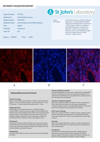
  • 1. ANTIBODY VALIDATION REPORT Report Number 90750-a Application Immunofluorescence Model Number STJ90750 Antibody Name Anti-Phospho-Akt (T308) antibody Host Rabbit Clonality Polyclonal Clone ID NA Species HUMAN Tissue LUNG Image Description Immunofluorescence analysis of Human lung tissue. 1: Akt (phospho Thr308) Polyclonal Antibody(red) was diluted at 1:200 (4 degree Celsius,overnight). 2: Cy3 labled Secondary antibody was diluted at 1:300 (room temperature, 50min).3: Picture B: DAPI(blue) 10min. Picture A:Target. Picture B: DAPI. Picture C: merge of A+B. Primary Antibody Incubation After blocking solution was removed a 1:200 primary antibody/PBS solution was added on the slide, and incubated overnight at 4°C (a small amount of distilled water was added into the incubation box to prevent evaporation of antibody). Secondary Antibody Incubation slides were washed with PBS on a shaker for 5min, and repeated 3 times. Shortly after the slides were dried and corresponding secondary antibody solution was added (HRP labelled), covering the tissues, and incubated at room temperature for 50min. DAPI Counter-Staining slides were washed with PBS on a shaker for 5min, repeated 3 times and then dried. DAPI staining solution was added inside the PAP circles and incubated for 10 min at room temperature without light exposure. Mounting Slides were washed with PBS on a shaker for 5min, and repeated 3 times. Shortly after slides were dried, anti-quench mountings were used to mount slides. Visualization The slides were observed and placed under a NIKON inverted fluorescence microscope (Ultra violet excitation 330-380nm, emission 420nm; FITC green excitation 465-495nm, emission 515- 555 nm; CY3 red excitation 510-560nm, emission 590nm) Immunofluorescence Protocol Tissue Processing Slides were incubated sequentially into: Xylene - 15min, Anhydrous ethanol – 15 min, Anhydrous ethanol – 5 min, 85% alcohol – 5 min, 75% alcohol – 5 min & washed with distilled water – 5 min. Antigen Retrieval Tissue slides were incubated with citric acid (PH6.0) antigen retrieval buffer, and microwaved for antigen retrieval (heated until boiled and then stop heating) for 8min. Slides were then heated with medium power for 7min. During this process slides are kept from drying out. After cooling down at room temperature, slides were washed with PBS on a shaker for 5min, and repeated 3 times. Anti-Quench shortly after slides were dried, a PAP pen was used to draw circles around the tissues (to prevent draining of the antibody). Inside the circles, anti-quench mountings were added and incubated for 5 min, and then flushed with water for 10min. BSA Blocking Inside the circles, BSA was used to cover the tissue evenly, blocking for 30min.
  • 2. ANTIBODY VALIDATION REPORT Report Number 90750-b Application Immunofluorescence Model Number STJ90750 Antibody Name Anti-Phospho-Akt (T308) antibody Host Rabbit Clonality Polyclonal Clone ID NA Species HUMAN Tissue LUNG Image Description Immunofluorescence analysis of Human lung tissue. 1: Akt (phospho Thr308) Polyclonal Antibody(red) was diluted at 1:200 (4 degree Celsius,overnight). 2: Cy3 labled Secondary antibody was diluted at 1:300 (room temperature, 50min).3: Picture B: DAPI(blue) 10min. Picture A:Target. Picture B: DAPI. Picture C: merge of A+B. Primary Antibody Incubation After blocking solution was removed a 1:200 primary antibody/PBS solution was added on the slide, and incubated overnight at 4°C (a small amount of distilled water was added into the incubation box to prevent evaporation of antibody). Secondary Antibody Incubation slides were washed with PBS on a shaker for 5min, and repeated 3 times. Shortly after the slides were dried and corresponding secondary antibody solution was added (HRP labelled), covering the tissues, and incubated at room temperature for 50min. DAPI Counter-Staining slides were washed with PBS on a shaker for 5min, repeated 3 times and then dried. DAPI staining solution was added inside the PAP circles and incubated for 10 min at room temperature without light exposure. Mounting Slides were washed with PBS on a shaker for 5min, and repeated 3 times. Shortly after slides were dried, anti-quench mountings were used to mount slides. Visualization The slides were observed and placed under a NIKON inverted fluorescence microscope (Ultra violet excitation 330-380nm, emission 420nm; FITC green excitation 465-495nm, emission 515- 555 nm; CY3 red excitation 510-560nm, emission 590nm) Immunofluorescence Protocol Tissue Processing Slides were incubated sequentially into: Xylene - 15min, Anhydrous ethanol – 15 min, Anhydrous ethanol – 5 min, 85% alcohol – 5 min, 75% alcohol – 5 min & washed with distilled water – 5 min. Antigen Retrieval Tissue slides were incubated with citric acid (PH6.0) antigen retrieval buffer, and microwaved for antigen retrieval (heated until boiled and then stop heating) for 8min. Slides were then heated with medium power for 7min. During this process slides are kept from drying out. After cooling down at room temperature, slides were washed with PBS on a shaker for 5min, and repeated 3 times. Anti-Quench shortly after slides were dried, a PAP pen was used to draw circles around the tissues (to prevent draining of the antibody). Inside the circles, anti-quench mountings were added and incubated for 5 min, and then flushed with water for 10min. BSA Blocking Inside the circles, BSA was used to cover the tissue evenly, blocking for 30min.
  • 3. ANTIBODY VALIDATION REPORT Report Number 90750-c Application Immunofluorescence Model Number STJ90750 Antibody Name Anti-Phospho-Akt (T308) antibody Host Rabbit Clonality Polyclonal Clone ID NA Species HUMAN Tissue STOMACH Image Description Immunofluorescence analysis of Human stomach tissue. 1: Akt (phospho Thr308) Polyclonal Antibody(red) was diluted at 1:200 (4 degree Celsius,overnight). 2: Cy3 labled Secondary antibody was diluted at 1:300 (room temperature, 50min).3: Picture B: DAPI(blue) 10min. Picture A:Target. Picture B: DAPI. Picture C: merge of A+B. Primary Antibody Incubation After blocking solution was removed a 1:200 primary antibody/PBS solution was added on the slide, and incubated overnight at 4°C (a small amount of distilled water was added into the incubation box to prevent evaporation of antibody). Secondary Antibody Incubation slides were washed with PBS on a shaker for 5min, and repeated 3 times. Shortly after the slides were dried and corresponding secondary antibody solution was added (HRP labelled), covering the tissues, and incubated at room temperature for 50min. DAPI Counter-Staining slides were washed with PBS on a shaker for 5min, repeated 3 times and then dried. DAPI staining solution was added inside the PAP circles and incubated for 10 min at room temperature without light exposure. Mounting Slides were washed with PBS on a shaker for 5min, and repeated 3 times. Shortly after slides were dried, anti-quench mountings were used to mount slides. Visualization The slides were observed and placed under a NIKON inverted fluorescence microscope (Ultra violet excitation 330-380nm, emission 420nm; FITC green excitation 465-495nm, emission 515- 555 nm; CY3 red excitation 510-560nm, emission 590nm) Immunofluorescence Protocol Tissue Processing Slides were incubated sequentially into: Xylene - 15min, Anhydrous ethanol – 15 min, Anhydrous ethanol – 5 min, 85% alcohol – 5 min, 75% alcohol – 5 min & washed with distilled water – 5 min. Antigen Retrieval Tissue slides were incubated with citric acid (PH6.0) antigen retrieval buffer, and microwaved for antigen retrieval (heated until boiled and then stop heating) for 8min. Slides were then heated with medium power for 7min. During this process slides are kept from drying out. After cooling down at room temperature, slides were washed with PBS on a shaker for 5min, and repeated 3 times. Anti-Quench shortly after slides were dried, a PAP pen was used to draw circles around the tissues (to prevent draining of the antibody). Inside the circles, anti-quench mountings were added and incubated for 5 min, and then flushed with water for 10min. BSA Blocking Inside the circles, BSA was used to cover the tissue evenly, blocking for 30min.
  • 4. ANTIBODY VALIDATION REPORT Report Number 90750-d Application Immunofluorescence Model Number STJ90750 Antibody Name Anti-Phospho-Akt (T308) antibody Host Rabbit Clonality Polyclonal Clone ID NA Species HUMAN Tissue STOMACH Image Description Immunofluorescence analysis of Human stomach tissue. 1: Akt (phospho Thr308) Polyclonal Antibody(red) was diluted at 1:200 (4 degree Celsius,overnight). 2: Cy3 labled Secondary antibody was diluted at 1:300 (room temperature, 50min).3: Picture B: DAPI(blue) 10min. Picture A:Target. Picture B: DAPI. Picture C: merge of A+B. Primary Antibody Incubation After blocking solution was removed a 1:200 primary antibody/PBS solution was added on the slide, and incubated overnight at 4°C (a small amount of distilled water was added into the incubation box to prevent evaporation of antibody). Secondary Antibody Incubation slides were washed with PBS on a shaker for 5min, and repeated 3 times. Shortly after the slides were dried and corresponding secondary antibody solution was added (HRP labelled), covering the tissues, and incubated at room temperature for 50min. DAPI Counter-Staining slides were washed with PBS on a shaker for 5min, repeated 3 times and then dried. DAPI staining solution was added inside the PAP circles and incubated for 10 min at room temperature without light exposure. Mounting Slides were washed with PBS on a shaker for 5min, and repeated 3 times. Shortly after slides were dried, anti-quench mountings were used to mount slides. Visualization The slides were observed and placed under a NIKON inverted fluorescence microscope (Ultra violet excitation 330-380nm, emission 420nm; FITC green excitation 465-495nm, emission 515- 555 nm; CY3 red excitation 510-560nm, emission 590nm) Immunofluorescence Protocol Tissue Processing Slides were incubated sequentially into: Xylene - 15min, Anhydrous ethanol – 15 min, Anhydrous ethanol – 5 min, 85% alcohol – 5 min, 75% alcohol – 5 min & washed with distilled water – 5 min. Antigen Retrieval Tissue slides were incubated with citric acid (PH6.0) antigen retrieval buffer, and microwaved for antigen retrieval (heated until boiled and then stop heating) for 8min. Slides were then heated with medium power for 7min. During this process slides are kept from drying out. After cooling down at room temperature, slides were washed with PBS on a shaker for 5min, and repeated 3 times. Anti-Quench shortly after slides were dried, a PAP pen was used to draw circles around the tissues (to prevent draining of the antibody). Inside the circles, anti-quench mountings were added and incubated for 5 min, and then flushed with water for 10min. BSA Blocking Inside the circles, BSA was used to cover the tissue evenly, blocking for 30min.
  • 5. ANTIBODY VALIDATION REPORT Report Number 90750-e Application Immunofluorescence Model Number STJ90750 Antibody Name Anti-Phospho-Akt (T308) antibody Host Rabbit Clonality Polyclonal Clone ID NA Species RAT Tissue KIDNEY Image Description Immunofluorescence analysis of Rat kidney tissue. 1: Akt (phospho Thr308) Polyclonal Antibody(red) was diluted at 1:200 (4 degree Celsius,overnight). 2: Cy3 labled Secondary antibody was diluted at 1:300 (room temperature, 50min).3: Picture B: DAPI(blue) 10min. Picture A:Target. Picture B: DAPI. Picture C: merge of A+B. Primary Antibody Incubation After blocking solution was removed a 1:200 primary antibody/PBS solution was added on the slide, and incubated overnight at 4°C (a small amount of distilled water was added into the incubation box to prevent evaporation of antibody). Secondary Antibody Incubation slides were washed with PBS on a shaker for 5min, and repeated 3 times. Shortly after the slides were dried and corresponding secondary antibody solution was added (HRP labelled), covering the tissues, and incubated at room temperature for 50min. DAPI Counter-Staining slides were washed with PBS on a shaker for 5min, repeated 3 times and then dried. DAPI staining solution was added inside the PAP circles and incubated for 10 min at room temperature without light exposure. Mounting Slides were washed with PBS on a shaker for 5min, and repeated 3 times. Shortly after slides were dried, anti-quench mountings were used to mount slides. Visualization The slides were observed and placed under a NIKON inverted fluorescence microscope (Ultra violet excitation 330-380nm, emission 420nm; FITC green excitation 465-495nm, emission 515- 555 nm; CY3 red excitation 510-560nm, emission 590nm) Immunofluorescence Protocol Tissue Processing Slides were incubated sequentially into: Xylene - 15min, Anhydrous ethanol – 15 min, Anhydrous ethanol – 5 min, 85% alcohol – 5 min, 75% alcohol – 5 min & washed with distilled water – 5 min. Antigen Retrieval Tissue slides were incubated with citric acid (PH6.0) antigen retrieval buffer, and microwaved for antigen retrieval (heated until boiled and then stop heating) for 8min. Slides were then heated with medium power for 7min. During this process slides are kept from drying out. After cooling down at room temperature, slides were washed with PBS on a shaker for 5min, and repeated 3 times. Anti-Quench shortly after slides were dried, a PAP pen was used to draw circles around the tissues (to prevent draining of the antibody). Inside the circles, anti-quench mountings were added and incubated for 5 min, and then flushed with water for 10min. BSA Blocking Inside the circles, BSA was used to cover the tissue evenly, blocking for 30min.
  • 6. ANTIBODY VALIDATION REPORT Report Number 90750-f Application Immunofluorescence Model Number STJ90750 Antibody Name Anti-Phospho-Akt (T308) antibody Host Rabbit Clonality Polyclonal Clone ID NA Species RAT Tissue KIDNEY Image Description Immunofluorescence analysis of Rat kidney tissue. 1: Akt (phospho Thr308) Polyclonal Antibody(red) was diluted at 1:200 (4 degree Celsius,overnight). 2: Cy3 labled Secondary antibody was diluted at 1:300 (room temperature, 50min).3: Picture B: DAPI(blue) 10min. Picture A:Target. Picture B: DAPI. Picture C: merge of A+B. Primary Antibody Incubation After blocking solution was removed a 1:200 primary antibody/PBS solution was added on the slide, and incubated overnight at 4°C (a small amount of distilled water was added into the incubation box to prevent evaporation of antibody). Secondary Antibody Incubation slides were washed with PBS on a shaker for 5min, and repeated 3 times. Shortly after the slides were dried and corresponding secondary antibody solution was added (HRP labelled), covering the tissues, and incubated at room temperature for 50min. DAPI Counter-Staining slides were washed with PBS on a shaker for 5min, repeated 3 times and then dried. DAPI staining solution was added inside the PAP circles and incubated for 10 min at room temperature without light exposure. Mounting Slides were washed with PBS on a shaker for 5min, and repeated 3 times. Shortly after slides were dried, anti-quench mountings were used to mount slides. Visualization The slides were observed and placed under a NIKON inverted fluorescence microscope (Ultra violet excitation 330-380nm, emission 420nm; FITC green excitation 465-495nm, emission 515- 555 nm; CY3 red excitation 510-560nm, emission 590nm) Immunofluorescence Protocol Tissue Processing Slides were incubated sequentially into: Xylene - 15min, Anhydrous ethanol – 15 min, Anhydrous ethanol – 5 min, 85% alcohol – 5 min, 75% alcohol – 5 min & washed with distilled water – 5 min. Antigen Retrieval Tissue slides were incubated with citric acid (PH6.0) antigen retrieval buffer, and microwaved for antigen retrieval (heated until boiled and then stop heating) for 8min. Slides were then heated with medium power for 7min. During this process slides are kept from drying out. After cooling down at room temperature, slides were washed with PBS on a shaker for 5min, and repeated 3 times. Anti-Quench shortly after slides were dried, a PAP pen was used to draw circles around the tissues (to prevent draining of the antibody). Inside the circles, anti-quench mountings were added and incubated for 5 min, and then flushed with water for 10min. BSA Blocking Inside the circles, BSA was used to cover the tissue evenly, blocking for 30min.