Immunofluorescence Antibody Validation Report for Anti-p70 S6 kinase α Antibody (STJ97443)

Serine/threonine-protein kinase that acts downstream of mTOR signaling in response to growth factors and nutrients to promote cell proliferation, cell growth and cell cycle progression. Regulates protein synthesis through phosphorylation of EIF4B, RPS6 and EEF2K, and contributes to cell survival by repressing the pro-apoptotic function of BAD. Under conditions of nutrient depletion, the inactive form associates with the EIF3 translation initiation complex. Upon mitogenic stimulation, phosphorylation by the mammalian target of rapamycin complex 1 (mTORC1) leads to dissociation from the EIF3 complex and activation. The active form then phosphorylates and activates several substrates in the pre-initiation complex, including the EIF2B complex and the cap-binding complex component EIF4B. Also controls translation initiation by phosphorylating a negative regulator of EIF4A, PDCD4, targeting it for ubiquitination and subsequent proteolysis. Anti-p70 S6 kinase α -http://www.stjohnslabs.com/p70-s6-kinase-a-antibody-p-99094 Join our Antibody Validation Project - http://www.stjohnslabs.com/services/antibody-validation

Recommended
Recommended
More Related Content
What's hot
What's hot (20)
Viewers also liked
Viewers also liked (20)
Similar to Immunofluorescence Antibody Validation Report for Anti-p70 S6 kinase α Antibody (STJ97443)
Similar to Immunofluorescence Antibody Validation Report for Anti-p70 S6 kinase α Antibody (STJ97443) (14)
More from St John's Laboratory Ltd
More from St John's Laboratory Ltd (20)
Recently uploaded
Recently uploaded (20)
Immunofluorescence Antibody Validation Report for Anti-p70 S6 kinase α Antibody (STJ97443)
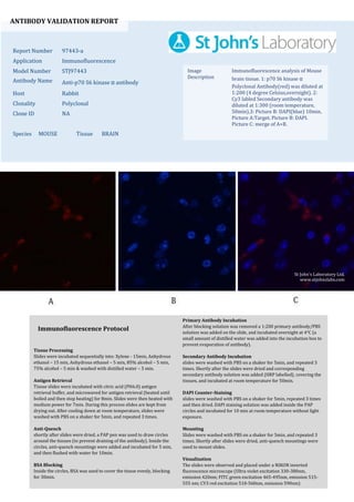
- 1. ANTIBODY VALIDATION REPORT Report Number 97443-a Application Immunofluorescence Model Number STJ97443 Antibody Name Anti-p70 S6 kinase α antibody Host Rabbit Clonality Polyclonal Clone ID NA Species MOUSE Tissue BRAIN Image Description Immunofluorescence analysis of Mouse brain tissue. 1: p70 S6 kinase α Polyclonal Antibody(red) was diluted at 1:200 (4 degree Celsius,overnight). 2: Cy3 labled Secondary antibody was diluted at 1:300 (room temperature, 50min).3: Picture B: DAPI(blue) 10min. Picture A:Target. Picture B: DAPI. Picture C: merge of A+B. Primary Antibody Incubation After blocking solution was removed a 1:200 primary antibody/PBS solution was added on the slide, and incubated overnight at 4°C (a small amount of distilled water was added into the incubation box to prevent evaporation of antibody). Secondary Antibody Incubation slides were washed with PBS on a shaker for 5min, and repeated 3 times. Shortly after the slides were dried and corresponding secondary antibody solution was added (HRP labelled), covering the tissues, and incubated at room temperature for 50min. DAPI Counter-Staining slides were washed with PBS on a shaker for 5min, repeated 3 times and then dried. DAPI staining solution was added inside the PAP circles and incubated for 10 min at room temperature without light exposure. Mounting Slides were washed with PBS on a shaker for 5min, and repeated 3 times. Shortly after slides were dried, anti-quench mountings were used to mount slides. Visualization The slides were observed and placed under a NIKON inverted fluorescence microscope (Ultra violet excitation 330-380nm, emission 420nm; FITC green excitation 465-495nm, emission 515- 555 nm; CY3 red excitation 510-560nm, emission 590nm) Immunofluorescence Protocol Tissue Processing Slides were incubated sequentially into: Xylene - 15min, Anhydrous ethanol – 15 min, Anhydrous ethanol – 5 min, 85% alcohol – 5 min, 75% alcohol – 5 min & washed with distilled water – 5 min. Antigen Retrieval Tissue slides were incubated with citric acid (PH6.0) antigen retrieval buffer, and microwaved for antigen retrieval (heated until boiled and then stop heating) for 8min. Slides were then heated with medium power for 7min. During this process slides are kept from drying out. After cooling down at room temperature, slides were washed with PBS on a shaker for 5min, and repeated 3 times. Anti-Quench shortly after slides were dried, a PAP pen was used to draw circles around the tissues (to prevent draining of the antibody). Inside the circles, anti-quench mountings were added and incubated for 5 min, and then flushed with water for 10min. BSA Blocking Inside the circles, BSA was used to cover the tissue evenly, blocking for 30min. St John's Laboratory Ltd. www.stjohnslabs.com
- 2. ANTIBODY VALIDATION REPORT Report Number 97443-b Application Immunofluorescence Model Number STJ97443 Antibody Name Anti-p70 S6 kinase α antibody Host Rabbit Clonality Polyclonal Clone ID NA Species MOUSE Tissue BRAIN Image Description Immunofluorescence analysis of Mouse brain tissue. 1: p70 S6 kinase α Polyclonal Antibody(red) was diluted at 1:200 (4 degree Celsius,overnight). 2: Cy3 labled Secondary antibody was diluted at 1:300 (room temperature, 50min).3: Picture B: DAPI(blue) 10min. Picture A:Target. Picture B: DAPI. Picture C: merge of A+B. Primary Antibody Incubation After blocking solution was removed a 1:200 primary antibody/PBS solution was added on the slide, and incubated overnight at 4°C (a small amount of distilled water was added into the incubation box to prevent evaporation of antibody). Secondary Antibody Incubation slides were washed with PBS on a shaker for 5min, and repeated 3 times. Shortly after the slides were dried and corresponding secondary antibody solution was added (HRP labelled), covering the tissues, and incubated at room temperature for 50min. DAPI Counter-Staining slides were washed with PBS on a shaker for 5min, repeated 3 times and then dried. DAPI staining solution was added inside the PAP circles and incubated for 10 min at room temperature without light exposure. Mounting Slides were washed with PBS on a shaker for 5min, and repeated 3 times. Shortly after slides were dried, anti-quench mountings were used to mount slides. Visualization The slides were observed and placed under a NIKON inverted fluorescence microscope (Ultra violet excitation 330-380nm, emission 420nm; FITC green excitation 465-495nm, emission 515- 555 nm; CY3 red excitation 510-560nm, emission 590nm) Immunofluorescence Protocol Tissue Processing Slides were incubated sequentially into: Xylene - 15min, Anhydrous ethanol – 15 min, Anhydrous ethanol – 5 min, 85% alcohol – 5 min, 75% alcohol – 5 min & washed with distilled water – 5 min. Antigen Retrieval Tissue slides were incubated with citric acid (PH6.0) antigen retrieval buffer, and microwaved for antigen retrieval (heated until boiled and then stop heating) for 8min. Slides were then heated with medium power for 7min. During this process slides are kept from drying out. After cooling down at room temperature, slides were washed with PBS on a shaker for 5min, and repeated 3 times. Anti-Quench shortly after slides were dried, a PAP pen was used to draw circles around the tissues (to prevent draining of the antibody). Inside the circles, anti-quench mountings were added and incubated for 5 min, and then flushed with water for 10min. BSA Blocking Inside the circles, BSA was used to cover the tissue evenly, blocking for 30min. St John's Laboratory Ltd. www.stjohnslabs.com