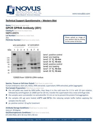
The antibody was tested by western blot on samples from human embryonic stem cells but did not detect the expected signal at the predicted size of around 100 kDa in either the stem cells or the positive control. The western blot procedure involved extracting protein from stem cell pellets and supernatant using RIPA buffer, running samples on an 8% SDS-PAGE gel and transferring to a membrane. The membrane was blocked for 1 hour before an overnight incubation with the primary antibody at a 1:500 dilution, followed by washing and a 1 hour incubation with a secondary antibody at a 1:10,000 dilution before detection. No band was observed at the expected molecular weight of around 100 kDa.
1. Technical Support Questionnaire – Western Blot
Name:Click here to enter text.
GPCR GPR48 Antibody (2D1)
Catalog #:Click here to enter text.
NBP2-00974
Lot Number:Click here to enter text.
A01 Please upload an image of
PO/Order Number:Click here to enter text.. your western blot by clicking
on the center of the box.
Species, Tissues or Cell Lines Tested:Click here to enter text.
human embryonic stem cell, hESCs; RIPA extracted, supernatant; RIPA extracted, pellet (aggregate)
Test Sample Preparation:Click here to enter text.
the cell pellet was lysed by RIPA buffer, then Keep it in the cold room for 1.5 hr with 10 rpm rotation,
followed by centrifugation at 13000 rpm for 20 min, transfer the supernatant into a new centrifuge tube.
the samples were sonicated(or no sonication)for 15 min on ice to prevent the protein fromaggregation
the samples (20 ug) were heated at37°C and 95°Cin the reducing sample buffer before applying the
samples into the well.
ps: positive control: 10 ug for treatment
Antibody Storage Conditions:Click here to enter text.
store at -20 degree
Gel/Electrophoresis Conditions:Click here to enter text.
8 % SDS-PAGE, 60 V 30 min/ 90 V, 90 min
2. Transfer Conditions:Click here to enter text.
24.7 mMTris base, 191.8 mM glycine and 10 % methanol; 45 V, O/N, then 100 V 30 min
Blocking Solution & Duration:Click here to enter text.
blocking at room temperature for 1 hr
blocking solution contents: 137.0 mMNaCl, 19.9 mMTris, 5 % milk, pH 7.6
Primary Antibody Diluent and Dilutions Tested:Click here to enter text.
dilute with TBST buffer (containing 0.1 % tween 20) in the ratio of 1:500
Primary Antibody Incubation Time and Temperature:Click here to enter text.
2hr at room temperature, then keep it in the cold room, O/N
Wash Solution Composition, Repetitions & Times:Click here to enter text.
wash the membrane with TBST buffer (19.9 mMTris, 137.0 mMNaCl, 0.1 % Tween 20, pH7.6) 3 times, 5 min for
each time
Secondary Antibody Manufacturer, Host Species, Dilution, & Diluent:Click here to enter text.
JacsonimmunoResearch, code No.: 115-035-008-100 ul, goat anti mouse IgG.
dilute with TBST (0.1 % tween 20) in the ratio of 1: 10000
Secondary Antibody Incubation Time & Temperature:Click here to enter text.
1 hr at room temperature
Wash Solution Composition, Repetitions, & Times:Click here to enter text.
wash the membrane with TBST buffer 5 times, 5 min for each time
(19.9 mMTris, 137.0 mMNaCl, 0.1 % Tween 20, pH7.6)
Detection System, Procedure & Development Time:Click here to enter text.
add 1000 ul of ECL substrate (thermo, femto) onto the membrane. then cover the membrane with a
transparent plastic membrane, followed by luminescence detection by FUJI FILM LAS-4000 with a exposure
time of 30 s
Molecular weight of band(s):Click here to enter text.
markers: thermo 26616
Controls:Click here to enter text.
positive control from Novus, NBP2-06791 Lot No.0812
Observations:Click here to enter text.
In human embryonic stem cell and positive control provided by your company (Novus), we can not see the
signal from the predicted size (about 100 kDa).