
Recommended
More Related Content
What's hot
What's hot (20)
Similar to monoclonal antibody production & hybridization and charecterization
Similar to monoclonal antibody production & hybridization and charecterization (20)
Recently uploaded
Recently uploaded (20)
monoclonal antibody production & hybridization and charecterization
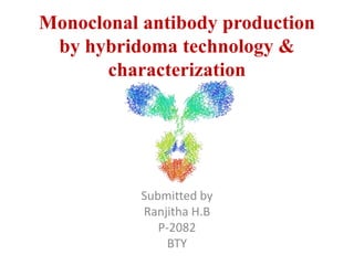
- 1. Monoclonal antibody production by hybridoma technology & characterization Submitted by Ranjitha H.B P-2082 BTY
- 2. • Antibody- Y-shaped protein produced mainly by plasma cells which neutralize pathogens such as bacteria and viruses. • Polyclonal antibodies- secreted by different B cell , each identify a different epitope • Monoclonal antibodies- made by identical immune cells, which bind to the same epitope
- 3. Monoclonal Antibodies • Class of highly specific antibodies produced by the clones of a single hybrid cell • Produced by fusing a B cell secreting the antibody with a myeloma cell capable of growing indefinitely - Hybridoma technology • Fused cell called hybridoma • Uses - Proteomics research, diagnosis of diseases & treatment of diseases (infections & cancer).
- 4. •The term hybridoma was coined by Leonard Herzenberg 1964- Littlefield developed a way to isolate hybrid cells from 2 parent cell lines using HAT selection media •Hybridoma technology- Kohler & Milstein in 1975 Discovery of hybridoma technology
- 6. HAT medium hypoxanthine, aminopterin & thymidine Nucleotide synthesis is essential for cell survival De nova pathway Salvage pathway De nova pathway is blocked in all cells Simple sugars nucleotides Aminopterin B cell HGPRT+ Myeloma cell HGPRT- Hybrid cells HGPRT+ Hypoxanthine Guanine HGPRT Myeloma cell HGPRT-
- 7. How hybrid cells are selected in HAT medium HAT medium Unfused B cells undergo normal cell death Large scale culture of hybrid cells. HGPRT+ from B cell Myeloma cells cannot grow in HAT medium Myeloma cells are HGPRT- & de nova pathway is blocked by aminopterin. So cannot grow in HAT medium
- 9. Materials Mice (Balb/c or A/J), 6 to 10 weeks old P3.653 myeloma Antigen (125 μg per mouse is optimal): Small antigens can be conjugated to keyhole limpet hemocyanin Adjuvants (alum or Freund’s adjuvant; complete and incomplete) TCD (T-Cell depletion) buffer: Hanks’s balanced salt solution + 10mM HEPES + 0.3% BSA NH4Cl, 0.16M Antimouse Thy 1.2 antibody 1:500 Rabbit complement- Reconstitute in 1mL of cold UPW, dilute 1:12 in TCD buffer, and filter sterilize serum-free medium: MEM
- 10. PEG (polyethylene glycol): Melt 10.5mL of PEG 1450 (Sigma) in a 56◦C water bath; add 19.5mL of warm sterile MEM(pH 8.3 to pH8.7) HAT medium: SCM (spleen-conditioned medium 8-Azaguanine stock, 10mM MEM, 10% fetal calf serum, 0.1mM 8-azaguanine (for maintenance of P3.653 myeloma)
- 11. Step 1: - Immunization Of Mice Antigen Mice (Balb/c or A/J), 6 to 10 weeks old Antigen: Intact cells, whole membranes, & microorganisms + Adjuvants Immunized every 2-3 weeks
- 12. Step 2: Screening of Mice for Antibody Production After several weeks of immunization Spleen cells Antigen Measurement of serum antibodies ELISA & Flow cytometry Antibody titer is high Cell fusion can be performed titer is too low Boost
- 14. • Step 3: Preparation of Myeloma Cells To ensure their sensitivity to hypoxanthine- aminopterin-thymidine (HAT) selection Maintain the P3.653 myeloma cell line in MEM + 10% fetal calf serum + 8- azaguanine- week before cell fusion
- 15. • Step 4: Fusion of Myeloma Cells with Immune Spleen Cells ratio of 4:1
- 16. Hybridoma Development • Single immunized spleen cells with myeloma cells in presence of PEG • Cells are plated on 96 well plates containing feeder cells derived from saline peritoneal wash of mice(murine BM derived MΦ)
- 17. • Step 5: Cloning of Hybridoma Cell Lines by “Limiting Dilution” or Expansion and Stabilization of Clones by Ascites Production
- 18. Screening • Selection in HAT medium • Limiting dilution • Clonal culture • Sub cloning
- 19. Production in cell culture ( In-vitro) Batch tissue culture method: • Grow hybridoma cells in batches • Purify Mabs from the culture media • Fetal bovine serum commonly used • low concentration (below 20 mg/ml) • denaturation during concentration Semi permeable membrane based system : • A barrier – hollow fibre or a membrane • Larger compartment containing culture media • Smaller chamber to isolate cells and Mabs • High concentrations (10-160 mg) • Method of choice for large scale production
- 20. Production in animals (in vivo) Mouse ascites method: • Hybridoma cells injected in mouse • Produce ascites • Fluid contains high concentration of Ab’s • No further concentration required • Purification required • Easy and inexpensive
- 21. Priming • Generation of ascites fluid • Most commonly used- Pristane (tetramethyl penta decane), others- Freund’s Incomplete Adjuvant (FIA) • Prime adult female mice of at least 6 weeks of age or retired breeders
- 22. Cont…… • If administering Pristane as the priming agent, do not exceed a volume of 0.2 ml • Administer FIA IP once (not to exceed 0.3 ml) • Standard interval between priming & inoculation of hybridoma cells is 10-14 days
- 23. Hybridoma Inoculation(In vitro) • Test hybridoma cells for presence of pathogens using PCR-based or species-specific antibody production assay • Inject primed mice 10^5- 10^7 hybridoma cells IP 0.1-0.5 ml • Monitor mice at least once daily for the first 7 days
- 24. Ascites/Tumor Growth • Abdominal distension will usually be noted 7 to 10 days after inoculation • 3 survival taps • weight gain should not exceed 20% of the original body weight • No signs of pain or distress (18-22G needle to remove excess fluid)
- 25. Ascites fluid collection • Each mouse will yield from 2.0 ml to over 5.0 ml of ascites fluid • Euthanize animals by cervical dislocation • Clean abdomen with 70% ethanol • Make an incision in the skin over the abdomen while holding the mouse over the collection container • Drain all the fluid into container and transfer into centrifuge tube
- 26. Cont… • Centrifuge ascites fluid at 1500 rpm for 5 minutes • Separate supernatant from blood cells and store at -80°C if necessary • Animals will be kept for a maximum of 30 days • If ascites has not developed within this timeframe, animals will be euthanized as per Rodent Euthanasia SOP
- 27. Ethical issues : • Freund’s complete adjuvant (FCA) (to enhance the immune response): painful lesions at the injection site. • Pristane as a "priming" agent - granulomatous reactions • Respiratory distress: due to ascites. • Shock - rapid fluid loss • FCA should not be used more than once in individual mice. • The volume of FCA and pristane used should not exceed 0.1ml and 0.2ml respectively. • Individual mice should not be inoculated with adjuvant more than 3 times. • Ascites fluid should only be harvested once at the time of euthanasia
- 29. Characterization of MABs Identification of class and sub class, antigen specificity
- 30. Antibody purification • Affinity chromatography: reversible interaction between a binding protein (e.g. antibody) and a ligand (e.g. antigen or Protein A) i) Fc binding protein purification ii) Antigen-specific purification
- 31. Methods for antibody characterization
- 32. Enzyme-linked immunosorbent assay, or ELISA • For the detection and quantification of MABs in supernatants
- 33. Western blotting • Identification of proteins in complex mixtures • validating that antibodies bind to a target protein of the expected size • posttranslational modifications
- 34. Immunohistochemistry • Information on protein expression in tissues, at both cellular and subcellular levels, can be provided by immunohistochemical (IHC) analysis
- 35. Immunofluorescence microscopy • Discrimination between nuclear or cytoplasmic staining is achievable and in some cases membranous staining can be recognized Fixation of cells permeabilization primary antibody secondary antibody conjugated with fluorescent tag immunofluoroscent microscopy
- 36. Epitope mapping • Method used to determine binding sites, or 'epitopes', of antibodies on their target antigens • X-ray crystallography • mutagenesis of the antigen • site-directed masking of epitopes
- 37. Isotyping of murine monoclonal antibodies • Qualitative determination of isotype of MABs IgG1, IgG2a, IgG2b, IgG3, IgM or IgA • By capture enzyme-linked immunosorbent assay (ELISA)
- 38. IsoStrip Mouse Monoclonal Antibody Isotyping Kit ( Sigma Aldrich) Pierce™ Rapid Antibody Isotyping Kit – Mouse ( Thermo Fisher Scientific) Mouse Monoclonal Antibody Isotyping Test Kit from AbD Serotec ( Bio-Rad ) Antibody Isotyping (eBioscience) Isotyping Kit for Mouse Monoclonal Antibody (Sino Biological Inc.) RapidYield Mouse Monoclonal Antibody Isotyping Kit (Crystal Chem's ) Mouse Antibody Isotyping Kit (Cell Biolabs, Inc.)
- 39. • THANK YOU
Editor's Notes
- that are all clones of a unique parent cell.
- antibody titer is high,. If the, mice can be boosted
- Fusion is accomplished by co-centrifuging freshly harvested spleen cells and myeloma cells in polyethylene glycol, a substance that causes cell membranes to fuse. The cells are then distributed to 96 well plates containing feeder cells derived from saline peritoneal washes of mice. Feeder cells are believed to supply growth factors that promote growth of the hybridoma cells Commercial preparations murine bone marrow-derived macrophages as feeder cells (
- Standard techniques could generate 103-104 clones in each experiment
- One of the interacting molecules is coupled to a solid support. The binding between antibodies and respective antigens involves a number of non-covalent interactions, including van der Waals forces, ionic bonds, hydrogen bonds and/or hydrophobic interactions. This enables elution of antibodies in a concentrated form by changing the conditions in the buffer (e.g. pH, ionic strength or polarity) used or introduction of a molecule that competes with the ligand for specific binding sites [