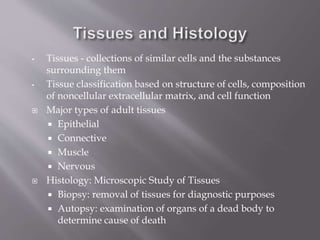
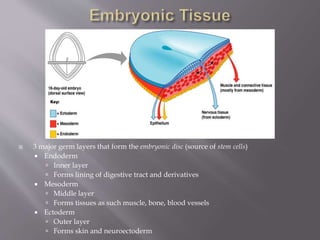
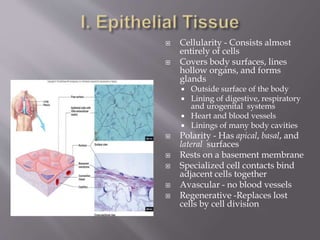
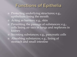
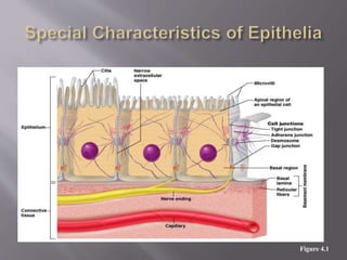
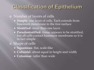
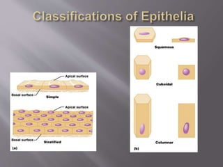
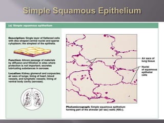
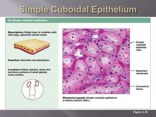
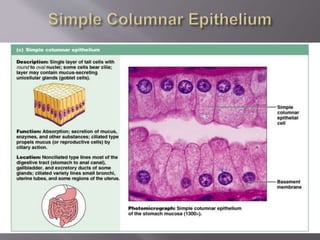
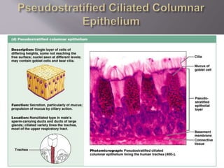
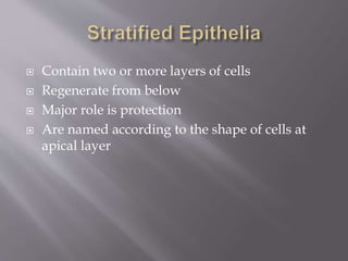
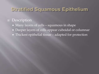
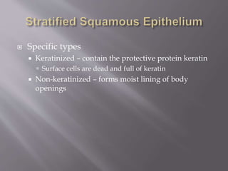
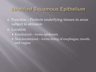
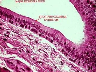
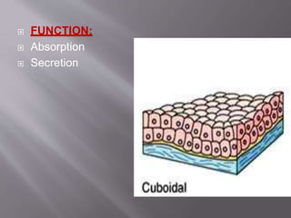
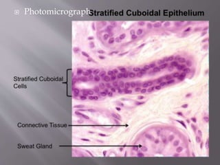
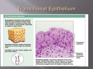
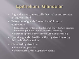
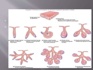
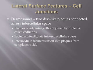
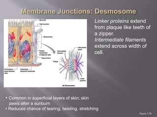
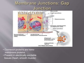
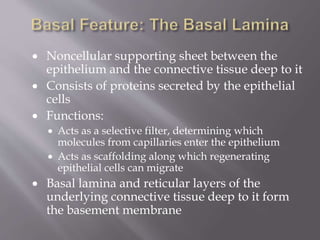
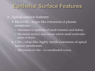
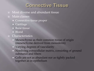
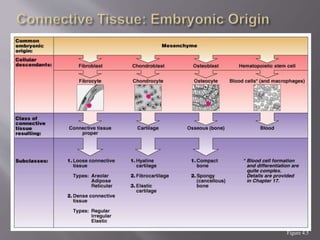
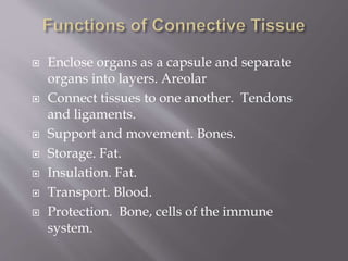
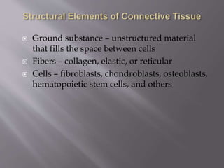
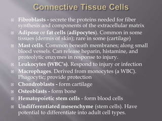
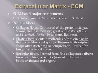
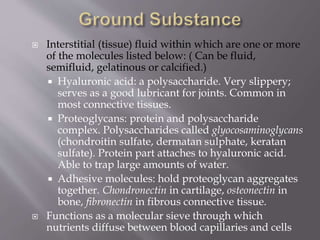
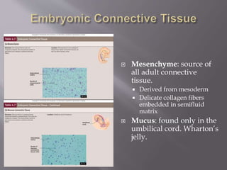
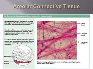
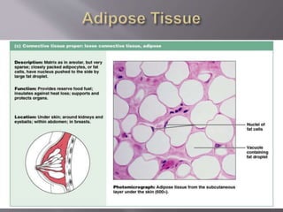
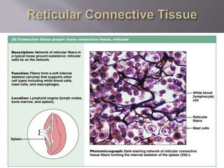
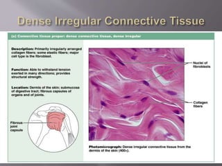
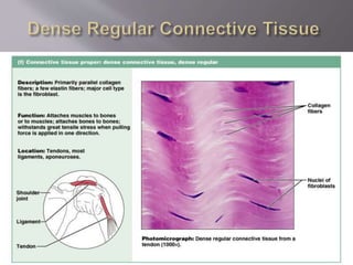
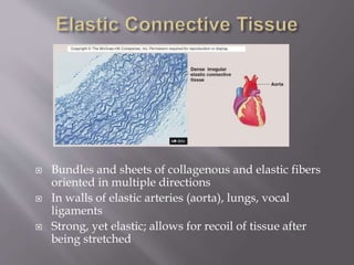
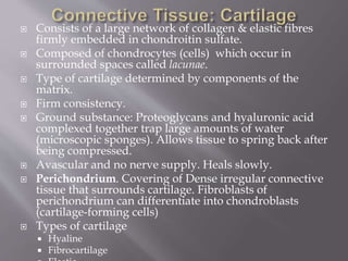
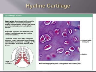
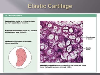
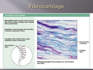
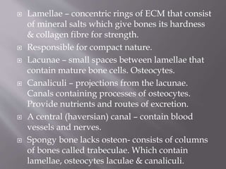
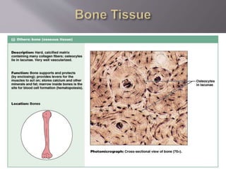
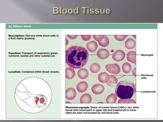
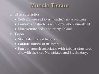
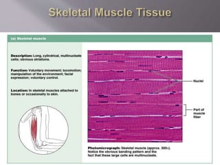
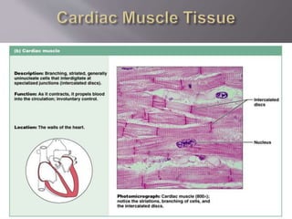
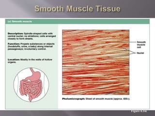
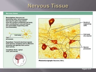
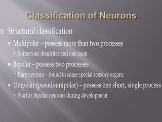
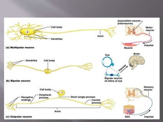
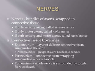
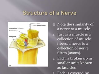
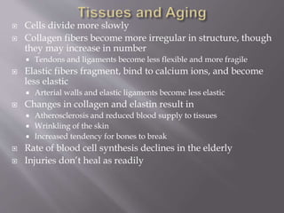
MR.AMADI.K.O
• Tissues - collections of similar cells and the substances
surrounding them
• Tissue classification based on structure of cells, composition
of noncellular extracellular matrix, and cell function
 Major types of adult tissues
 Epithelial
 Connective
 Muscle
 Nervous
 Histology: Microscopic Study of Tissues
 Biopsy: removal of tissues for diagnostic purposes
 Autopsy: examination of organs of a dead body to
determine cause of death
 3 major germ layers that form the embryonic disc (source of stem cells)
 Endoderm
 Inner layer
 Forms lining of digestive tract and derivatives
 Mesoderm
 Middle layer
 Forms tissues as such muscle, bone, blood vessels
 Ectoderm
 Outer layer
 Forms skin and neuroectoderm
 Cellularity - Consists almost
entirely of cells
 Covers body surfaces, lines
hollow organs, and forms
glands
 Outside surface of the body
 Lining of digestive, respiratory
and urogenital systems
 Heart and blood vessels
 Linings of many body cavities
 Polarity - Has apical, basal, and
lateral surfaces
 Rests on a basement membrane
 Specialized cell contacts bind
adjacent cells together
 Avascular - no blood vessels
 Regenerative -Replaces lost
cells by cell division
 Protecting underlying structures; e.g.,
epithelium lining the mouth
 Acting as barriers; e.g., skin
 Permitting the passage of substances; e.g.,
cells lining air sacs in lungs and nephrons in
kidney
 Secreting substances; e.g., pancreatic cells
 Absorbing substances; e.g., lining of
stomach and small intestine
Figure 4.1
 Number of layers of cells
 Simple- one layer of cells. Each extends from
basement membrane to the free surface
 Stratified- more than one layer.
 Pseudostratified- tissue appears to be stratified,
but all cells contact basement membrane so it is
in fact simple
 Shape of cells
 Squamous- flat, scale-like
 Cuboidal- about equal in height and width
 Columnar- taller than wide
Figure 4.3a
Figure 4.3b
Figure 4.3c
Figure 4.3d
 Contain two or more layers of cells
 Regenerate from below
 Major role is protection
 Are named according to the shape of cells at
apical layer
 Description
 Many layers of cells – squamous in shape
 Deeper layers of cells appear cuboidal or columnar
 Thickest epithelial tissue – adapted for protection
 Specific types
 Keratinized – contain the protective protein keratin
 Surface cells are dead and full of keratin
 Non-keratinized – forms moist lining of body
openings
 Function – Protects underlying tissues in areas
subject to abrasion
 Location
 Keratinized – forms epidermis
 Non-keratinized – forms lining of esophagus, mouth,
and vagina
Figure 4.3e
 It is a rare type of epithelial tissues composed
of column shaped cells arranged in multiple
layers
STRATIFIED CUBOIDAL EPITHELIUM
 Thin, stratified epithelium which usually consists of only
two or three layers of cuboidal or low columnar cells.
 Not involved in significant absorptive or secretory activity
PRESENT IN:
Ducts of sweat glands
Large ducts of exocrine glands
Anorectal junction
 FUNCTION:
 Absorption
 Secretion
 Photomicrograph
Figure 4.3h
 A gland is one or more cells that makes and secretes
an aqueous fluid
 Two types of glands formed by infolding of
epithelium:
 Endocrine: no contact with exterior of body; ductless; produce
hormones (pituitary, thyroid, adrenals, pancreas)
 Exocrine: open to exterior of body via ducts (sweat, oil)
 Exocrine glands classified either by structure or by
the method of secretion
 Classified by structure
 Unicellular: goblet cells
 Multicellular: sweat, oil, pituitary, adrenal
 Classified on the basis of types
of ducts or mode of secretion
 Types of ducts
 Simple: ducts with few
branches
 Compound: ducts with
many branches
 If ducts end in tubules or
sac-like structures: acini.
Pancreas
 If ducts end in simple sacs:
alveoli. Lungs
 Tight junctions
 Desmosomes
 Gap junctions
• Integral proteins of adjacent
cells fuse together
• Completely encircle the cell
and form an adhesion belt.
• Form an impermeable
junction.
• Common near apical region
 Desmosomes – two disc-like plaques connected
across intercellular space
 Plaques of adjoining cells are joined by proteins
called cadherins
 Proteins interdigitate into extracellular space
 Intermediate filaments insert into plaques from
cytoplasmic side
Figure 3.5b
Linker proteins extend
from plaque like teeth of
a zipper.
Intermediate filaments
extend across width of
cell.
• Common in superficial layers of skin; skin
peels after a sunburn
• Reduces chance of tearing, twisting, stretching
• Connexon proteins are trans-
membrane proteins.
• Present in electrically excitable
tissues (heart, smooth muscle)
 Noncellular supporting sheet between the
epithelium and the connective tissue deep to it
 Consists of proteins secreted by the epithelial
cells
 Functions:
 Acts as a selective filter, determining which
molecules from capillaries enter the epithelium
 Acts as scaffolding along which regenerating
epithelial cells can migrate
 Basal lamina and reticular layers of the
underlying connective tissue deep to it form
the basement membrane
 Apical surface features
 Microvilli – finger-like extensions of plasma
membrane
 Abundant in epithelia of small intestine and kidney
 Maximize surface area across which small molecules
enter or leave
 Cilia – whip-like, highly motile extensions of apical
surface membranes
 Movement of cilia – in coordinated waves
 Most diverse and abundant tissue
 Main classes
 Connective tissue proper
 Cartilage
 Bone tissue
 Blood
 Characteristics
 Mesenchyme as their common tissue of origin
(mesenchyme derived from mesoderm)
 Varying degrees of vascularity
 Nonliving extracellular matrix, consisting of ground
substance and fibers
 Cells are not as abundant nor as tightly packed
together as in epithelium
Figure 4.5
 Enclose organs as a capsule and separate
organs into layers. Areolar
 Connect tissues to one another. Tendons
and ligaments.
 Support and movement. Bones.
 Storage. Fat.
 Insulation. Fat.
 Transport. Blood.
 Protection. Bone, cells of the immune
system.
 Ground substance – unstructured material
that fills the space between cells
 Fibers – collagen, elastic, or reticular
 Cells – fibroblasts, chondroblasts, osteoblasts,
hematopoietic stem cells, and others
 Fibroblasts - secrete the proteins needed for fiber
synthesis and components of the extracellular matrix
 Adipose or fat cells (adipocytes). Common in some
tissues (dermis of skin); rare in some (cartilage)
 Mast cells. Common beneath membranes; along small
blood vessels. Can release heparin, histamine, and
proteolytic enzymes in response to injury.
 Leukocytes (WBC’s). Respond to injury or infection
 Macrophages. Derived from monocytes (a WBC).
Phagocytic; provide protection
 Chondroblasts - form cartilage
 Osteoblasts - form bone
 Hematopoietic stem cells - form blood cells
 Undifferentiated mesenchyme (stem cells). Have
potential to differentiate into adult cell types.
 ECM has 3 major components
1. Protein fibers 2. Ground substance 3. Fluid
 Protein fibers
 Collagen fibers. Composed of the protein collagen.
Strong, flexible, inelastic; great tensile strength (i.e.
resist stretch). Perfect for tendons, ligaments
 Elastic fibers. Contain molecules of protein elastin
that resemble coiled springs. Returns to its original
shape after stretching or compression. Perfect for
lungs, large blood vessels
 Reticular fibers. Formed from fine collagenous fibers;
form branching networks (stroma). Fill spaces
between tissues and organs.
 Interstitial (tissue) fluid within which are one or more
of the molecules listed below: ( Can be fluid,
semifluid, gelatinous or calcified.)
 Hyaluronic acid: a polysaccharide. Very slippery;
serves as a good lubricant for joints. Common in
most connective tissues.
 Proteoglycans: protein and polysaccharide
complex. Polysaccharides called glyocosaminoglycans
(chondroitin sulfate, dermatan sulphate, keratan
sulfate). Protein part attaches to hyaluronic acid.
Able to trap large amounts of water.
 Adhesive molecules: hold proteoglycan aggregates
together. Chondronectin in cartilage, osteonectin in
bone, fibronectin in fibrous connective tissue.
 Functions as a molecular sieve through which
nutrients diffuse between blood capillaries and cells
 Mesenchyme: source of
all adult connective
tissue.
 Derived from mesoderm
 Delicate collagen fibers
embedded in semifluid
matrix
 Mucus: found only in the
umbilical cord. Wharton’s
jelly.
Figure 4.12b
Figure 4.12c
Figure 4.12d
Figure 4.12e
Figure 4.12f
 Bundles and sheets of collagenous and elastic fibers
oriented in multiple directions
 In walls of elastic arteries (aorta), lungs, vocal
ligaments
 Strong, yet elastic; allows for recoil of tissue after
being stretched
 Consists of a large network of collagen & elastic fibres
firmly embedded in chondroitin sulfate.
 Composed of chondrocytes (cells) which occur in
surrounded spaces called lacunae.
 Type of cartilage determined by components of the
matrix.
 Firm consistency.
 Ground substance: Proteoglycans and hyaluronic acid
complexed together trap large amounts of water
(microscopic sponges). Allows tissue to spring back after
being compressed.
 Avascular and no nerve supply. Heals slowly.
 Perichondrium. Covering of Dense irregular connective
tissue that surrounds cartilage. Fibroblasts of
perichondrium can differentiate into chondroblasts
(cartilage-forming cells)
 Types of cartilage
 Hyaline
 Fibrocartilage
Figure 4.12g
Figure 4.12h
Figure 4.12i
 Bone tissue
 Cartilage, joints and bones make up the skeletal
system.
 Bones store calcium & phosphorous, house the
red bone marrow& yellow marrow.
 Bones are organs composed of several
connective tissue eg bone(osseus) tissue,
periosteum, red & yellow marrow and
endosteum.
 Bone is classified as either compact or spongy
depending on the arrangement ECM.
 Compact bone basic unit is an osteon or
haversian system and have four parts.
 Lamellae – concentric rings of ECM that consist
of mineral salts which give bones its hardness
& collagen fibre for strength.
 Responsible for compact nature.
 Lacunae – small spaces between lamellae that
contain mature bone cells. Osteocytes.
 Canaliculi – projections from the lacunae.
Canals containing processes of osteocytes.
Provide nutrients and routes of excretion.
 A central (haversian) canal – contain blood
vessels and nerves.
 Spongy bone lacks osteon- consists of columns
of bones called trabeculae. Which contain
lamellae, osteocytes laculae & canaliculi.
Figure 4.12j
Figure 4.12k
 Characteristics
 Cells are referred to as muscle fibers or myocytes
 Contracts or shortens with force when stimulated
 Moves entire body and pumps blood
 Types
 Skeletal: attached to bones
 Cardiac: muscle of the heart.
 Smooth: muscle associated with tubular structures
and with the skin. Nonstriated and involuntary.
Figure 4.14a
Figure 4.14b
Figure 4.14c
Special characteristics of neurons
 Longevity – can live and function for a lifetime
 Do not divide (amitotic) – fetal neurons lose their
ability to undergo mitosis; neural stem cells are an
exception
 High metabolic rate – require abundant oxygen
and glucose
Parts of the Neuron
Cell Body. Aka Soma or Perikaryon
Contains usual organelles plus
other structures
Nissl bodies = chromatophilic
substance = rough E.R: primary
site of protein synthesis
Cytoskeleton of neurofilaments
and neurotubules
No centrioles (hence its amitotic
nature)
Major biosynthetic center
Most neuronal cell bodies
Located within CNS
Ganglia - clusters of cell bodies
that lie along nerves in PNS
Tapers to form axon hillock
Figure 4.15
 Nerves - bundles of axons wrapped in
connective tissue
 If only sensory axons, called sensory nerves
 If only motor axons, called motor nerves
 If both sensory and motor axons, called mixed nerves
 Connective Tissue Coverings
 Endoneurium – layer of delicate connective tissue
surrounding the axon
 Nerve fascicles – groups of axons bound into bundles
 Perineurium – connective tissue wrapping
surrounding a nerve fascicle
 Epineurium – whole nerve is surrounded by tough
fibrous sheath
 Note the similarity of
a nerve to a muscle
1. Just as a muscle is a
collection of muscle
fibers, a nerve is a
collection of nerve
fibers (axons).
2. Each is broken up in
smaller units known
as fascicles
 Cells divide more slowly
 Collagen fibers become more irregular in structure, though
they may increase in number
 Tendons and ligaments become less flexible and more fragile
 Elastic fibers fragment, bind to calcium ions, and become
less elastic
 Arterial walls and elastic ligaments become less elastic
 Changes in collagen and elastin result in
 Atherosclerosis and reduced blood supply to tissues
 Wrinkling of the skin
 Increased tendency for bones to break
 Rate of blood cell synthesis declines in the elderly
 Injuries don’t heal as readily

tissue and histology.pptx

  • 1.
  • 2.
    • Tissues -collections of similar cells and the substances surrounding them • Tissue classification based on structure of cells, composition of noncellular extracellular matrix, and cell function  Major types of adult tissues  Epithelial  Connective  Muscle  Nervous  Histology: Microscopic Study of Tissues  Biopsy: removal of tissues for diagnostic purposes  Autopsy: examination of organs of a dead body to determine cause of death
  • 3.
     3 majorgerm layers that form the embryonic disc (source of stem cells)  Endoderm  Inner layer  Forms lining of digestive tract and derivatives  Mesoderm  Middle layer  Forms tissues as such muscle, bone, blood vessels  Ectoderm  Outer layer  Forms skin and neuroectoderm
  • 4.
     Cellularity -Consists almost entirely of cells  Covers body surfaces, lines hollow organs, and forms glands  Outside surface of the body  Lining of digestive, respiratory and urogenital systems  Heart and blood vessels  Linings of many body cavities  Polarity - Has apical, basal, and lateral surfaces  Rests on a basement membrane  Specialized cell contacts bind adjacent cells together  Avascular - no blood vessels  Regenerative -Replaces lost cells by cell division
  • 5.
     Protecting underlyingstructures; e.g., epithelium lining the mouth  Acting as barriers; e.g., skin  Permitting the passage of substances; e.g., cells lining air sacs in lungs and nephrons in kidney  Secreting substances; e.g., pancreatic cells  Absorbing substances; e.g., lining of stomach and small intestine
  • 6.
  • 7.
     Number oflayers of cells  Simple- one layer of cells. Each extends from basement membrane to the free surface  Stratified- more than one layer.  Pseudostratified- tissue appears to be stratified, but all cells contact basement membrane so it is in fact simple  Shape of cells  Squamous- flat, scale-like  Cuboidal- about equal in height and width  Columnar- taller than wide
  • 9.
  • 10.
  • 11.
  • 12.
  • 13.
     Contain twoor more layers of cells  Regenerate from below  Major role is protection  Are named according to the shape of cells at apical layer
  • 14.
     Description  Manylayers of cells – squamous in shape  Deeper layers of cells appear cuboidal or columnar  Thickest epithelial tissue – adapted for protection
  • 15.
     Specific types Keratinized – contain the protective protein keratin  Surface cells are dead and full of keratin  Non-keratinized – forms moist lining of body openings
  • 16.
     Function –Protects underlying tissues in areas subject to abrasion  Location  Keratinized – forms epidermis  Non-keratinized – forms lining of esophagus, mouth, and vagina
  • 17.
  • 18.
     It isa rare type of epithelial tissues composed of column shaped cells arranged in multiple layers
  • 20.
    STRATIFIED CUBOIDAL EPITHELIUM Thin, stratified epithelium which usually consists of only two or three layers of cuboidal or low columnar cells.  Not involved in significant absorptive or secretory activity PRESENT IN: Ducts of sweat glands Large ducts of exocrine glands Anorectal junction
  • 22.
  • 23.
  • 24.
  • 25.
     A glandis one or more cells that makes and secretes an aqueous fluid  Two types of glands formed by infolding of epithelium:  Endocrine: no contact with exterior of body; ductless; produce hormones (pituitary, thyroid, adrenals, pancreas)  Exocrine: open to exterior of body via ducts (sweat, oil)  Exocrine glands classified either by structure or by the method of secretion  Classified by structure  Unicellular: goblet cells  Multicellular: sweat, oil, pituitary, adrenal
  • 26.
     Classified onthe basis of types of ducts or mode of secretion  Types of ducts  Simple: ducts with few branches  Compound: ducts with many branches  If ducts end in tubules or sac-like structures: acini. Pancreas  If ducts end in simple sacs: alveoli. Lungs
  • 27.
     Tight junctions Desmosomes  Gap junctions
  • 28.
    • Integral proteinsof adjacent cells fuse together • Completely encircle the cell and form an adhesion belt. • Form an impermeable junction. • Common near apical region
  • 29.
     Desmosomes –two disc-like plaques connected across intercellular space  Plaques of adjoining cells are joined by proteins called cadherins  Proteins interdigitate into extracellular space  Intermediate filaments insert into plaques from cytoplasmic side
  • 30.
    Figure 3.5b Linker proteinsextend from plaque like teeth of a zipper. Intermediate filaments extend across width of cell. • Common in superficial layers of skin; skin peels after a sunburn • Reduces chance of tearing, twisting, stretching
  • 31.
    • Connexon proteinsare trans- membrane proteins. • Present in electrically excitable tissues (heart, smooth muscle)
  • 32.
     Noncellular supportingsheet between the epithelium and the connective tissue deep to it  Consists of proteins secreted by the epithelial cells  Functions:  Acts as a selective filter, determining which molecules from capillaries enter the epithelium  Acts as scaffolding along which regenerating epithelial cells can migrate  Basal lamina and reticular layers of the underlying connective tissue deep to it form the basement membrane
  • 33.
     Apical surfacefeatures  Microvilli – finger-like extensions of plasma membrane  Abundant in epithelia of small intestine and kidney  Maximize surface area across which small molecules enter or leave  Cilia – whip-like, highly motile extensions of apical surface membranes  Movement of cilia – in coordinated waves
  • 34.
     Most diverseand abundant tissue  Main classes  Connective tissue proper  Cartilage  Bone tissue  Blood  Characteristics  Mesenchyme as their common tissue of origin (mesenchyme derived from mesoderm)  Varying degrees of vascularity  Nonliving extracellular matrix, consisting of ground substance and fibers  Cells are not as abundant nor as tightly packed together as in epithelium
  • 35.
  • 36.
     Enclose organsas a capsule and separate organs into layers. Areolar  Connect tissues to one another. Tendons and ligaments.  Support and movement. Bones.  Storage. Fat.  Insulation. Fat.  Transport. Blood.  Protection. Bone, cells of the immune system.
  • 37.
     Ground substance– unstructured material that fills the space between cells  Fibers – collagen, elastic, or reticular  Cells – fibroblasts, chondroblasts, osteoblasts, hematopoietic stem cells, and others
  • 38.
     Fibroblasts -secrete the proteins needed for fiber synthesis and components of the extracellular matrix  Adipose or fat cells (adipocytes). Common in some tissues (dermis of skin); rare in some (cartilage)  Mast cells. Common beneath membranes; along small blood vessels. Can release heparin, histamine, and proteolytic enzymes in response to injury.  Leukocytes (WBC’s). Respond to injury or infection  Macrophages. Derived from monocytes (a WBC). Phagocytic; provide protection  Chondroblasts - form cartilage  Osteoblasts - form bone  Hematopoietic stem cells - form blood cells  Undifferentiated mesenchyme (stem cells). Have potential to differentiate into adult cell types.
  • 39.
     ECM has3 major components 1. Protein fibers 2. Ground substance 3. Fluid  Protein fibers  Collagen fibers. Composed of the protein collagen. Strong, flexible, inelastic; great tensile strength (i.e. resist stretch). Perfect for tendons, ligaments  Elastic fibers. Contain molecules of protein elastin that resemble coiled springs. Returns to its original shape after stretching or compression. Perfect for lungs, large blood vessels  Reticular fibers. Formed from fine collagenous fibers; form branching networks (stroma). Fill spaces between tissues and organs.
  • 40.
     Interstitial (tissue)fluid within which are one or more of the molecules listed below: ( Can be fluid, semifluid, gelatinous or calcified.)  Hyaluronic acid: a polysaccharide. Very slippery; serves as a good lubricant for joints. Common in most connective tissues.  Proteoglycans: protein and polysaccharide complex. Polysaccharides called glyocosaminoglycans (chondroitin sulfate, dermatan sulphate, keratan sulfate). Protein part attaches to hyaluronic acid. Able to trap large amounts of water.  Adhesive molecules: hold proteoglycan aggregates together. Chondronectin in cartilage, osteonectin in bone, fibronectin in fibrous connective tissue.  Functions as a molecular sieve through which nutrients diffuse between blood capillaries and cells
  • 41.
     Mesenchyme: sourceof all adult connective tissue.  Derived from mesoderm  Delicate collagen fibers embedded in semifluid matrix  Mucus: found only in the umbilical cord. Wharton’s jelly.
  • 42.
  • 43.
  • 44.
  • 45.
  • 46.
  • 47.
     Bundles andsheets of collagenous and elastic fibers oriented in multiple directions  In walls of elastic arteries (aorta), lungs, vocal ligaments  Strong, yet elastic; allows for recoil of tissue after being stretched
  • 48.
     Consists ofa large network of collagen & elastic fibres firmly embedded in chondroitin sulfate.  Composed of chondrocytes (cells) which occur in surrounded spaces called lacunae.  Type of cartilage determined by components of the matrix.  Firm consistency.  Ground substance: Proteoglycans and hyaluronic acid complexed together trap large amounts of water (microscopic sponges). Allows tissue to spring back after being compressed.  Avascular and no nerve supply. Heals slowly.  Perichondrium. Covering of Dense irregular connective tissue that surrounds cartilage. Fibroblasts of perichondrium can differentiate into chondroblasts (cartilage-forming cells)  Types of cartilage  Hyaline  Fibrocartilage
  • 49.
  • 50.
  • 51.
  • 52.
     Bone tissue Cartilage, joints and bones make up the skeletal system.  Bones store calcium & phosphorous, house the red bone marrow& yellow marrow.  Bones are organs composed of several connective tissue eg bone(osseus) tissue, periosteum, red & yellow marrow and endosteum.  Bone is classified as either compact or spongy depending on the arrangement ECM.  Compact bone basic unit is an osteon or haversian system and have four parts.
  • 53.
     Lamellae –concentric rings of ECM that consist of mineral salts which give bones its hardness & collagen fibre for strength.  Responsible for compact nature.  Lacunae – small spaces between lamellae that contain mature bone cells. Osteocytes.  Canaliculi – projections from the lacunae. Canals containing processes of osteocytes. Provide nutrients and routes of excretion.  A central (haversian) canal – contain blood vessels and nerves.  Spongy bone lacks osteon- consists of columns of bones called trabeculae. Which contain lamellae, osteocytes laculae & canaliculi.
  • 54.
  • 55.
  • 56.
     Characteristics  Cellsare referred to as muscle fibers or myocytes  Contracts or shortens with force when stimulated  Moves entire body and pumps blood  Types  Skeletal: attached to bones  Cardiac: muscle of the heart.  Smooth: muscle associated with tubular structures and with the skin. Nonstriated and involuntary.
  • 57.
  • 58.
  • 59.
  • 60.
    Special characteristics ofneurons  Longevity – can live and function for a lifetime  Do not divide (amitotic) – fetal neurons lose their ability to undergo mitosis; neural stem cells are an exception  High metabolic rate – require abundant oxygen and glucose
  • 61.
    Parts of theNeuron Cell Body. Aka Soma or Perikaryon Contains usual organelles plus other structures Nissl bodies = chromatophilic substance = rough E.R: primary site of protein synthesis Cytoskeleton of neurofilaments and neurotubules No centrioles (hence its amitotic nature) Major biosynthetic center Most neuronal cell bodies Located within CNS Ganglia - clusters of cell bodies that lie along nerves in PNS Tapers to form axon hillock
  • 62.
  • 66.
     Nerves -bundles of axons wrapped in connective tissue  If only sensory axons, called sensory nerves  If only motor axons, called motor nerves  If both sensory and motor axons, called mixed nerves  Connective Tissue Coverings  Endoneurium – layer of delicate connective tissue surrounding the axon  Nerve fascicles – groups of axons bound into bundles  Perineurium – connective tissue wrapping surrounding a nerve fascicle  Epineurium – whole nerve is surrounded by tough fibrous sheath
  • 67.
     Note thesimilarity of a nerve to a muscle 1. Just as a muscle is a collection of muscle fibers, a nerve is a collection of nerve fibers (axons). 2. Each is broken up in smaller units known as fascicles
  • 68.
     Cells dividemore slowly  Collagen fibers become more irregular in structure, though they may increase in number  Tendons and ligaments become less flexible and more fragile  Elastic fibers fragment, bind to calcium ions, and become less elastic  Arterial walls and elastic ligaments become less elastic  Changes in collagen and elastin result in  Atherosclerosis and reduced blood supply to tissues  Wrinkling of the skin  Increased tendency for bones to break  Rate of blood cell synthesis declines in the elderly  Injuries don’t heal as readily