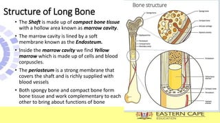
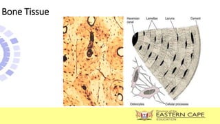
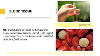
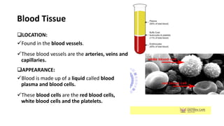
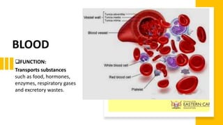
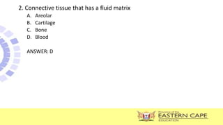
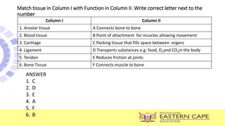
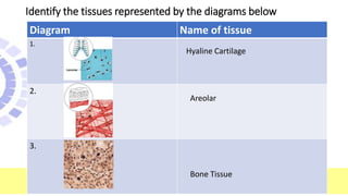
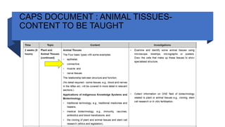
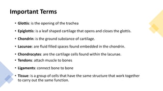
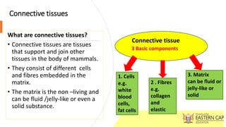
The document covers connective tissues, particularly focusing on their terminology, structure, and functions, including tendons, ligaments, cartilage, bone, and blood. It describes the basic components of connective tissues, their various types, and specific examples such as areolar and fibrous connective tissues. Additionally, it outlines the structure of long bones and the unique characteristics of blood as a connective tissue.


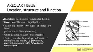
![AREOLAR TISSUE:
Location, structure and function continued
Functions of areolar tissue:
• Serves as an insulating material-
prevent heat loss.
• Protects the organs by serving as a
packing tissue, e.g. around the kidneys.
• Connects the skin to underlying layers.
• It serves as a packing tissue that fills
the spaces between organs, blood
vessels, nerves and muscles.
• NB: [when large amount of fat are
stored in this tissue, it is known as
adipose/fat tissue]](https://image.slidesharecdn.com/00lifesciencesgrade10pptconnectivetissues-240620154255-3ffde7cf/85/00-Life-Sciences-Grade-10-PPT-Connective-Tissues-pptx-9-320.jpg)