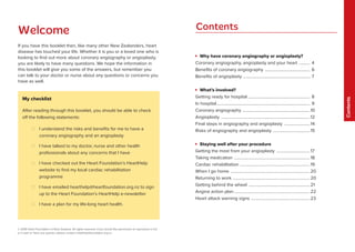
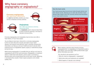
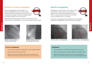
Coronary angiography and angioplasty are procedures used to examine and treat narrowed or blocked coronary arteries that supply blood to the heart. The document provides information on:
1) Why these procedures are performed - to determine if narrowing or blockages exist in the arteries and potentially treat them by opening the arteries to improve blood flow.
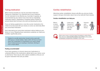
2) What is involved - including preparing for the hospital visit, the procedures which use catheters and sometimes stents, and recovering at home while resuming medications and lifestyle changes.
3) Risks and benefits are outlined to help patients understand the procedures and make informed decisions with their healthcare team. Ongoing management focuses on medication adherence and heart-healthy
Peripheral angiography is a radiological procedure used to examine arteries and veins after injecting contrast media. It involves puncturing an artery such as the femoral artery using the Seldinger technique and threading a catheter over a guidewire to inject contrast media and obtain images. The procedure is used to diagnose and treat various vascular conditions. Precise positioning, sterile equipment and contrast injection are needed to obtain diagnostic images of the peripheral vasculature.
This document summarizes coronary stents, including their purpose, types, history, procedure for placement, and future advancements. Stents are small mesh devices that are placed in arteries to keep them open and improve blood flow after balloon angioplasty. There are bare metal stents and drug-eluting stents, which combat re-narrowing after placement. The first coronary stent was placed in 1986, and drug-eluting stents were introduced in 2001 to further reduce re-narrowing. Placement involves threading a catheter through an artery to the blockage and expanding a balloon and stent to open the artery. Future advancements include bioresorbable polymers that dissolve after artery healing is complete.
Stents are used to support the inside of a tubular passage or lumen, such as coronary and peripheral arteries, oesophagus, biliary duct, colon or an airway. They are placed temporarily or permanently in the strictured region where the lumen’s diameter is narrowed due to a disease. This presentation describes design characteristics of self-expandable stents.
This document provides an overview of percutaneous coronary intervention (PCI) including:
- A brief history of PCI and the development of stents.
- Clinical factors that can influence PCI outcomes like diabetes, kidney disease, and ability to tolerate dual antiplatelet therapy.
- Equipment used in PCI like guide catheters, guide wires, and balloon catheters.
- Medications given during and after PCI like aspirin, P2Y12 inhibitors, and GP IIb/IIIa inhibitors to prevent clotting and restenosis.
- Considerations for PCI in different clinical scenarios like UA/NSTEMI, STEMI, and adjunctive diagnostic devices that can
This document discusses abdominal aortic aneurysms (AAAs) and their endovascular repair (EVAR). It defines AAAs as a dilatation of the abdominal aorta over 3cm in diameter. EVAR involves inserting a folded graft through the femoral artery which expands to exclude the aneurysm sac from blood flow and pressure. The benefits of EVAR over open repair include lower peri-operative mortality and complications. Proper patient assessment including vascular anatomy and medical comorbidities is important for determining candidacy for EVAR. The procedure involves deploying graft components in the aorta and iliac arteries under imaging guidance. Post-operative surveillance with imaging is needed to monitor for complications like endoleaks.
This document provides information about renal angiography including:
- Renal angiography involves injecting contrast into the renal arteries to detect abnormalities in blood supply to the kidneys.
- It describes materials, catheters, and techniques used including the Seldinger technique for vascular access.
- Potential complications are also outlined. Indications for renal angiography include renal artery stenosis, aneurysms, tumors and other conditions.
- Case examples are provided to illustrate different pathologies visualized on renal angiography such as fibromuscular dysplasia, angiomyolipomas and their treatment with angioplasty or embolization.
interventional cardiology, Guiding catheters, wires, and balloons equipment...salman habeeb
This document provides an overview of guiding catheters, guide wires, and balloons which are core equipment used in percutaneous coronary interventions (PCI). It describes the design characteristics, advantages, and disadvantages of over-the-wire and rapid exchange balloon catheters. Key attributes of balloons like entry, tracking, and compliance are defined. Guiding catheters are discussed in terms of size, shape, and selection for accessing different coronary arteries. Finally, guide wire features such as core material, coating, and tip design are reviewed alongside common wire types used in various clinical scenarios.
This document discusses different designs of coronary stents. It begins by providing background on the development of coronary stents and their approval for use. It then describes some of the earliest stent designs, including the Gianturco-Roubin coil stent and the Palmaz-Schatz slotted tube stent. The document goes on to discuss various aspects of stent design that can impact performance, such as the geometric configuration, materials used, coatings, and drug-eluting capabilities. Key design considerations like strut thickness, number of struts, and mechanical properties are also reviewed.
This presentation will be talking about coronary angiography which is a part of a general group of procedures known as (heart) cardiac catherization .this is performed for both diagnostic and interventional (treatment) purposes
This document provides an overview of cardiac catheterization procedures. It discusses indications, contraindications, techniques, views obtained, and interpretation of pressure waveforms. Key points include that cardiac catheterization guides treatment decisions by measuring pressures, outputs, and obtaining images. It is now often used therapeutically for procedures like angioplasty and device closures. The document outlines patient preparation, access methods, catheters used, views obtained, and complications that can occur.
Angiography is the process of radiography of blood or lymph vessels, carried out after introduction of a radiopaque substance. Medifee Find the hospitals and clinics that offer Angiography in major Indian cities along with their prices.
Angiography is a medical imaging technique that uses radiography to visualize blood vessels and organs after injecting a contrast medium. It can detect diseases of the arteries and veins like atherosclerosis, aneurysms, and internal bleeding. The procedure involves a team inserting a catheter into the blood vessels and injecting iodinated contrast dye before capturing x-ray images. Risks are generally low but can include minor bleeding, vessel damage, and allergic reactions to the contrast medium. Advances in digital subtraction angiography now make angiography the gold standard for assessing vascular diseases when other imaging modalities are inconclusive.
Arteriography and interventional radiologyMilan Silwal
Angiography involves the radiologic examination of blood vessels after injection of iodinated contrast medium. Arteriography specifically examines arteries, while venography examines veins. Techniques include non-invasive ultrasound and MRI, minimally invasive CT or MRI with contrast, and invasive catheterization. Catheters and guide wires are used to access vessels and inject contrast medium. Potential complications include contrast reactions, embolism, infection, and vessel damage. Indications for arteriography include evaluating congenital anomalies, aneurysms, stenoses, arteritis, trauma, embolism, vascular malformations, fistulas, hemorrhage, and masses.
This document provides a summary of angiography and arterial anatomy. It introduces Dr. Mustafa Zuhair Mahmoud and gives a quick overview of the circulatory system including the heart, arteries, capillaries, and veins. It then describes the arteries of the head and neck, brain, abdomen, and angiographic techniques including contrast agents, injection methods, equipment, and projections used. The purpose, patient preparation, and indications for arterial imaging are outlined. Diagrams demonstrate selective catheterization of various arteries like the celiac, mesenteric, renal, and iliac arteries.
Temporary cardiac pacing is used to treat acute bradyarrhythmias or tachyarrhythmias until the underlying condition resolves or permanent pacing can be initiated. It aims to re-establish normal hemodynamics compromised by abnormal heart rates. Transvenous pacing is the preferred method, involving insertion of endocardial leads through veins to the heart. Precise lead placement is important and is confirmed with imaging. Pacing parameters like threshold, rate and sensing are optimized. Complications include those related to vascular access and device malfunction requiring troubleshooting. Close monitoring is needed to ensure proper pacing and detect any issues.
Technique of peripheral angiogram and complicationMai Parachy
The document discusses techniques for peripheral angiograms and potential complications. It covers operating room preparation including equipment such as needles, guide wires, sheaths, and catheters. Access site selection is discussed including the common femoral, popliteal, tibial, brachial, subclavian, and radial arteries. The angiogram procedure is outlined including artery puncture, sheath placement, guidewire insertion, catheter selection, contrast injection, and closure techniques such as manual compression or closure devices. Complications from the procedure are also mentioned.
- Coronary CT angiography uses x-rays and contrast material to examine the coronary arteries with high spatial and temporal resolution. It is non-invasive compared to traditional coronary angiography.
- Key factors for cardiac imaging include high temporal resolution (<250ms), spatial resolution (<0.75mm), and synchronization with the cardiac cycle using ECG gating. Prospective and retrospective gating, partial and multi-segment reconstruction, and low pitch values (<0.5) help achieve this.
- Advances in multi-detector CT scanners, faster gantry rotation times (<330ms), and improved reconstruction algorithms now allow temporal resolutions as low as 80ms for coronary CT angiography.
This document discusses contrast media used to enhance visualization of structures in radiographic examinations. It begins by outlining the objectives and introducing contrast agents. It then differentiates between positive and negative contrast media, with positive agents appearing bright and negative agents appearing dark. The two main positive agents discussed are barium sulfate and iodinated compounds. Barium sulfate is insoluble and used to coat gastrointestinal structures, while iodinated compounds can be water-soluble or oil-based depending on the exam. The document outlines the properties, uses, and considerations of both contrast agent types.
The document provides an overview of basic pacing concepts including:
- Types of pacemakers such as single chamber, dual chamber, and triple chamber systems.
- Components of a pacemaker system including the pulse generator, leads, and electrical concepts such as voltage, current, and impedance.
- Factors that can affect pacing thresholds and how to test the pacemaker circuit including identifying high and low impedance conditions.
The document discusses coronary angioplasty, a minimally invasive procedure to open blocked coronary arteries. It involves inserting a catheter into the heart and inflating a small balloon to widen narrowed arteries. A stent may also be inserted to keep the artery open. The document outlines the indications, contraindications, procedure steps, risks, postoperative care and complications of coronary angioplasty and stent placement.
Coronary angiography remains the gold standard for detecting coronary artery disease. The technique was first performed in 1958 and is used to visualize the coronary arteries and assess for stenosis. It can determine treatment options and prognosis. Complications are rare but include vascular injury and contrast reactions. Proper angiographic views are important for evaluating different coronary artery segments.
This document discusses coronary guidewires used in percutaneous coronary intervention (PCI). It begins by outlining the history of angioplasty and guidewire development. It then covers the purpose, components, classifications, and appropriate uses of guidewires. The main components include the core, tip, coils, covers, and coatings. Guidewires are classified based on flexibility, device support, and clinical usage. Complications like vessel perforation, pseudolesions, and entrapment are also discussed. Proper guidewire manipulation and strategies for difficult lesions are outlined to maximize safety and efficacy.
CT angiography uses x-rays and iodine contrast dye to produce detailed images of blood vessels and tissues. A CT scan is performed after the contrast dye is injected into the bloodstream. CT angiography can be used to diagnose and evaluate diseases of the blood vessels like injuries, aneurysms, and blockages. It provides more precise anatomical detail than MRI for small blood vessels. Potential risks include radiation exposure and allergic reaction to the contrast dye.
Defibrillation is a treatment for cardiac arrest that uses electric shocks to restore a normal heart rhythm. It has evolved significantly since its first use on humans in 1947. Key developments include pre-hospital defibrillation by paramedics in the 1960s-70s and the introduction of automated external defibrillators (AEDs) for public use. Defibrillation works by depolarizing the heart and interrupting abnormal rhythms, allowing the heart's natural pacemaker to resume control. It is most effective for shockable rhythms like ventricular fibrillation when administered quickly after cardiac arrest. Modern defibrillators use biphasic waveforms that deliver lower peak currents, reducing risk of injury compared to older
Cardiac catheterization is a test that uses a thin tube inserted into blood vessels to examine the heart. It can check for blockages in coronary arteries, assess heart function and valves, and diagnose congenital heart defects. The procedure involves inserting a catheter into the groin or wrist artery and guiding it to the heart while injecting dye for imaging. Potential risks include bleeding, infection, arrhythmias, and reactions to dye, though complications are rare. Post-procedure care involves monitoring for symptoms and restricting activity to allow puncture sites to heal.
This document provides information on complications that can occur after femoral access for catheterization procedures. The major complications discussed include hematoma, pseudoaneurysm, arteriovenous fistula, and arterial occlusion. For each complication, the summary describes typical physical findings and recommended nursing interventions. Studies comparing different approaches to hemostasis and sheath removal are also summarized, finding no significant difference in vascular complication rates between sandbag compression and bandage dressings.
Coronary artery bypass grafting (CABG) surgery is performed to treat coronary artery disease by creating bypasses around blockages in the coronary arteries to improve blood flow to the heart. The document provides details on:
1) What coronary artery disease and CABG surgery are
2) How CABG surgery is performed
3) Post-operative recovery, including returning to normal activities and long-term precautions like controlling risk factors
Dr. Suraj Ingole's clinic is best cardiologist, angiography,angioplasty,Echocardiologists,Cardioversion Doctors,Angioplasty Bypass Surgeons in pune,Bhekrai Nagar,SasaneNagar,sayyed nagar,magarpatta,phursungi,vaiduwadi,mundwa,ramtekdi,Kale Padal,hadapsar
Cardiac catheterization is an invasive imaging procedure used to evaluate heart function and diagnose coronary artery disease. During the procedure, a catheter is inserted into an artery and threaded to the heart to take images of blood flow and heart function. It can be used to diagnose issues, evaluate blockages, and place stents. After the procedure, patients need to rest and avoid activity to prevent bleeding complications. Recovery may involve medications and cardiac rehabilitation to improve heart health.
Cardiac catheterization is a procedure where a thin tube is inserted into blood vessels and threaded to the heart to examine coronary arteries and cardiac structures. It is used to diagnose conditions like coronary artery disease and congenital heart defects. During the procedure, dye is injected through the catheter to take x-ray images of the heart and coronary arteries. Potential complications include bleeding, arrhythmias, kidney damage, and myocardial infarction. Nurses monitor the patient during and after the procedure and assess the insertion site for complications.
Coronary artery disease (CAD) occurs when the arteries that supply blood to the heart become narrowed due to plaque buildup within the arteries over many years. This reduces blood flow and oxygen to the heart muscle. CAD is diagnosed using tests like electrocardiograms, stress tests, echocardiograms, and heart scans. Treatment options include medications to improve blood flow, angioplasty to widen narrowed arteries using a balloon catheter, stents to prop open the arteries, or atherectomy using a tiny drill to remove plaque. Lifestyle changes like a healthy diet, exercise, weight control, and managing conditions like diabetes and high blood pressure can help prevent and manage CAD.
Coronary artery disease (CAD) occurs when the arteries that supply blood to the heart become narrowed due to plaque buildup within the arteries over many years. This reduces blood flow and oxygen delivery to the heart muscle. CAD has certain unmodifiable risk factors like age, gender and family history, but also modifiable ones like high blood pressure, high cholesterol, diabetes, obesity, physical inactivity and smoking. Diagnostic tests like electrocardiograms, stress tests, echocardiograms and heart scans can detect CAD. Cardiac catheterization involves threading a catheter into the heart to take pictures of the coronary arteries and measure pressure. Angioplasty and stenting can then be used to open blocked arteries and improve blood
The document describes various types of echocardiograms (also known as cardiac ultrasounds), which are noninvasive tests that use ultrasound to image the heart. It discusses how echocardiograms work, what information they provide about heart size and function, when they may be ordered, and what patients experience during the different types of echocardiogram tests.
What are the signs of a heart attack and what should you doBiocon
Heart attacks and strokes can strike suddenly and be fatal if assistance is not sought immediately. The document outlines signs of heart attacks and strokes and advises that if any signs are detected, emergency medical help should be contacted without delay. Treatment options are described that may help restore blood flow and minimize damage, but quick action is critical to survival. Lifestyle changes after the event can also help prevent future attacks.
The document describes the anatomy of the heart and how blood flows through its four chambers - the right and left atria and ventricles. It then provides instructions for what to do if experiencing a heart attack alone, advising repeated deep coughing to keep blood circulating and help the heart regain its rhythm until help arrives. Finally, it discusses different types of heart surgery like coronary artery bypass grafting and valve repair or replacement, and how newer minimally invasive techniques are reducing risks compared to traditional open-heart surgery.
Institute of Cardiac Science and Research (Cardiology - AIG Hospitals)AIG Hospitals
AIG Institute of Cardiac Sciences offers comprehensive cardiac care services supported by the latest technology, advanced infrastructure, and expertise in
treating rare and complex heart
conditions.
The multidisciplinary approach to cardiac care involves renowned senior cardiologists, electrophysiologists, cardiac
surgeons, critical care intensivists,
anesthetists, cardiac technicians, and nurses.
Our cardiologists are leaders in research, diagnosis, and therapies for people with heart conditions.
Common Heart Diseases:
Heart attack is usually what strikes most people when they think of heart disease. However, there are a number of diseases that affect the heart or blood vessels of a
person. Some of them are mentioned below:
Coronary Artery Disease (CAD)/Ischemic heart disease
Arrhythmias and Palpitations
Inflammatory heart disease
Hypertensive heart disease
Heart failure
Valvular heart disease
Rheumatic heart disease
Causes of Heart Diseases:
In order to prevent heart diseases, controlling various risk factors that cause heart problems is essential.
Factors that cause heart diseases are:
Abnormal blood cholesterol (dyslipidemia)
High blood pressure (hypertension)
Smoking and other forms of tobacco use
Diabetes
Being overweight or obese
Physical inactivity
Age over 55 (for men) or 65 (for women)
Family history of CAD
Psychological stress
Who needs Heart Surgery?
People need heart surgery for different reasons. The most common reasons are
Coronary Artery Disease
Damaged Heart Valves
Enlarged Aortic artery
Understanding the facts about Coronary Angiography and Angioplasty
Cardiologists at AIG Hospitals have expertise in using latest stent
technology such Drug Eluting Stent and Bioabsorbable stents
Coronary artery bypass grafting (CABG)
Coronary artery bypass grafting is a type of surgery that
improves blood ow to the heart. This surgery is used to treat
coronary artery disease (CAD). CABG also is known as bypass
surgery, coronary artery bypass surgery, and open-heart surgery.
Coronary artery bypass grafting (CABG) involves taking a vein or
artery from another part of the body, usually the chest or leg, and
attaching it to the coronary artery above and below the
narrowed area or blockage. This replacement vein or artery is
called a graft and allows blood to bypass the narrowing or block-
age, improving blood supply to the heart muscle.
Common medical tests that are performed for the diagnosis of a heart condition
ECG (Electrocardiogram)
Blood tests, including tests to see whether cardiac enzymes are high, can be a sign of heart damage.
Cardiology & Cardiothoracic Procedures performed here at AIG Hospitals:
Non-invasive Cardiology
ECG
2D- Echo, Dobutamine stress echo
TMT / Cardiac stress test
Ambulatory Blood Pressure
Nuclear stress test
Holter Monitoring
CT Angiogram
Cardiac MRI
Pulmonary Function Test (PFT)
X-ray Chest
PET-CT Scan
Cardiac catheterization is a medical procedure where a thin catheter is inserted into an artery or vein and threaded into the heart. It allows physicians to diagnose and treat various heart conditions. During the procedure, the catheter is used to locate blockages, measure heart pressures and function, and take tissue samples. Patients undergo preparation including fasting and consent. The procedure involves inserting the catheter while the patient is sedated and obtaining diagnostic images and measurements. Patients then recover in the hospital for observation before being discharged if there are no complications, which can include bruising, infection, or in rare cases, heart attack or stroke.
Desun Hospital Health Insights : Modern Approach on AngioplastyDESUN Hospital
The document discusses angiography and angioplasty procedures. Angiography uses X-rays to examine blood vessels and determine if they are blocked or damaged. Angioplasty is a procedure to open blocked coronary arteries and improve blood flow to the heart. It can help reduce chest pain and the risk of heart attack. The document provides details on how the procedures are performed, the equipment involved, and post-procedure care for patients.
This document provides information about pulmonary embolism and vena cava filters. It discusses what pulmonary embolism is, its causes and symptoms. It then describes how vena cava filters trap blood clots in the inferior vena cava to prevent pulmonary embolisms and details the types of filters available from Cook Medical, including the Günther Tulip and Celect filters. It outlines the risks and benefits of filter placement and removal and provides an overview of the implantation and retrieval procedures.
Heart attack symptoms vary. Not all heart attacks begin with a sudden, . The warning signs and symptoms of a heart attack aren't the same for everyone. Many heart attacks start slowly as mild pain or discomfort. Some people don’t have symptoms at all (this is called a silent heart attack).
Looking for the best cardiologists in Shalimar Bagh Fortis Hospital Delhi? Dr. Nityanand Tripathi is one of the best cardiologist in Delhi Shalimar Bagh.
Exercise stress testing involves walking on a treadmill or biking while monitoring the heart to determine how it responds to exertion. It is used to evaluate heart health, check for abnormal heart rhythms, determine the need for further testing, and help develop a safe exercise program. Cardiac catheterization threads a thin tube into the heart to perform diagnostic tests and treatments. It is used to check blood flow and pressure in the heart chambers and arteries to diagnose conditions like coronary artery disease. Angiography uses an injected dye to make arteries visible on x-rays, allowing detection of blood vessel problems and evaluation of blood flow.
Similar to angiography-and-angioplasty-2018.pdf (20)
The document discusses Teconnex's plans to increase sales and implement alternative fuel technologies across various regions and market sectors from 2021-2035. It provides sales forecasts by product group and market sector for the UK, Europe, India, Mexico, and globally. Key areas of focus include improving fuel efficiency, reducing emissions, and moving to biofuels and hydrogen fuel cells to support decarbonization goals.
The document provides an overview of a lecture on project management. It includes:
- Details of the lecture such as the chapter, topic, and dates
- A brief description of topics covered in the previous lecture
- An explanation of different demand forecasting methods like jury of executive opinion, Delphi method, and chain ratio method
- Examples of how to calculate income and price elasticity of demand
- An overview of other demand forecasting techniques like Bass diffusion method and use of leading indicators
This document outlines a 5-year strategic quality plan for an organization. The plan focuses on improving quality across supplier quality, plant quality, customer quality, quality management systems, process performance, preventative approaches, and customer satisfaction. Specific goals include achieving ISO and IATF certifications, reducing internal and customer PPM, implementing new technologies like industry 4.0, and gaining new customers and customer awards annually through exceeding requirements. Key performance indicators like internal PPM, COPQ, sigma levels, and customer PPM will be tracked annually with the goal of reaching six sigma levels by 2026.
Teconnex Ltd conducted an anti-bribery and corruption risk assessment that identified several risks and gaps. Key risks included a lack of an overarching compliance framework and awareness, risks in overseas operations, and risks around payment processes and conflicts of interest. The assessment recommended developing a robust compliance program, enhancing controls for high-risk areas and payments, implementing a conflicts of interest policy, and reviewing whistleblowing and due diligence procedures.
The document discusses the balanced scorecard framework created by Robert Kaplan. It describes the balanced scorecard as a strategic planning and management system that is used to align business activities to the vision and strategy of the organization, improve internal and external communications, and monitor organization performance against strategic goals. The balanced scorecard addresses four key perspectives: financial, customer, internal business processes, and learning and growth. It allows organizations to translate their mission and strategy into objectives and measures across these four perspectives and use strategic feedback and learning to improve strategic management.
The document outlines the quality management structure of an organization. It lists the quality roles and responsibilities including the quality manager who oversees overall quality, quality system, audits, and supplier management. The plant quality engineer supports with in-process quality, audits, and supplier quality. Additional roles include quality technicians who conduct inspections and audits.
This document contains a request for cashless hospitalization for a medical insurance policy. It includes forms for the insured/patient, treating doctor, and hospital to fill out with details of the hospitalization such as:
- Patient information like name, age, policy details
- Doctor and hospital details
- Diagnosis and treatment details
- Expected costs for room rent, investigations, surgery, medicines, etc.
- Past medical history and comorbidities
- Declarations to be signed by patient, doctor and hospital regarding terms and conditions.
This document contains a job application from Rakesh Sonawane for the role of Sourcing/Purchasing Manager. It includes a job objective, profile summary, education details, core competencies, managerial skills, notable accomplishments, career timeline, and personal information. The applicant has over 15 years of experience in purchasing operations, supplier management, negotiation, cost reduction, and strategic sourcing. He is seeking a reputed organization where he can apply his analytical and leadership skills.
1. The document provides a summary of SCM metrics for October 2022, including customer and supplier delivery performance, premium freight, inventory status, material price movements, and current SCM updates and challenges.
2. Key points discussed include zero customer premium freight in October, supplier delivery issues and rejections, sudden increases in material schedule and prices, and plans to develop new local fastener suppliers in India.
3. Updates on localization efforts provided timelines for developing a new fastener supplier and auditing potential new raw material suppliers.
RISM_Course Outline of Area Compulsory Course for Module II (EFPM 2022 Batch)...SURESH SIVADASAN
This document outlines the course details for a strategic management module offered to executive fellows. It includes information on the three faculty teaching the course, their contact details and consultation times. The course aims to provide foundational knowledge in key aspects of strategic management through a series of seminars. Students will analyze assigned readings each week and submit a term paper. The course covers various theories related to economics, sociology, behavior, learning and leadership as they apply to strategic management. Student performance will be evaluated based on class participation and the term paper.
Culture is a shared system of symbols, beliefs, attitudes, values, expectations, and norms of behavior that is learned and shared by a group of people. It influences how people view the world, make sense of what they see, and express themselves. There are multiple dimensions of culture, including individualism vs collectivism, power distance, uncertainty avoidance, and time orientation, that impact communication styles across cultures. Understanding cultural differences can help reduce bias and improve intercultural communication and relationships.
This document discusses intrapersonal and interpersonal communication. It defines concepts like self-concept, identity scripts, attachment styles, the generalized other, and the multidimensional self. The document also discusses the Johari window model of self-awareness and awareness of how others see you. Finally, it examines different communication styles like aggression, passivity, and behavioral bidding, outlining reasons for and impacts of each style on self and others.
This document discusses non-verbal communication and its various aspects. It defines non-verbal communication as communication transmitted without words. It then covers different types of non-verbal communication like kinesics, paralanguage, artifacts, proxemics, and chronemics. It discusses functions and characteristics of non-verbal communication. Specific aspects covered in more detail include paralanguage, proxemics, artifactual communication, and kinesics. The document emphasizes that non-verbal communication carries cultural biases and should be interpreted in context.
Effective communication skills can greatly increase lifetime earnings and earning power. Communication is the process of managing messages to create meaning, and operates on two levels involving both verbal and nonverbal elements. The basics of good communication include clarity, completeness, conciseness, concreteness, consideration, courtesy, correctness, and credibility.
The document discusses various theories and concepts related to group communication and team dynamics. It covers:
- Differences between groups and teams
- Situational theory, social exchange theory, and symbolic convergence theory for understanding group communication
- Communication accommodation theory and expectancy violation theory
- Five stages of team building: forming, storming, norming, performing
- Key roles in teams like the leader and strategies for effective listening, conflict resolution, and giving feedback.
Basavarajeeyam is a Sreshta Sangraha grantha (Compiled book ), written by Neelkanta kotturu Basavaraja Virachita. It contains 25 Prakaranas, First 24 Chapters related to Rogas& 25th to Rasadravyas.
Cell Therapy Expansion and Challenges in Autoimmune DiseaseHealth Advances
There is increasing confidence that cell therapies will soon play a role in the treatment of autoimmune disorders, but the extent of this impact remains to be seen. Early readouts on autologous CAR-Ts in lupus are encouraging, but manufacturing and cost limitations are likely to restrict access to highly refractory patients. Allogeneic CAR-Ts have the potential to broaden access to earlier lines of treatment due to their inherent cost benefits, however they will need to demonstrate comparable or improved efficacy to established modalities.
In addition to infrastructure and capacity constraints, CAR-Ts face a very different risk-benefit dynamic in autoimmune compared to oncology, highlighting the need for tolerable therapies with low adverse event risk. CAR-NK and Treg-based therapies are also being developed in certain autoimmune disorders and may demonstrate favorable safety profiles. Several novel non-cell therapies such as bispecific antibodies, nanobodies, and RNAi drugs, may also offer future alternative competitive solutions with variable value propositions.
Widespread adoption of cell therapies will not only require strong efficacy and safety data, but also adapted pricing and access strategies. At oncology-based price points, CAR-Ts are unlikely to achieve broad market access in autoimmune disorders, with eligible patient populations that are potentially orders of magnitude greater than the number of currently addressable cancer patients. Developers have made strides towards reducing cell therapy COGS while improving manufacturing efficiency, but payors will inevitably restrict access until more sustainable pricing is achieved.
Despite these headwinds, industry leaders and investors remain confident that cell therapies are poised to address significant unmet need in patients suffering from autoimmune disorders. However, the extent of this impact on the treatment landscape remains to be seen, as the industry rapidly approaches an inflection point.
Adhd Medication Shortage Uk - trinexpharmacy.comreignlana06
The UK is currently facing a Adhd Medication Shortage Uk, which has left many patients and their families grappling with uncertainty and frustration. ADHD, or Attention Deficit Hyperactivity Disorder, is a chronic condition that requires consistent medication to manage effectively. This shortage has highlighted the critical role these medications play in the daily lives of those affected by ADHD. Contact : +1 (747) 209 – 3649 E-mail : sales@trinexpharmacy.com
Histololgy of Female Reproductive System.pptxAyeshaZaid1
Dive into an in-depth exploration of the histological structure of female reproductive system with this comprehensive lecture. Presented by Dr. Ayesha Irfan, Assistant Professor of Anatomy, this presentation covers the Gross anatomy and functional histology of the female reproductive organs. Ideal for students, educators, and anyone interested in medical science, this lecture provides clear explanations, detailed diagrams, and valuable insights into female reproductive system. Enhance your knowledge and understanding of this essential aspect of human biology.
Local Advanced Lung Cancer: Artificial Intelligence, Synergetics, Complex Sys...Oleg Kshivets
Overall life span (LS) was 1671.7±1721.6 days and cumulative 5YS reached 62.4%, 10 years – 50.4%, 20 years – 44.6%. 94 LCP lived more than 5 years without cancer (LS=2958.6±1723.6 days), 22 – more than 10 years (LS=5571±1841.8 days). 67 LCP died because of LC (LS=471.9±344 days). AT significantly improved 5YS (68% vs. 53.7%) (P=0.028 by log-rank test). Cox modeling displayed that 5YS of LCP significantly depended on: N0-N12, T3-4, blood cell circuit, cell ratio factors (ratio between cancer cells-CC and blood cells subpopulations), LC cell dynamics, recalcification time, heparin tolerance, prothrombin index, protein, AT, procedure type (P=0.000-0.031). Neural networks, genetic algorithm selection and bootstrap simulation revealed relationships between 5YS and N0-12 (rank=1), thrombocytes/CC (rank=2), segmented neutrophils/CC (3), eosinophils/CC (4), erythrocytes/CC (5), healthy cells/CC (6), lymphocytes/CC (7), stick neutrophils/CC (8), leucocytes/CC (9), monocytes/CC (10). Correct prediction of 5YS was 100% by neural networks computing (error=0.000; area under ROC curve=1.0).
Osteoporosis - Definition , Evaluation and Management .pdfJim Jacob Roy
Osteoporosis is an increasing cause of morbidity among the elderly.
In this document , a brief outline of osteoporosis is given , including the risk factors of osteoporosis fractures , the indications for testing bone mineral density and the management of osteoporosis
Does Over-Masturbation Contribute to Chronic Prostatitis.pptxwalterHu5
In some case, your chronic prostatitis may be related to over-masturbation. Generally, natural medicine Diuretic and Anti-inflammatory Pill can help mee get a cure.
Muktapishti is a traditional Ayurvedic preparation made from Shoditha Mukta (Purified Pearl), is believed to help regulate thyroid function and reduce symptoms of hyperthyroidism due to its cooling and balancing properties. Clinical evidence on its efficacy remains limited, necessitating further research to validate its therapeutic benefits.
Clinic ^%[+27633867063*Abortion Pills For Sale In Tembisa Central19various
Clinic ^%[+27633867063*Abortion Pills For Sale In Tembisa Central Clinic ^%[+27633867063*Abortion Pills For Sale In Tembisa CentralClinic ^%[+27633867063*Abortion Pills For Sale In Tembisa CentralClinic ^%[+27633867063*Abortion Pills For Sale In Tembisa CentralClinic ^%[+27633867063*Abortion Pills For Sale In Tembisa Central
share - Lions, tigers, AI and health misinformation, oh my!.pptxTina Purnat
• Pitfalls and pivots needed to use AI effectively in public health
• Evidence-based strategies to address health misinformation effectively
• Building trust with communities online and offline
• Equipping health professionals to address questions, concerns and health misinformation
• Assessing risk and mitigating harm from adverse health narratives in communities, health workforce and health system
3. 4 5
Why
have
it?
Why
have
it?
When making a decision about having coronary
angiography and / or angioplasty, it is important to talk to
your doctor, nurse and other health professionals to get
all the information you need.
You may like to ask questions such as:
• Why do I need an angiography / angioplasty?
• What other tests and treatments are available?
• What happens if I decide to do nothing?
Coronary angiography
also called cardiac catheterisation
is a test that identifies whether you have
narrow or blocked coronary artieries.
How the heart works
Your heart pumps blood around your body through arteries and
other blood vessels, allowing you to walk, talk and think. Heart
disease often begins when the coronary arteries that feed blood
to your heart start to narrow.
Coronary angiography and angioplasty may be done soon after a
heart attack, or if you have angina.
You are likely to have been referred for a coronary angiography
to find out more about the severity and extent of your heart
disease, and measure how well your heart is working. Knowing this
information can help you and your healthcare team to decide what
treatment (such as angioplasty, bypass surgery or medications) would
be best for you.
While having angiography it is possible that you will go on to have an
angioplasty as part of the same procedure. Angioplasty is a treatment
where a metal mesh tube (stent) is used to open a narrowed or
blocked artery and increase blood flow to the heart muscle. Widening
a narrowed artery results in fewer angina symptoms and a better
quality of life.
Heart disease
Plaque builds up in an
artery
Angina
Heart attack
It is harder
for blood to
get through
the artery
Plaque cracks and a
blood clot blocks the
artery
Angioplasty
also called percutaneous coronary
intervention (PCI)
is a treatment used to improve blood flow
to heart muscle by opening a narrowed or
blocked coronary artery.
Why have coronary
angiography or angioplasty?
4. 6 7
Why
have
it?
Why
have
it?
7
6
Coronary angiography is used to take x-ray
pictures (angiograms) of the coronary arteries.
These pictures show the extent and location of any
narrowing or blockage that may be stopping blood
from flowing easily to your heart muscle.
Depending on what the pictures show, you may need to have
treatment with an angioplasty or coronary artery bypass graft surgery.
Coronary angiography
• A test to find narrowing or blockages in your coronary arteries.
• Gives you pictures of your arteries.
• Helps you and your healthcare team gather information to
make informed decisions about what your next step may be.
Angioplasty
• The treatment for a narrowed or blocked coronary artery.
• Opens a narrowed artery using a balloon and usually a stent.
• Improves blood flow to your heart, so you may experience
fewer or no angina symptoms and enjoy a better quality of life.
Coronary artery – post-angioplasty
Angioplasty is used to widen a narrowing or open
up a blocked coronary artery using a special
balloon and a metal mesh tube (stent). In the long
term, this should improve blood flow to the heart
muscle, resulting in less or no angina.
Benefits of coronary angiography
Normal coronary arteries on
the left side of the heart
Normal coronary arteries on
the right side of the heart
Benefits of angioplasty
Coronary artery – pre-angioplasty
If needed, an angioplasty will normally be done immediately
following an angiography, as part of the same procedure.
5. 8 9
What’s
involved?
What’s
involved?
9
8
You may be instructed by your doctor to stop some
medications prior to the test.
Tell your doctor or nurse if you have an allergy to contrast
fluid (sometimes referred to as dye), iodine or seafood.
It is important to talk about the medications you are taking,
including:
• blood thinning medication, e.g. warfarin, clopidogrel,
dabigatran, ticagrelor, aspirin
• medication for diabetes, including insulin
• diuretics (water pills)
•supplements and over-the-counter medications.
Getting ready for hospital
You may have a coronary angiography to check on the health of your
heart rather than to immediately fix a problem.
Depending on the urgency of your situation, you may have a
coronary angiography and / or angioplasty done while you are in
hospital following a heart attack or unstable angina. Otherwise
you may have the procedure(s) as an outpatient for more stable
symptoms. In this case, you will usually be seen at a pre-admission
clinic some days or weeks before the procedure. At the clinic, or
when you arrive for the coronary angiography, you may be asked
to have heart tests like an electrocardiogram, a chest x-ray, and / or
blood tests.
In hospital
When you go to hospital the doctor or nurse will explain the
procedure, along with the risks, and can answer any questions you
and your family / whānau may have.
Occasionally other tests may be done to measure your blood
pressure within the heart chambers, check your heart valves are
working and make sure your heart is pumping properly.
Before the procedure, you will have an intravenous (IV) cannula
placed in a vein on the back of your hand or in your arm (medications
are given through this as required). You will be asked to change into
a hospital gown, and an area on your arm or groin will be shaved to
allow for insertion of the cardiac catheter.
You will be given a sedative, which will help you to relax. This will
make you a little drowsy, but you will still be able to follow the
doctor’s instructions.
Think about your medical history, allergies and medications.
You will be asked about these before your procedure.
Ask a friend or relative to drive you home and stay with
you as you should not be alone on the night following your
procedure. You may not be able to drive for several days.
If you live in another city, you will need to stay overnight
near the hospital. Talk to your doctor or nurse about this and
arrange any necessary accommodation.
Talk to your doctor or nurse about whether or not you may
need to stop taking any medications before your procedure,
especially if you take blood thinners. Also check how long
you should stop eating and drinking before the procedure.
Bring all your medications with you to hospital
Before going to hospital you should:
What’s involved?
6. 10 11
What’s
involved?
What’s
involved?
10 11
Coronary angiography
Coronary angiography usually takes about 30 minutes. Your cardiologist
and medical team will be monitoring you and the progress of the
procedure on a television screen.
Most people find having an angiogram is
easier than they expect and often say
they hardly felt anything during
the procedure. Any pain or
discomfort you may feel will
be closely monitored by the
clinical team throughout the
procedure. It is normal to feel
slight pressure as the catheter is
inserted into your artery, but not
inside your blood vessels.
1. Preparing the injection site
You will lie under an x-ray
camera in an angiography
theatre (cath lab). The catheter
insertion site in your arm or
groin will be cleaned and
covered in sterile sheets.
The doctor will inject local
anaesthetic into this area.
2. Inserting the sheath
When this area is numb, a
narrow tube (sheath) is inserted
into the artery. Sometimes an
extra tube may be inserted into
a vein in the groin to test the
pressure on the right side of the
heart.
3. Inserting the catheter
A thin, flexible plastic tube
called a cardiac catheter will be
threaded through the sheath.
The catheter is guided through
the artery until it reaches the
part of the aorta immediately
outside the heart, where the
coronary arteries begin.
4. Making coronary arteries
visible
A special x-ray contrast fluid is
injected through the catheter.
Coronary arteries do not
show up on normal x-rays, so
this contrast fluid is used to
highlight the blood flow in your
coronary arteries and show any
narrowed areas.
5. Taking the x-rays
As x-rays are being done, it is
important to lie flat and still.
You may be asked to hold your
breath while x-rays are taken
of your heart and coronary
arteries.
7. 12 13
What’s
involved?
What’s
involved?
13
12
Angioplasty
1. The balloon and stent
are positioned in the
narrowed part of the
artery.
2. The balloon is
inflated and the stent
expands, pushing the
plaque back against
the artery wall.
3. The balloon is then
deflated and removed,
leaving the stent
propping open the
artery.
4. The widened artery
improves blood flow to
the heart muscle.
If the pictures from your angiography show a narrowing or blockage
that can be treated immediately, your cardiologist may decide to
continue and perform an angioplasty at once.
The procedure for an angioplasty is very similar to that for an
angiography, with a balloon and stent usually inserted into the same
artery.
It acts as an internal support
framework to keep the artery
open by continuing to press
plaque back against the artery
wall.
Often the stent is coated
with medication which is slowly
released into the surrounding area,
helping to lower the chance of a
clot forming and the artery blocking.
This is called a drug eluding stent.
What does a stent do?
A stent is a metal mesh tube that can be inserted into the narrowed artery.
8. 14 15
What’s
involved?
What’s
involved?
14 15
Final steps in coronary angiography and
angioplasty
The sheath in your arm or groin will be taken out at the end of the
procedure. To stop any bleeding and seal the insertion site, either a
tiny plug will be inserted into the artery or pressure will be applied
to the area for up to 20 minutes. Often a pressure band is used to
seal the radial artery in your arm. A pressure band is like an inflatable
bracelet and can be pumped up so that it sits tightly on your arm and
applies enough pressure to prevent bleeding.
Risks of angiography and angioplasty
As with all medical procedures, there are both risks and benefits
associated with having a coronary angiography and angioplasty.
You are more likely to develop complications based on:
• your age – the older you are, the higher your risk
• if the procedure was planned or is emergency treatment –
emergency treatment is always riskier because there is less
time to plan it and the patient is unwell to start with
• if you have kidney disease – the dye used during an
angiography can occasionally cause further damage to your
kidneys
• if you have one or more blocked coronary arteries
• if you have a history of serious heart disease.
Minor complications may include:
• Minor oozing from the insertion site – a band aid can be applied.
If more obvious bleeding, swelling or pain occurs you must call
111, as this is a serious complication.
• Bruising at the catheter entry point – this is relatively common.
You should not be overly concerned unless the area becomes
painful, or appears to be getting worse. Bruising discolouration
may stay for a few weeks.
• Allergy to the contrast dye used, causing symptoms such as a
rash – you should discuss any allergies that you have with your
cardiologist before having the procedure.
Unless the catheter was inserted in your groin, you should be able to
sit up and walk soon after the procedure ends. However, if the groin
was used, you will need to lie flat for a few hours. You will either go
home that evening or the following day. Arrange for someone to take
you home, because you cannot drive yourself.
Before going home, a nurse will teach you how to check the site for
bleeding and explain what to do if this happens. If the groin was the
site used, you may be asked to avoid heavy lifting and straining for
a week. If the wrist was used, avoid pulling and pushing doors and
such like for 48 hours.
A pressure band on
your wrist
9. 17
Staying
well
16
What’s
involved?
17
16
More serious complications are uncommon, but may include:
• serious bleeding, needs immediate attention call 111
• damage to the artery in the arm or groin from the catheter,
possibly affecting blood supply to the limb
• heart attack
• stroke
• damage to the kidneys caused by the contrast dye
• tissue damage caused by x-ray radiation if the procedure is
prolonged
• death.
Talk to your doctor, nurse and other health
professional about the risks and benefits for you,
and any concerns you may have. Your healthcare
team can give you more information about your
individual circumstances and level of risk.
Getting the most from your angioplasty
An angioplasty is NOT a cure for heart disease.
An angioplasty can help to control your symptoms, but it does not fix
the underlying heart disease that caused the symptoms in the first
place. To lower your chance of further heart problems, you will need
to make and maintain changes to your lifestyle.
Choices you can make to lower your risk of heart attack and stroke
Make heart healthy
eating and drinking
choices
Stop smoking Take
medications
Move more Lose weight
After an angioplasty
In a small percentage of cases, the coronary artery may narrow again
in the same place and cause angina symptoms. If so, the recurrence
will generally happen in the first six months. The coronary artery may
also narrow in another location. It is important to take your medication
as prescribed and maintain a heart healthy lifestyle to reduce the
chance of this occurring.
If you do develop angina or chest pain after an angioplasty, you
can follow the angina action plan (see page 22). Make sure that you
talk to your doctor within 24 hours of an angina attack, so that you
receive appropriate treatment.
Staying well
10. 18 19
Staying
well
Staying
well
18
Before leaving hospital you may be prescribed medication.
Taking your medication is an important part of your treatment.
For the medication to be effective you must take it regularly, as
recommended by your doctor. If you have had an angioplasty,
medication (aspirin, clopidogrel or ticagrelor) will be needed to
prevent blood clots forming on the stent and blocking the artery.
If you are taking supplements or over-the-counter medications this
needs to be discussed with your doctor, as some can adversely affect
your prescribed medication.
Don’t stop taking your prescribed medication without talking to your
doctor or nurse. Stopping these medications suddenly can make your
condition significantly worse.
Taking medication
Tablets for erectile dysfunction should not be used with
GTN spray or nitrate tablets. Taken within 24 hours of each
other, this combination of medications can cause a large
drop in blood pressure resulting in dizziness, fainting or
even a heart attack.
Improve
your
level of
fitness
Return
to work
and other
activities
sooner
Lower your
chances of
having chest
pain and
anxiety
Talk to your nurse or visit the Heart Foundation’s online Heart
Help directory www.heartfoundation.org.nz/hhd to find cardiac
rehabilitation programmes available in your area.
Cardiac rehabilitation
Attending cardiac rehabilitation classes will offer you and your family
support, and give information about how to live well with heart disease.
Cardiac rehabilitation can help you:
Feeling uncomfortable?
If you have questions or concerns about your medications or
unwanted effects of the medications, please talk to your doctor, nurse
or pharmacist. There may be other options available so that you can
be given medication that suits you better.
11. 20 21
Staying
well
Staying
well
21
20
When I go home
After a coronary angiography or angioplasty, most
people resume normal everyday activities within one
to two days of returning home. Depending on your condition, it may
take you more or less time to resume everyday activities. You may
like to discuss this with your cardiologist.
Returning to work
Many people can return to work after two to five days,
although this may vary depending on the nature of
your work and any other heart events. Talk to your cardiologist,
nurse or other health professional about what you can expect.
Getting behind the wheel
The New Zealand Transport Agency (NZTA) guidelines
state that you must not drive a car for at least two days
after an angioplasty. If you have had an angiography, you should also
wait 48 hours after the procedure before driving, to prevent possible
bleeding complications.
If you have complications arising from the procedure, or you have
had a heart attack resulting in angioplasty, you must not drive until
you have been given medical clearance which is generally two
weeks for a car licence.
If you hold a vocational licence and, for example, drive passenger
vehicles, trucks, fork lifts, courier vans or fly aeroplanes, then
different rules apply. Your licence needs to be approved by a
cardiologist or specialist who will examine you and make sure it
is safe for you to drive. It also pays to check with your insurance
company to ensure you are fully covered.
For more information, contact the NZTA,
phone 0800 822 422 or visit www.nzta.govt.nz.
For more Heart Foundation information about heart disease and
staying well in the future visit www.hearthelp.org.nz
12. 22
If your symptoms are relieved, you can resume your activities
gently
IMPORTANT - if your angina becomes more frequent, severe,
lasts longer or happens when you are doing very little or
resting, see your doctor in the next 24 hours
Stop what you are doing and rest now
Tell someone how you are feeling
Take 1 puff of your GTN spray, or 1
tablet under your tongue
After another 5 minutes if you still have
symptoms, treat it as a heart attack -
dial 111 and ask for an ambulance
If instructed to by a paramedic, chew
an aspirin
After 5 minutes if your symptoms
have not been relieved, take 1 more
puff of your GTN spray, or 1 more
tablet under your tongue
ANGINA ACTION PLAN
Stop and rest now.
Tell someone how
you feel.
If you take angina
medication and the
symptoms have not
been relieved within
10 minutes or if the
symptoms are severe
or getting worse.
Call 111 and ask
for an ambulance.
If instructed and
aspirin is available,
chew one.
TIGHTNESS
PRESSURE
DISCOMFORT
/PAIN
HEAVINESS
ARE YOU EXPERIENCING…
Heart Attack Warning Signs
You may also
experience:
NAUSEA
FATIGUE
SWEATING
DIZZINESS
SHORTNESS
OF BREATH
In ANY of
these areas:
JAW
NECK
SHOULDER
CHEST
BACK (PAIN
IN MID BACK)
ARM
10
min 03
02
01 STOP
YES
22
13. Hearts fit for life
The Heart Foundation is the charity that works to stop all people in New Zealand
dying prematurely from heart disease and enable people with heart disease to live
full lives.
Contact your local branch to:
• join information and support sessions
• share your story
• ask questions
If you would like to help people living with heart disease,
please consider donating
To donate:
Visit: heartfoundation.org.nz/donate
Phone: 0800 830 100
Heart Foundation, PO Box 17160, Greenlane, Auckland 1546
T 09 571 9191 F 09 571 9190 E info@heartfoundation.org.nz
Revised and printed August 2018. Information is correct at time of printing.
ISBN 978-1-927263-67-9 (print) ISBN 978-1-927263-68-6 (pdf)
The Heart Foundation of New Zealand is a registered charity (CC23052) under the Charities
Act 2005
As a charity, we thank our generous donors
for helping bring this resource to life.