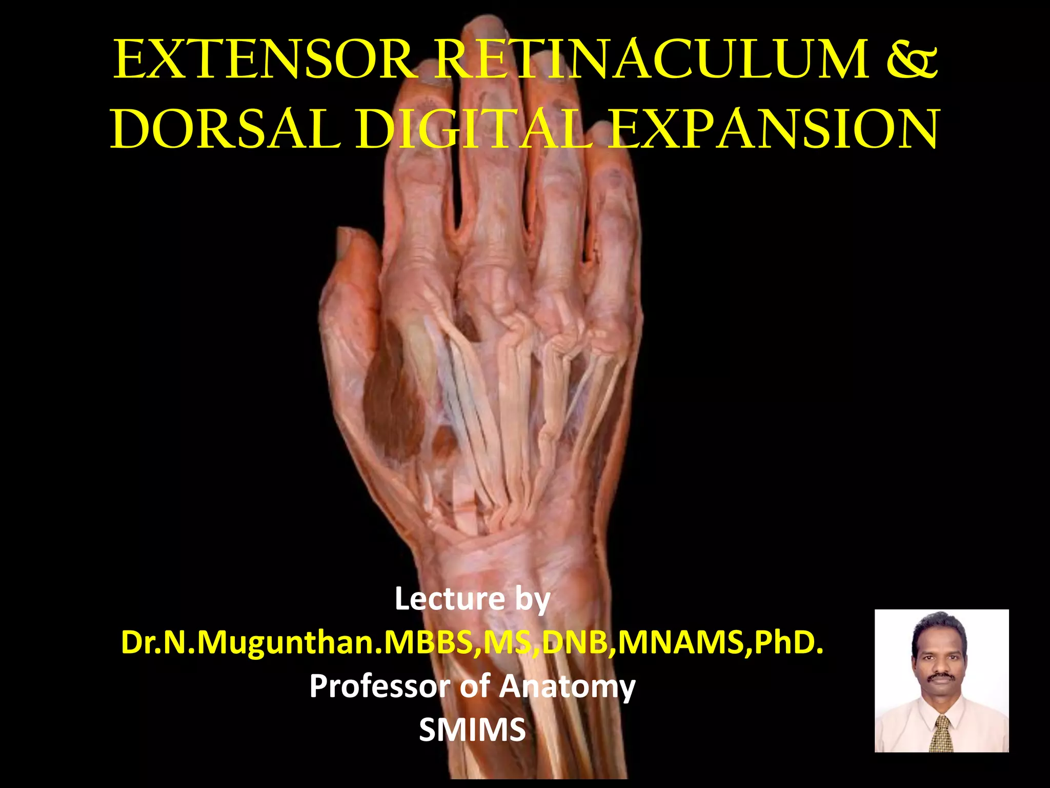
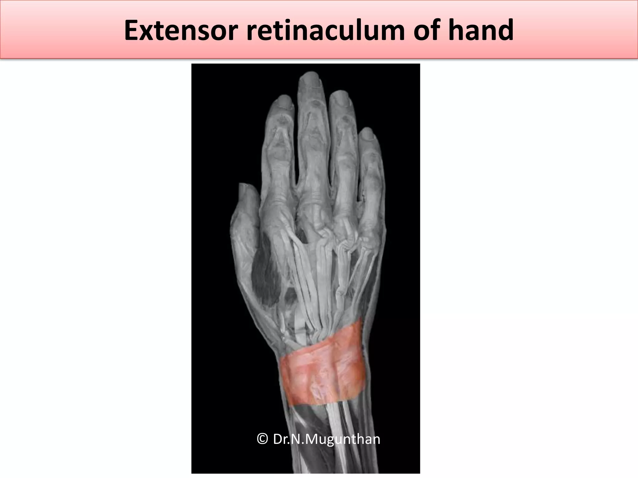
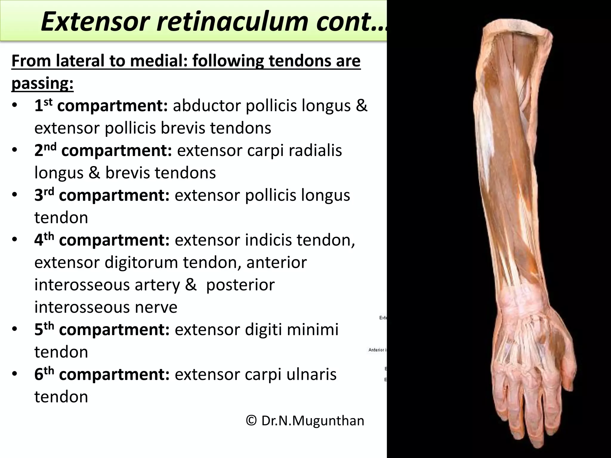
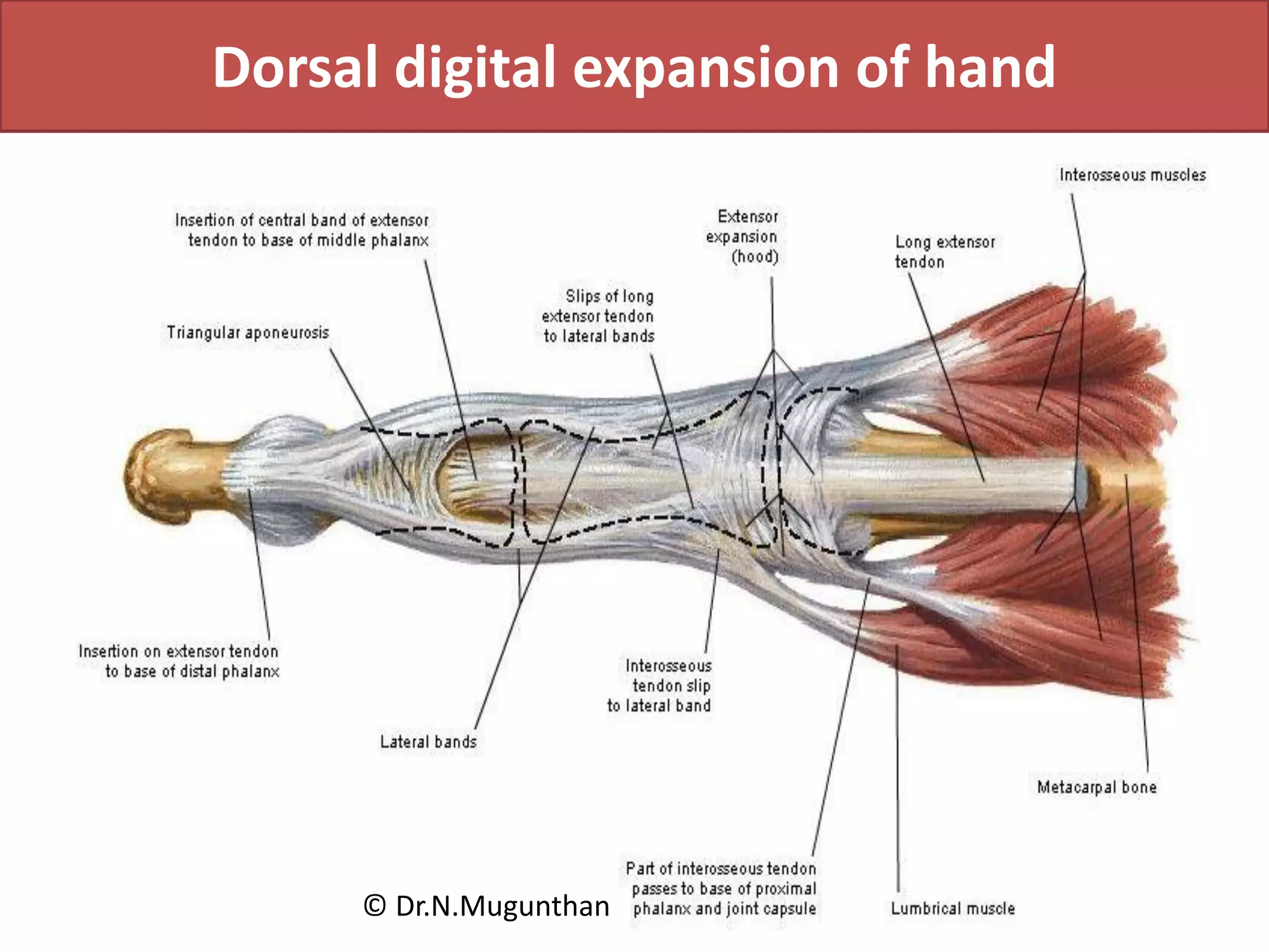
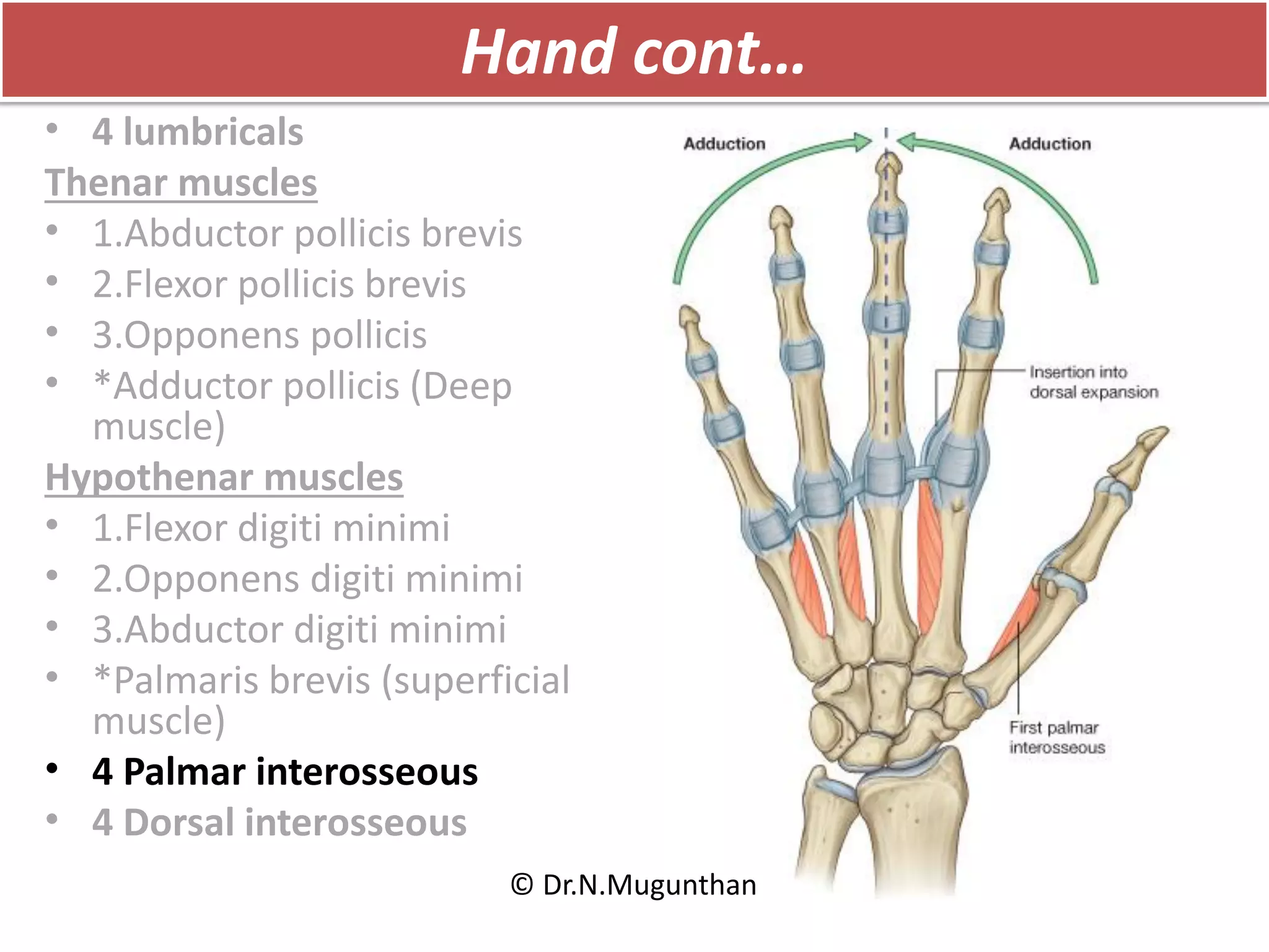
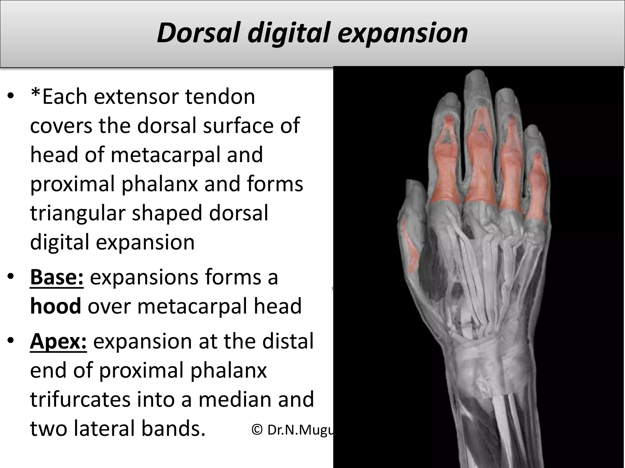
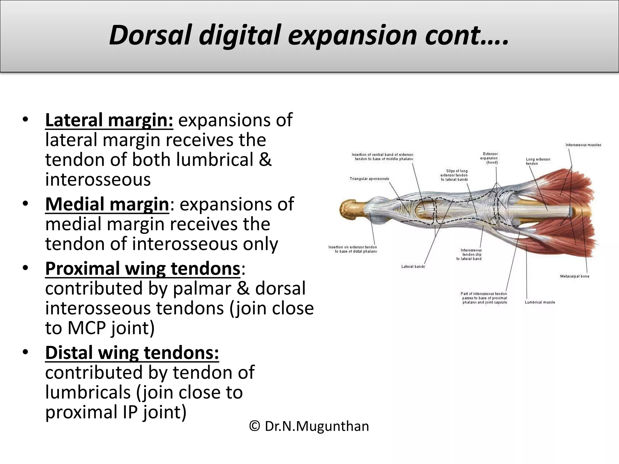
EXTENSOR RETINACULUM &
DORSAL DIGITAL EXPANSION
Lecture by
Dr.N.Mugunthan.MBBS,MS,DNB,MNAMS,PhD.
Professor of Anatomy
SMIMS
Learning objectives
Extensor retinaculum
• Attachments
• 6 compartments
• Structures passing deep to it
Dorsal digital expansion
• Formation
© Dr.N.Mugunthan
Extensor retinaculum of hand
© Dr.N.Mugunthan
Back of forearm
Superficial group:
• *Brachioradialis
• *Anconeus
• *Extensor carpi radialis longus
• Extensor carpi radialis brevis
• Extensor digitorum
• Extensor digiti minimi
• Extensor carpi ulnaris
Deep group:
• Supinator
• Abductor pollicis longus
• Extensor pollicis longus
• Extensor pollicis brevis
• Extensor indicis
© Dr.N.Mugunthan
Back of forearm cont…
Superficial group:
• *Brachioradialis
• *Anconeus
• *Extensor carpi radialis longus
• Extensor carpi radialis brevis
• Extensor digitorum
• Extensor digiti minimi
• Extensor carpi ulnaris
Deep group:
• Supinator
• Abductor pollicis longus
• Extensor pollicis longus
• Extensor pollicis brevis
• Extensor indicis
© Dr.N.Mugunthan
Extensor retinaculum
Attachment:
• Medial end: styloid process of
ulna, triquetral and pisiform
• Lateral end: lower part of anterior
border of radius
© Dr.N.Mugunthan
Extensor retinaculum cont…
From lateral to medial: following tendons are
passing:
• 1st compartment: abductor pollicis longus &
extensor pollicis brevis tendons
• 2nd compartment: extensor carpi radialis
longus & brevis tendons
• 3rd compartment: extensor pollicis longus
tendon
• 4th compartment: extensor indicis tendon,
extensor digitorum tendon, anterior
interosseous artery & posterior
interosseous nerve
• 5th compartment: extensor digiti minimi
tendon
• 6th compartment: extensor carpi ulnaris
tendon
© Dr.N.Mugunthan
Anatomical snuff box
Boundaries:
• Medial: tendon of extensor
pollicis longus
• Lateral: tendon of abductor
pollicis longus & extensor
pollicis brevis
• Floor : styloid process of
radius, scaphoid & base of
1st metacarpal
• Roof : skin , superficial
fascia which contains
cephalic vein & deep fascia
Contents :
• Terminal part of radial
artery © Dr.N.Mugunthan
Dorsal digital expansion of hand
© Dr.N.Mugunthan
Hand
• 4 lumbricals
Thenar muscles
• 1.Abductor pollicis brevis
• 2.Flexor pollicis brevis
• 3.Opponens pollicis
• *Adductor pollicis (Deep
muscle)
Hypothenar muscles
• 1.Flexor digiti minimi
• 2.Opponens digiti minimi
• 3.Abductor digiti minimi
• *Palmaris brevis (superficial
muscle)
• 4 Palmar interosseous
• 4 Dorsal interosseous
© Dr.N.Mugunthan
Hand cont…
• 4 lumbricals
Thenar muscles
• 1.Abductor pollicis brevis
• 2.Flexor pollicis brevis
• 3.Opponens pollicis
• *Adductor pollicis (Deep
muscle)
Hypothenar muscles
• 1.Flexor digiti minimi
• 2.Opponens digiti minimi
• 3.Abductor digiti minimi
• *Palmaris brevis (superficial
muscle)
• 4 Palmar interosseous
• 4 Dorsal interosseous
© Dr.N.Mugunthan
Dorsal digital expansion
• *Each extensor tendon
covers the dorsal surface of
head of metacarpal and
proximal phalanx and forms
triangular shaped dorsal
digital expansion
• Base: expansions forms a
hood over metacarpal head
• Apex: expansion at the distal
end of proximal phalanx
trifurcates into a median and
two lateral bands. © Dr.N.Mugunthan
• Lateral margin: expansions of
lateral margin receives the
tendon of both lumbrical &
interosseous
• Medial margin: expansions of
medial margin receives the
tendon of interosseous only
• Proximal wing tendons:
contributed by palmar & dorsal
interosseous tendons (join close
to MCP joint)
• Distal wing tendons:
contributed by tendon of
lumbricals (join close to
proximal IP joint)
Dorsal digital expansion cont….
© Dr.N.Mugunthan
Summary
Extensor retinaculum
• Attachments
• 6 compartments
• Structures passing deep to it
Dorsal digital expansion
• Formation
© Dr.N.Mugunthan
Extensor retinaculum & dorsal digital expansion Dr.N.Mugunthan

Extensor retinaculum & dorsal digital expansion Dr.N.Mugunthan

  • 1.
    EXTENSOR RETINACULUM & DORSALDIGITAL EXPANSION Lecture by Dr.N.Mugunthan.MBBS,MS,DNB,MNAMS,PhD. Professor of Anatomy SMIMS
  • 2.
    Learning objectives Extensor retinaculum •Attachments • 6 compartments • Structures passing deep to it Dorsal digital expansion • Formation © Dr.N.Mugunthan
  • 3.
    Extensor retinaculum ofhand © Dr.N.Mugunthan
  • 4.
    Back of forearm Superficialgroup: • *Brachioradialis • *Anconeus • *Extensor carpi radialis longus • Extensor carpi radialis brevis • Extensor digitorum • Extensor digiti minimi • Extensor carpi ulnaris Deep group: • Supinator • Abductor pollicis longus • Extensor pollicis longus • Extensor pollicis brevis • Extensor indicis © Dr.N.Mugunthan
  • 5.
    Back of forearmcont… Superficial group: • *Brachioradialis • *Anconeus • *Extensor carpi radialis longus • Extensor carpi radialis brevis • Extensor digitorum • Extensor digiti minimi • Extensor carpi ulnaris Deep group: • Supinator • Abductor pollicis longus • Extensor pollicis longus • Extensor pollicis brevis • Extensor indicis © Dr.N.Mugunthan
  • 6.
    Extensor retinaculum Attachment: • Medialend: styloid process of ulna, triquetral and pisiform • Lateral end: lower part of anterior border of radius © Dr.N.Mugunthan
  • 7.
    Extensor retinaculum cont… Fromlateral to medial: following tendons are passing: • 1st compartment: abductor pollicis longus & extensor pollicis brevis tendons • 2nd compartment: extensor carpi radialis longus & brevis tendons • 3rd compartment: extensor pollicis longus tendon • 4th compartment: extensor indicis tendon, extensor digitorum tendon, anterior interosseous artery & posterior interosseous nerve • 5th compartment: extensor digiti minimi tendon • 6th compartment: extensor carpi ulnaris tendon © Dr.N.Mugunthan
  • 8.
    Anatomical snuff box Boundaries: •Medial: tendon of extensor pollicis longus • Lateral: tendon of abductor pollicis longus & extensor pollicis brevis • Floor : styloid process of radius, scaphoid & base of 1st metacarpal • Roof : skin , superficial fascia which contains cephalic vein & deep fascia Contents : • Terminal part of radial artery © Dr.N.Mugunthan
  • 9.
    Dorsal digital expansionof hand © Dr.N.Mugunthan
  • 10.
    Hand • 4 lumbricals Thenarmuscles • 1.Abductor pollicis brevis • 2.Flexor pollicis brevis • 3.Opponens pollicis • *Adductor pollicis (Deep muscle) Hypothenar muscles • 1.Flexor digiti minimi • 2.Opponens digiti minimi • 3.Abductor digiti minimi • *Palmaris brevis (superficial muscle) • 4 Palmar interosseous • 4 Dorsal interosseous © Dr.N.Mugunthan
  • 11.
    Hand cont… • 4lumbricals Thenar muscles • 1.Abductor pollicis brevis • 2.Flexor pollicis brevis • 3.Opponens pollicis • *Adductor pollicis (Deep muscle) Hypothenar muscles • 1.Flexor digiti minimi • 2.Opponens digiti minimi • 3.Abductor digiti minimi • *Palmaris brevis (superficial muscle) • 4 Palmar interosseous • 4 Dorsal interosseous © Dr.N.Mugunthan
  • 12.
    Hand cont… • 4lumbricals Thenar muscles • 1.Abductor pollicis brevis • 2.Flexor pollicis brevis • 3.Opponens pollicis • *Adductor pollicis (Deep muscle) Hypothenar muscles • 1.Flexor digiti minimi • 2.Opponens digiti minimi • 3.Abductor digiti minimi • *Palmaris brevis (superficial muscle) • 4 Palmar interosseous • 4 Dorsal interosseous © Dr.N.Mugunthan
  • 13.
    Hand cont… • 4lumbricals Thenar muscles • 1.Abductor pollicis brevis • 2.Flexor pollicis brevis • 3.Opponens pollicis • *Adductor pollicis (Deep muscle) Hypothenar muscles • 1.Flexor digiti minimi • 2.Opponens digiti minimi • 3.Abductor digiti minimi • *Palmaris brevis (superficial muscle) • 4 Palmar interosseous • 4 Dorsal interosseous © Dr.N.Mugunthan
  • 14.
    Hand cont… • 4lumbricals Thenar muscles • 1.Abductor pollicis brevis • 2.Flexor pollicis brevis • 3.Opponens pollicis • *Adductor pollicis (Deep muscle) Hypothenar muscles • 1.Flexor digiti minimi • 2.Opponens digiti minimi • 3.Abductor digiti minimi • *Palmaris brevis (superficial muscle) • 4 Palmar interosseous • 4 Dorsal interosseous © Dr.N.Mugunthan
  • 15.
    Dorsal digital expansion •*Each extensor tendon covers the dorsal surface of head of metacarpal and proximal phalanx and forms triangular shaped dorsal digital expansion • Base: expansions forms a hood over metacarpal head • Apex: expansion at the distal end of proximal phalanx trifurcates into a median and two lateral bands. © Dr.N.Mugunthan
  • 16.
    • Lateral margin:expansions of lateral margin receives the tendon of both lumbrical & interosseous • Medial margin: expansions of medial margin receives the tendon of interosseous only • Proximal wing tendons: contributed by palmar & dorsal interosseous tendons (join close to MCP joint) • Distal wing tendons: contributed by tendon of lumbricals (join close to proximal IP joint) Dorsal digital expansion cont…. © Dr.N.Mugunthan
  • 17.
    Summary Extensor retinaculum • Attachments •6 compartments • Structures passing deep to it Dorsal digital expansion • Formation © Dr.N.Mugunthan