Bootcamp Muscle Tables.pdf with origin insertion and action
1.
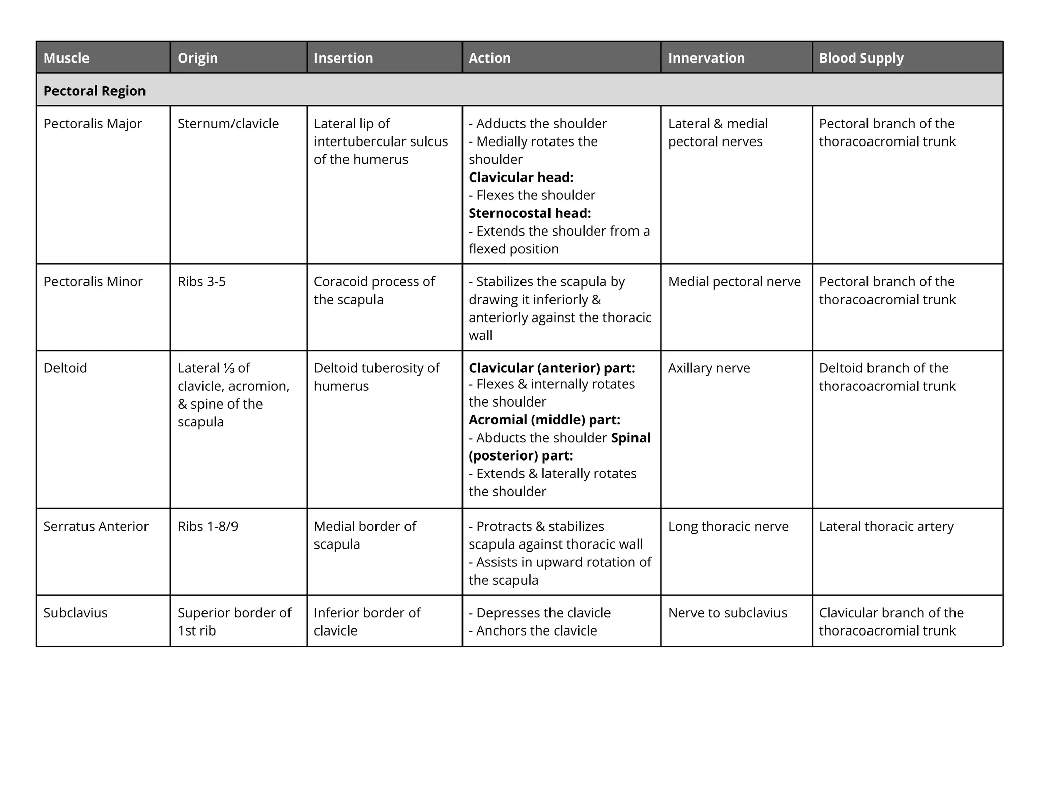
Muscle Origin InsertionAction Innervation Blood Supply
Pectoral Region
Pectoralis Major Sternum/clavicle Lateral lip of
intertubercular sulcus
of the humerus
- Adducts the shoulder
- Medially rotates the
shoulder
Clavicular head:
- Flexes the shoulder
Sternocostal head:
- Extends the shoulder from a
flexed position
Lateral & medial
pectoral nerves
Pectoral branch of the
thoracoacromial trunk
Pectoralis Minor Ribs 3-5 Coracoid process of
the scapula
- Stabilizes the scapula by
drawing it inferiorly &
anteriorly against the thoracic
wall
Medial pectoral nerve Pectoral branch of the
thoracoacromial trunk
Deltoid Lateral ⅓ of
clavicle, acromion,
& spine of the
scapula
Deltoid tuberosity of
humerus
Clavicular (anterior) part:
- Flexes & internally rotates
the shoulder
Acromial (middle) part:
- Abducts the shoulder Spinal
(posterior) part:
- Extends & laterally rotates
the shoulder
Axillary nerve Deltoid branch of the
thoracoacromial trunk
Serratus Anterior Ribs 1-8/9 Medial border of
scapula
- Protracts & stabilizes
scapula against thoracic wall
- Assists in upward rotation of
the scapula
Long thoracic nerve Lateral thoracic artery
Subclavius Superior border of
1st rib
Inferior border of
clavicle
- Depresses the clavicle
- Anchors the clavicle
Nerve to subclavius Clavicular branch of the
thoracoacromial trunk
2.
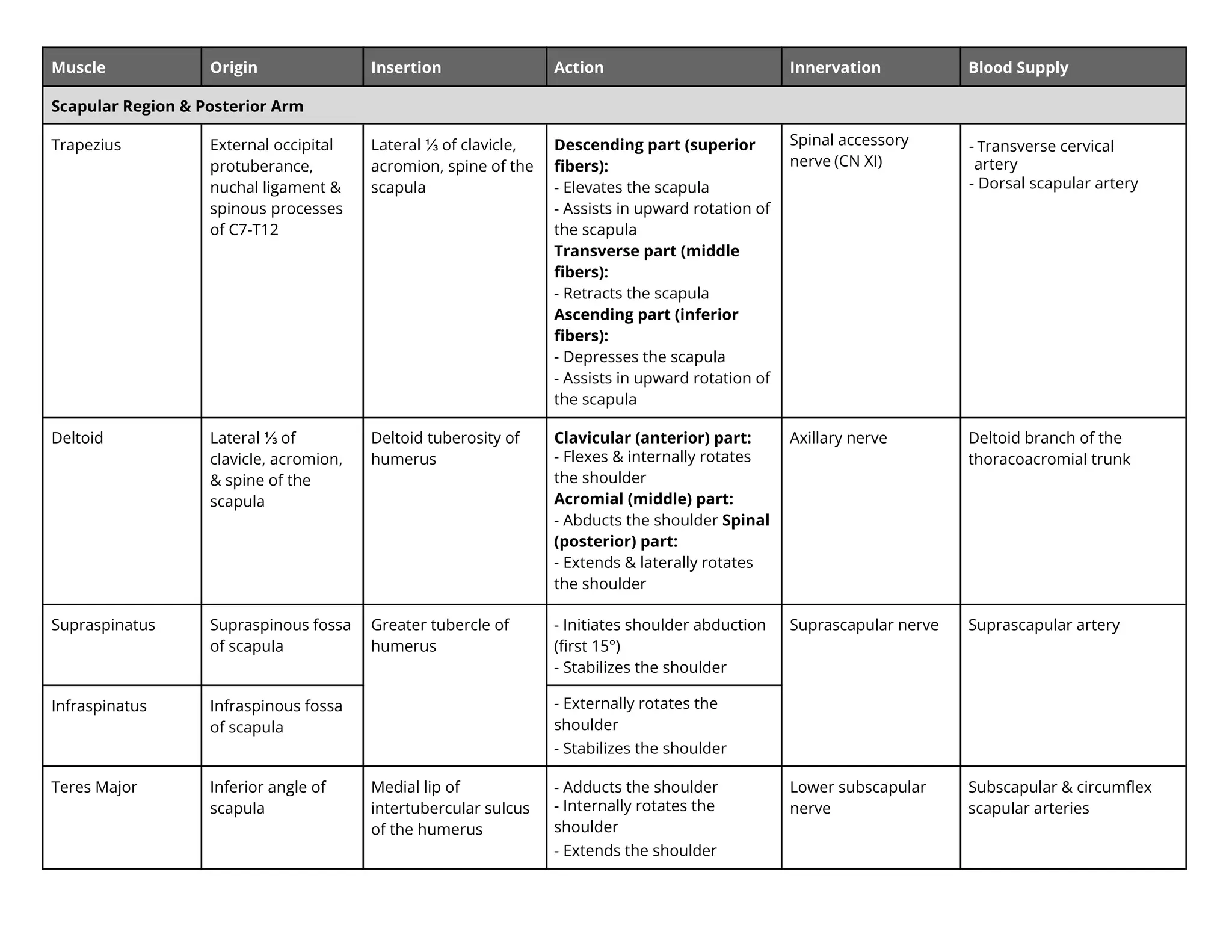
Muscle Origin InsertionAction Innervation Blood Supply
Scapular Region & Posterior Arm
Trapezius External occipital
protuberance,
nuchal ligament &
spinous processes
of C7-T12
Lateral ⅓ of clavicle,
acromion, spine of the
scapula
Descending part (superior
fibers):
- Elevates the scapula
- Assists in upward rotation of
the scapula
Transverse part (middle
fibers):
- Retracts the scapula
Ascending part (inferior
fibers):
- Depresses the scapula
- Assists in upward rotation of
the scapula
Spinal accessory
nerve (CN XI)
- Transverse cervical
artery
- Dorsal scapular artery
Deltoid Lateral ⅓ of
clavicle, acromion,
& spine of the
scapula
Deltoid tuberosity of
humerus
Clavicular (anterior) part:
- Flexes & internally rotates
the shoulder
Acromial (middle) part:
- Abducts the shoulder Spinal
(posterior) part:
- Extends & laterally rotates
the shoulder
Axillary nerve Deltoid branch of the
thoracoacromial trunk
Supraspinatus Supraspinous fossa
of scapula
Greater tubercle of
humerus
- Initiates shoulder abduction
(first 15°)
- Stabilizes the shoulder
Suprascapular nerve Suprascapular artery
Infraspinatus Infraspinous fossa
of scapula
- ternally rotates the
shoulder
- Stabilizes the shoulder
Teres Major Inferior angle of
scapula
Medial lip of
intertubercular sulcus
of the humerus
- Adducts the shoulder
- nternally rotates the
shoulder
- Extends the shoulder
Lower subscapular
nerve
Subscapular & circumflex
scapular arteries
3.
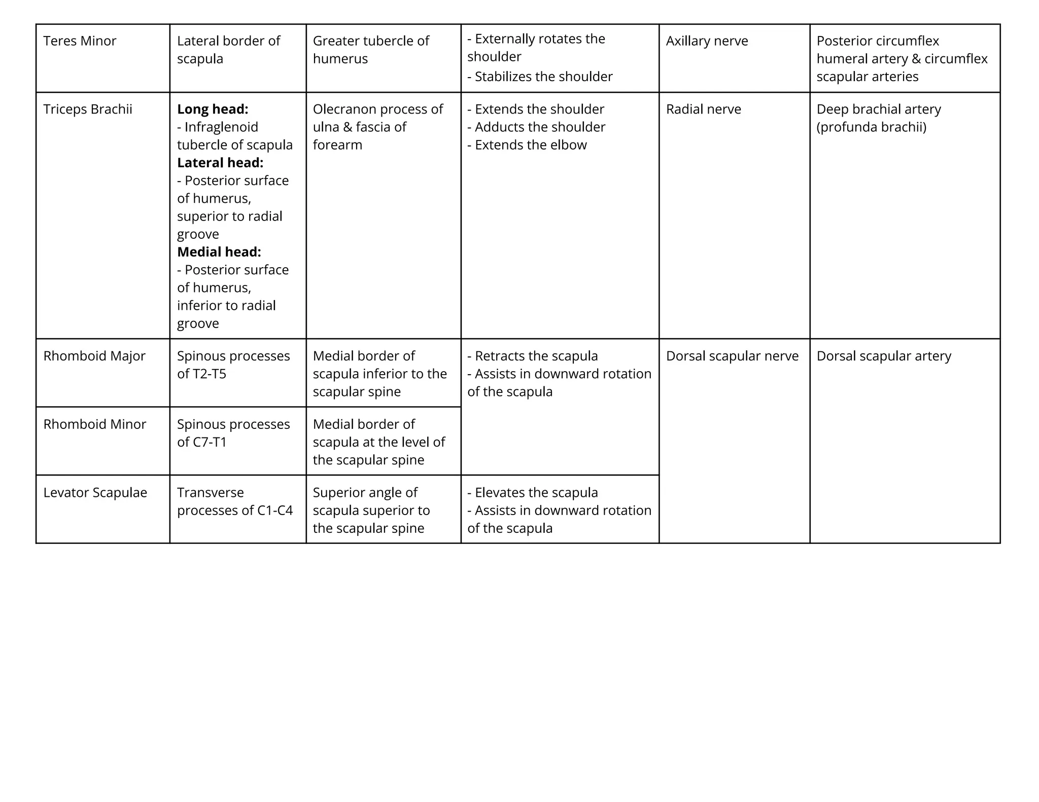
Teres Minor Lateralborder of
scapula
Greater tubercle of
humerus
- ternally rotates the
shoulder
- Stabilizes the shoulder
Axillary nerve Posterior circumflex
humeral artery & circumflex
scapular arteries
Triceps Brachii Long head:
- Infraglenoid
tubercle of scapula
Lateral head:
- Posterior surface
of humerus,
superior to radial
groove
Medial head:
- Posterior surface
of humerus,
inferior to radial
groove
Olecranon process of
ulna & fascia of
forearm
- Extends the shoulder
- Adducts the shoulder
- Extends the elbow
Radial nerve Deep brachial artery
(profunda brachii)
Rhomboid Major Spinous processes
of T2-T5
Medial border of
scapula inferior to the
scapular spine
- Retracts the scapula
- Assists in downward rotation
of the scapula
Dorsal scapular nerve Dorsal scapular artery
Rhomboid Minor Spinous processes
of C7-T1
Medial border of
scapula at the level of
the scapular spine
Levator Scapulae Transverse
processes of C1-C4
Superior angle of
scapula superior to
the scapular spine
- Elevates the scapula
- Assists in downward rotation
of the scapula
4.
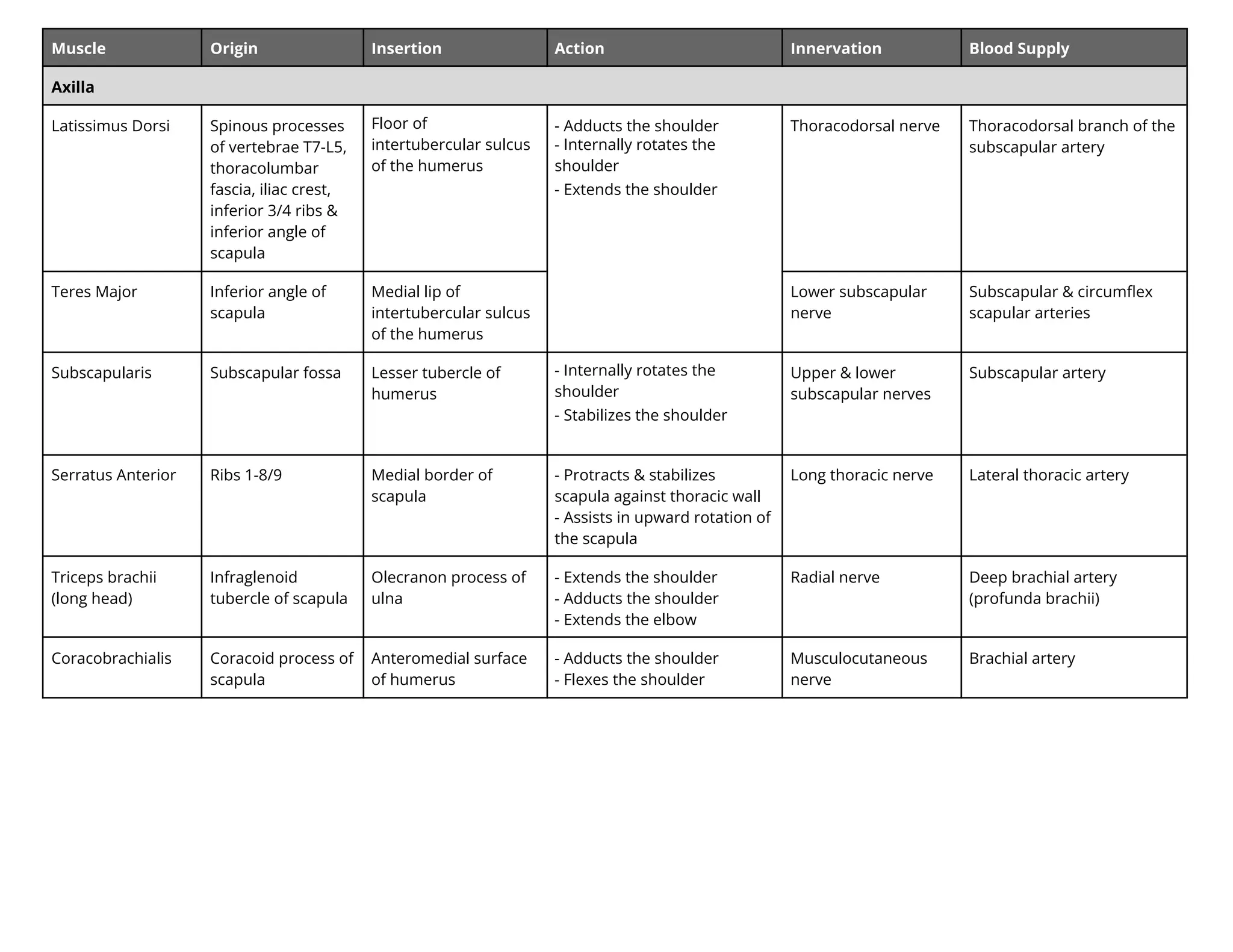
Muscle Origin InsertionAction Innervation Blood Supply
Axilla
Latissimus Dorsi Spinous processes
of vertebrae T7-L5,
thoracolumbar
fascia, iliac crest,
inferior 3/4 ribs &
inferior angle of
scapula
Floor of
intertubercular sulcus
of the humerus
- Adducts the shoulder
- nternally rotates the
shoulder
- Extends the shoulder
Thoracodorsal nerve Thoracodorsal branch of the
subscapular artery
Teres Major Inferior angle of
scapula
Medial lip of
intertubercular sulcus
of the humerus
Lower subscapular
nerve
Subscapular & circumflex
scapular arteries
Subscapularis Subscapular fossa Lesser tubercle of
humerus
- nternally rotates the
shoulder
- Stabilizes the shoulder
Upper & lower
subscapular nerves
Subscapular artery
Serratus Anterior Ribs 1-8/9 Medial border of
scapula
- Protracts & stabilizes
scapula against thoracic wall
- Assists in upward rotation of
the scapula
Long thoracic nerve Lateral thoracic artery
Triceps brachii
(long head)
Infraglenoid
tubercle of scapula
Olecranon process of
ulna
- Extends the shoulder
- Adducts the shoulder
- Extends the elbow
Radial nerve Deep brachial artery
(profunda brachii)
Coracobrachialis Coracoid process of
scapula
Anteromedial surface
of humerus
- Adducts the shoulder
- Flexes the shoulder
Musculocutaneous
nerve
Brachial artery
5.
Muscle Origin InsertionAction Innervation Blood Supply
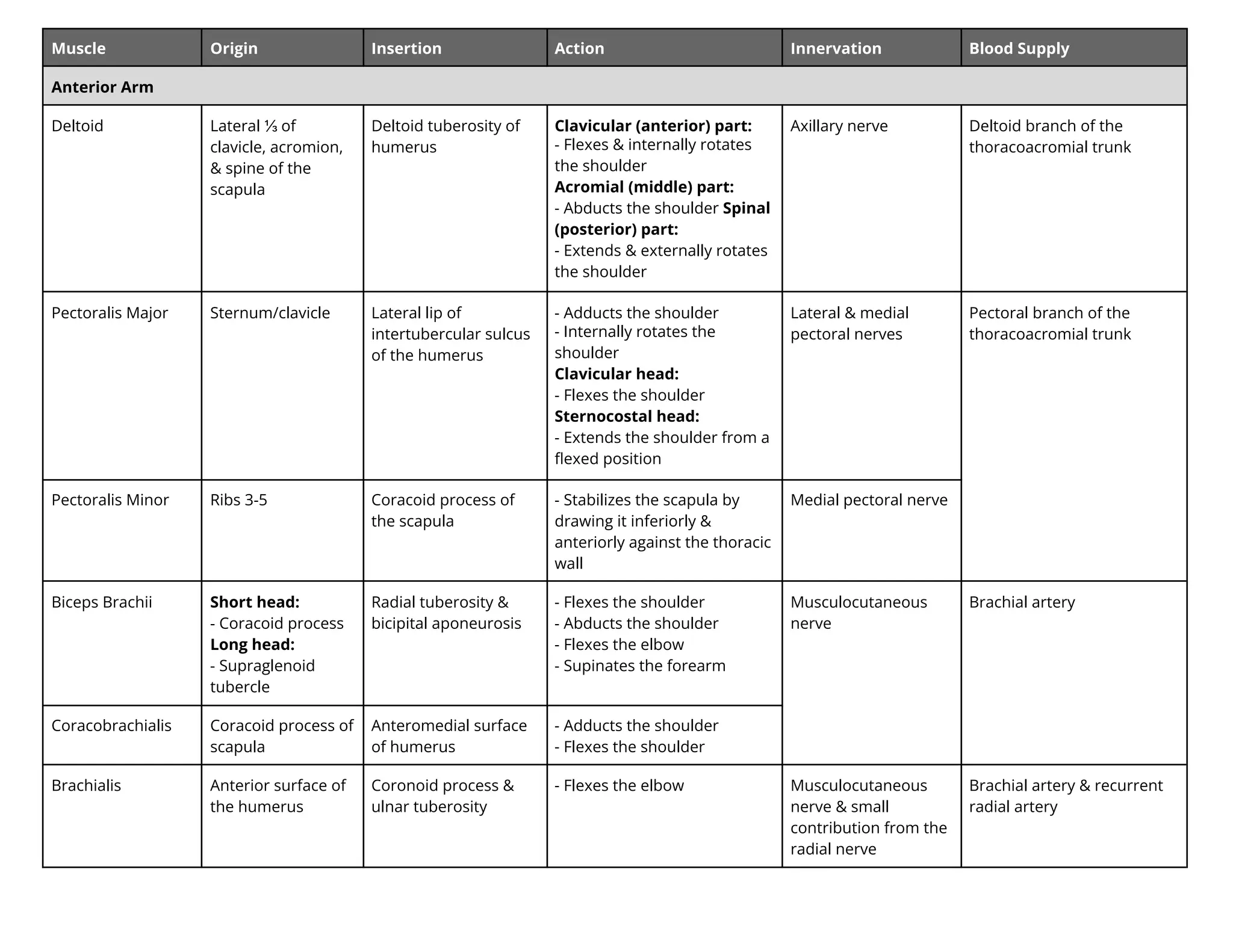
Anterior Arm
Deltoid Lateral ⅓ of
clavicle, acromion,
& spine of the
scapula
Deltoid tuberosity of
humerus
Clavicular (anterior) part:
- Flexes & internally rotates
the shoulder
Acromial (middle) part:
- Abducts the shoulder Spinal
(posterior) part:
- Extends & e ternally rotates
the shoulder
Axillary nerve Deltoid branch of the
thoracoacromial trunk
Pectoralis Major Sternum/clavicle Lateral lip of
intertubercular sulcus
of the humerus
- Adducts the shoulder
- nternally rotates the
shoulder
Clavicular head:
- Flexes the shoulder
Sternocostal head:
- Extends the shoulder from a
flexed position
Lateral & medial
pectoral nerves
Pectoral branch of the
thoracoacromial trunk
Pectoralis Minor Ribs 3-5 Coracoid process of
the scapula
- Stabilizes the scapula by
drawing it inferiorly &
anteriorly against the thoracic
wall
Medial pectoral nerve
Biceps Brachii Short head:
- Coracoid process
Long head:
- Supraglenoid
tubercle
Radial tuberosity &
bicipital aponeurosis
- Flexes the shoulder
- Abducts the shoulder
- Flexes the elbow
- Supinates the forearm
Musculocutaneous
nerve
Brachial artery
Coracobrachialis Coracoid process of
scapula
Anteromedial surface
of humerus
- Adducts the shoulder
- Flexes the shoulder
Brachialis Anterior surface of
the humerus
Coronoid process &
ulnar tuberosity
- Flexes the elbow Musculocutaneous
nerve & small
contribution from the
radial nerve
Brachial artery & recurrent
radial artery
6.
Muscle Origin InsertionAction Innervation Blood Supply
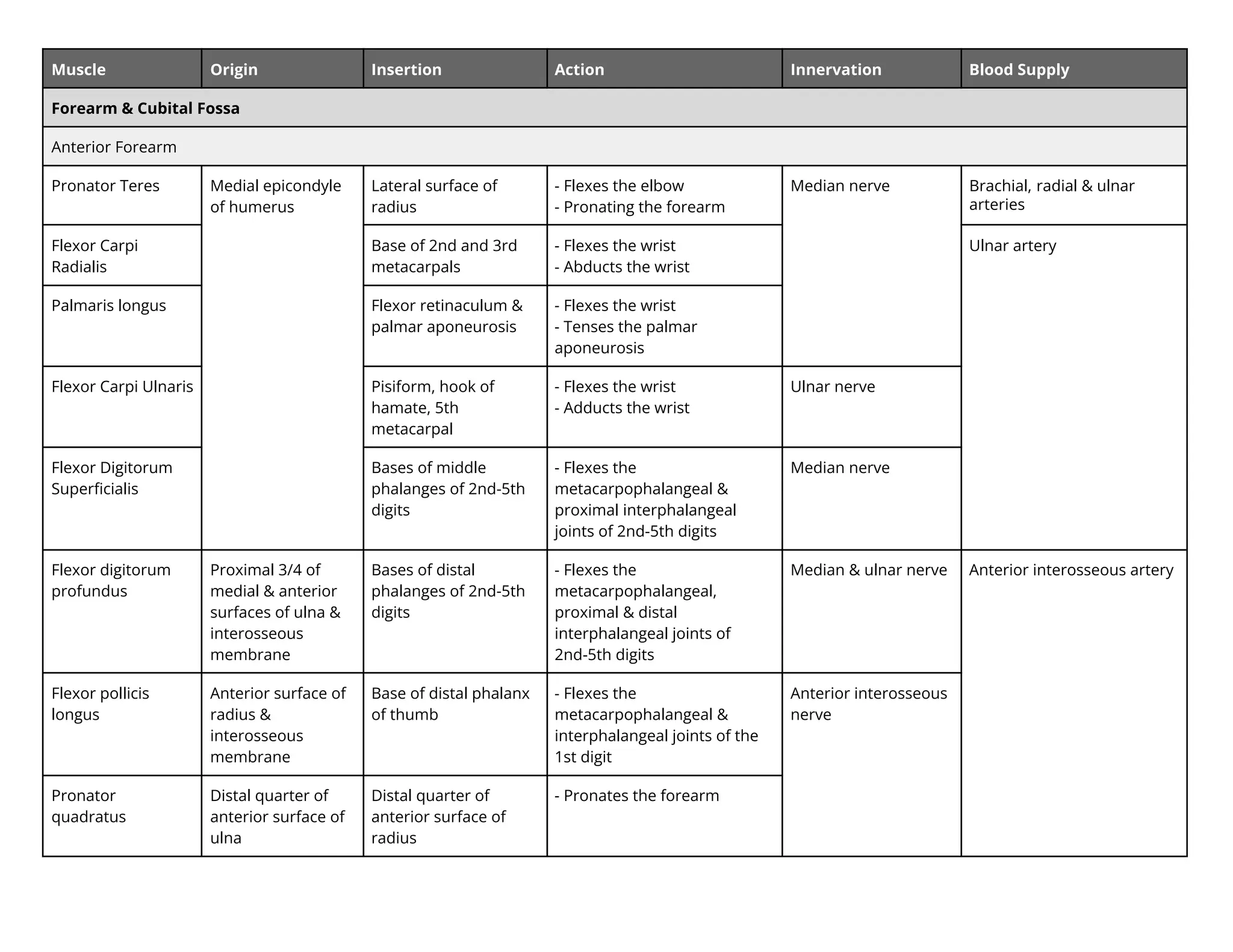
Forearm & Cubital Fossa
Anterior Forearm
Pronator Teres Medial epicondyle
of humerus
Lateral surface of
radius
- Flexes the elbow
- Pronating the forearm
Median nerve Brachial, radial & ulnar
arteries
Flexor Carpi
Radialis
Base of 2nd an r
metacarpal
- Flexes the wrist
- Abducts the wrist
Ulnar artery
Palmaris longus Flexor retinaculum &
palmar aponeurosis
- Flexes the wrist
- Tenses the palmar
aponeurosis
Flexor Carpi Ulnaris Pisiform, hook of
hamate, 5th
metacarpal
- Flexes the wrist
- Adducts the wrist
Ulnar nerve
Flexor Digitorum
Superficialis
Bases of middle
phalanges of 2nd-5th
digits
- Flexes the
metacarpophalangeal &
proximal interphalangeal
joints of 2nd-5th digits
Median nerve
Flexor digitorum
profundus
Proximal 3/4 of
medial & anterior
surfaces of ulna &
interosseous
membrane
Bases of distal
phalanges of 2nd-5th
digits
- Flexes the
metacarpophalangeal,
proximal & distal
interphalangeal joints of
2nd-5th digits
Median & ulnar nerve Anterior interosseous artery
Flexor pollicis
longus
Anterior surface of
radius &
interosseous
membrane
Base of distal phalanx
of thumb
- Flexes the
metacarpophalangeal &
interphalangeal joints of the
1st digit
Anterior interosseous
nerve
Pronator
quadratus
Distal quarter of
anterior surface of
ulna
Distal quarter of
anterior surface of
radius
- Pronates the forearm
7.
Posterior Forearm
Supinator Lateralepicondyle
of humerus
Lateral, posterior, &
anterior surfaces of
proximal 1/3 of radius
- Supinates the forearm Deep branch of radial
nerve
Recurrent radial artery
Brachioradialis Proximal 2/3 of
supra-epicondylar
ridge of humerus
Lateral surface of
distal end of radius
proximal to styloid
process
- Flexes the elbow Radial nerve
Extensor Carpi
Radialis Longus
Lateral
supra-epicondyler
ridge of humerus
Dorsal aspect of base
of 2nd metacarpal
- Extends the wrist
- Abducts the wrist
Radial artery
Extensor Carpi
Radialis Brevis
Lateral epicondyle
of humerus
Dorsal aspect of base
of 3rd metacarpal
Deep branch of radial
nerve
Abductor Pollicis
Longus
Posterior surface of
proximal halves of
ulna, radius &
interosseous
membrane
Base of 1st metacarpal - Abducts the thumb at the
carpometacarpal joint
Posterior
interosseous nerve
Posterior interosseous
artery
Extensor Pollicis
Longus
Posterior surface of
middle 1/3 of ulna
& interosseous
membrane
Dorsal aspect of base
of distal phalanx of
thumb
- Extends the interphalangeal,
metacarpophalangeal &
carpometacarpal joints of
thumb
Extensor Pollicis
Brevis
Posterior surface of
distal 1/3 of radius
& interosseous
membrane
Dorsal aspect of base
of proximal phalanx of
thumb
- Extends the
metacarpophalangeal &
carpometacarpal joints of the
thumb
Extensor Indicis Posterior surface of
distal 1/3 of ulna &
interosseous
membrane
Extensor expansion of
2nd digit
- Extends the 2nd digit
8.
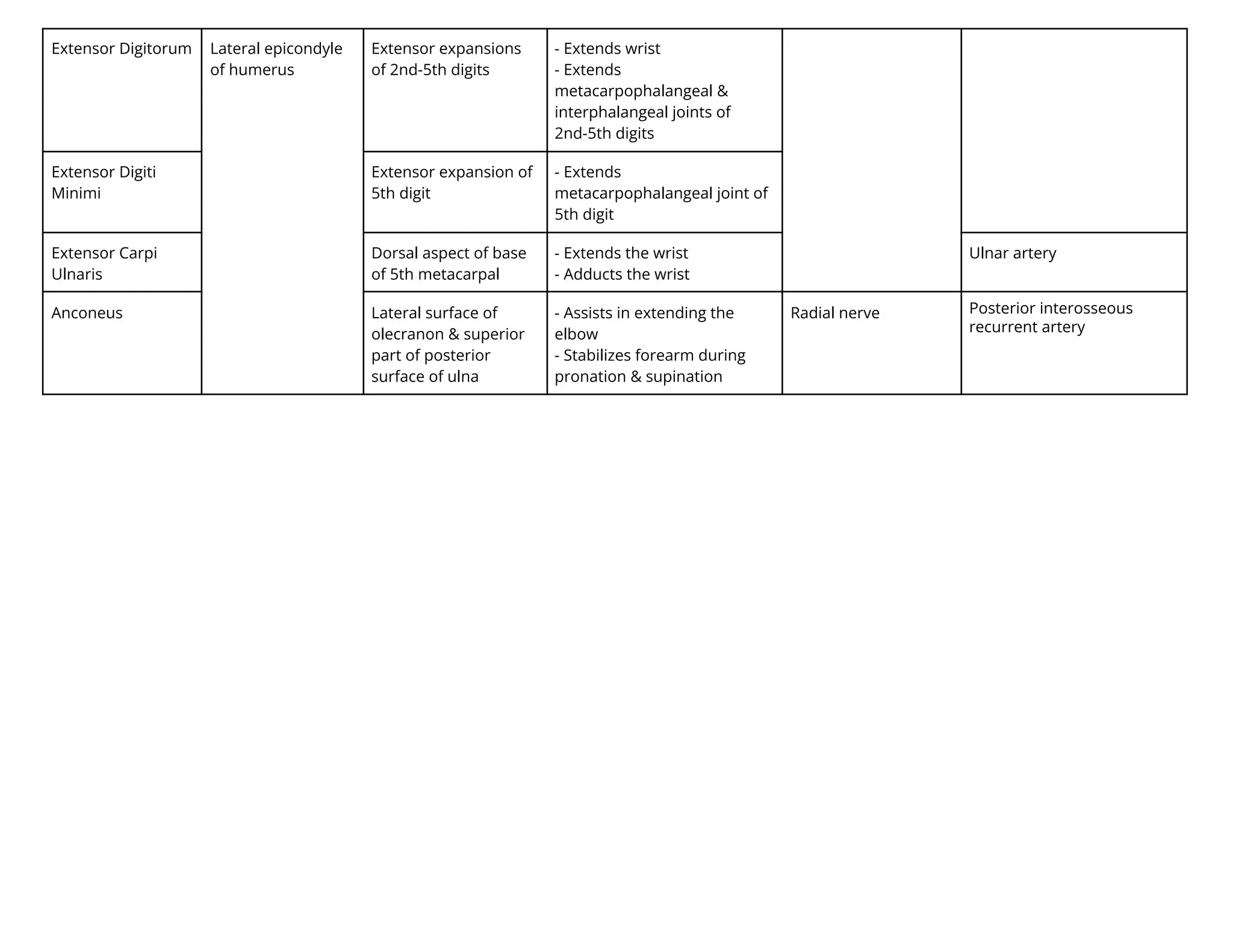
Extensor Digitorum Lateralepicondyle
of humerus
Extensor expansions
of 2nd-5th digits
- Extends wrist
- Extends
metacarpophalangeal &
interphalangeal joints of
2nd-5th digits
Extensor Digiti
Minimi
Extensor expansion of
5th digit
- Extends
metacarpophalangeal joint of
5th digit
Extensor Carpi
Ulnaris
Dorsal aspect of base
of 5th metacarpal
- Extends the wrist
- Adducts the wrist
Ulnar artery
Anconeus Lateral surface of
olecranon & superior
part of posterior
surface of ulna
- Assists in extending the
elbow
- Stabilizes forearm during
pronation & supination
Radial nerve Posterior interosseous
recurrent artery
9.
Muscle Origin InsertionAction Innervation Blood Supply
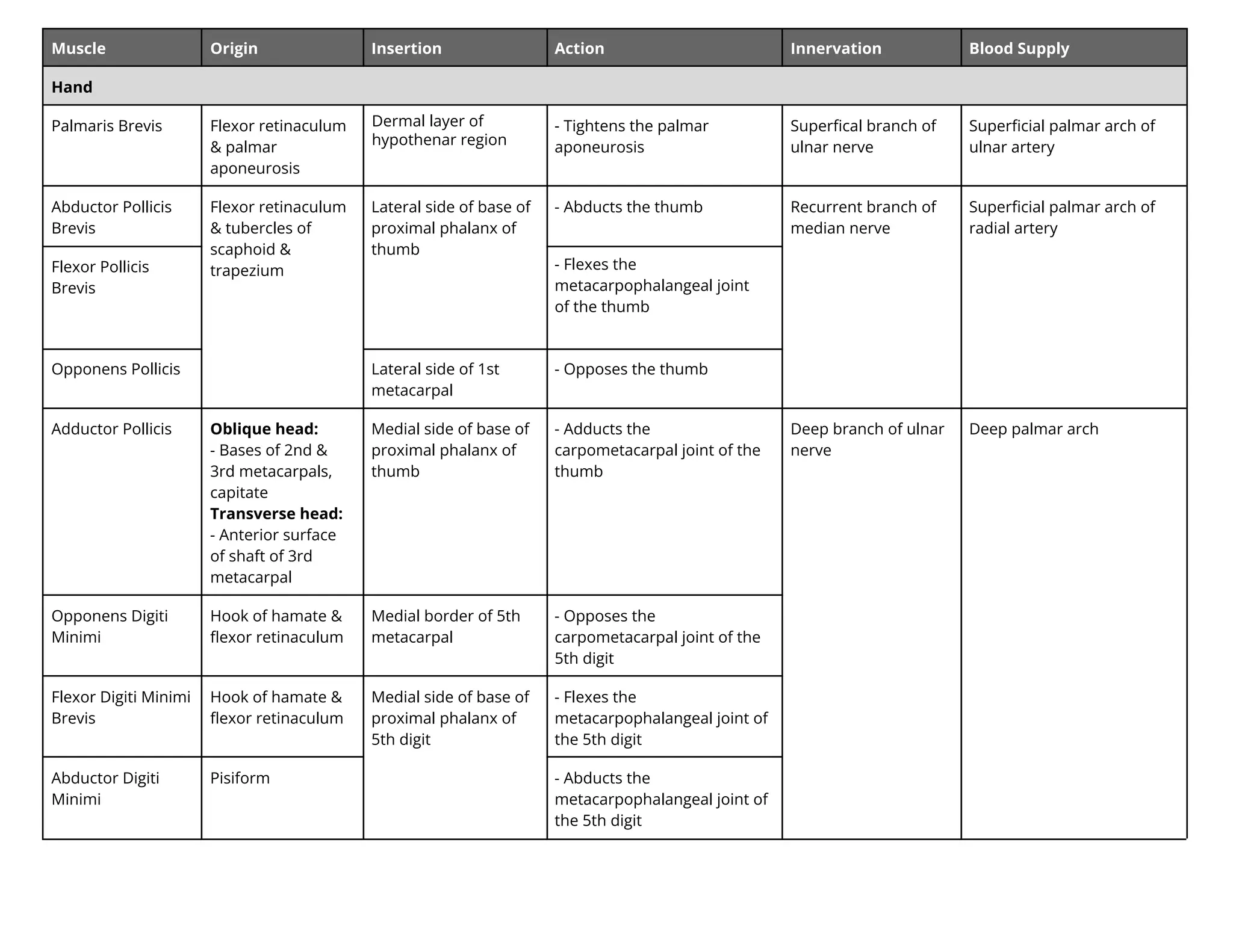
Hand
Palmaris Brevis Flexor retinaculum
& palmar
aponeurosis
er al layer
y t enar re i n
- Tightens the palmar
aponeurosis
Superfical branch of
ulnar nerve
Superficial palmar arch of
ulnar artery
Abductor Pollicis
Brevis
Flexor retinaculum
& tubercles of
scaphoid &
trapezium
Lateral side of base of
proximal phalanx of
thumb
- Abducts the thumb Recurrent branch of
median nerve
Superficial palmar arch of
radial artery
Flexor Pollicis
Brevis
- Flexes the
metacarpophalangeal joint
of the thumb
Opponens Pollicis Lateral side of 1st
metacarpal
- Opposes the thumb
Adductor Pollicis Oblique head:
- Bases of 2nd &
3rd metacarpals,
capitate
Transverse head:
- Anterior surface
of shaft of 3rd
metacarpal
Medial side of base of
proximal phalanx of
thumb
- Adducts the
carpometacarpal joint of the
thumb
Deep branch of ulnar
nerve
Deep palmar arch
Opponens Digiti
Minimi
Hook of hamate &
flexor retinaculum
Medial border of 5th
metacarpal
- Opposes the
carpometacarpal joint of the
5th digit
Flexor Digiti Minimi
Brevis
Hook of hamate &
flexor retinaculum
Medial side of base of
proximal phalanx of
5th digit
- Flexes the
metacarpophalangeal joint of
the 5th digit
Abductor Digiti
Minimi
Pisiform - Abducts the
metacarpophalangeal joint of
the 5th digit
10.
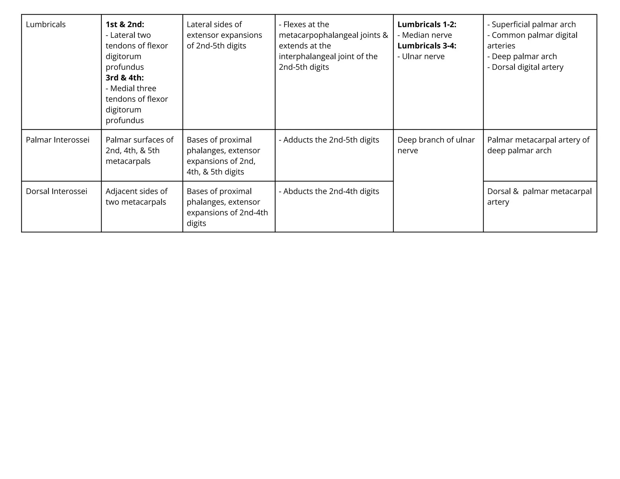
Lumbricals 1st &2nd:
- Lateral two
tendons of flexor
digitorum
profundus
3rd & 4th:
- Medial three
tendons of flexor
digitorum
profundus
Lateral sides of
extensor expansions
of 2nd-5th digits
- Flexes at the
metacarpophalangeal joints &
extends at the
interphalangeal joint of the
2nd-5th digits
Lumbricals 1-2:
- Median nerve
Lumbricals 3-4:
- Ulnar nerve
- Superficial palmar arch
- Common palmar digital
arteries
- Deep palmar arch
- Dorsal digital artery
Palmar Interossei Palmar surfaces of
2nd, 4th, & 5th
metacarpals
Bases of proximal
phalanges, extensor
expansions of 2nd,
4th, & 5th digits
- Adducts the 2nd-5th digits Deep branch of ulnar
nerve
Palmar metacarpal artery of
deep palmar arch
Dorsal Interossei Adjacent sides of
two metacarpals
Bases of proximal
phalanges, extensor
expansions of 2nd-4th
digits
- A ducts the 2nd-4th digits Dorsal & palmar metacarpal
artery
11.
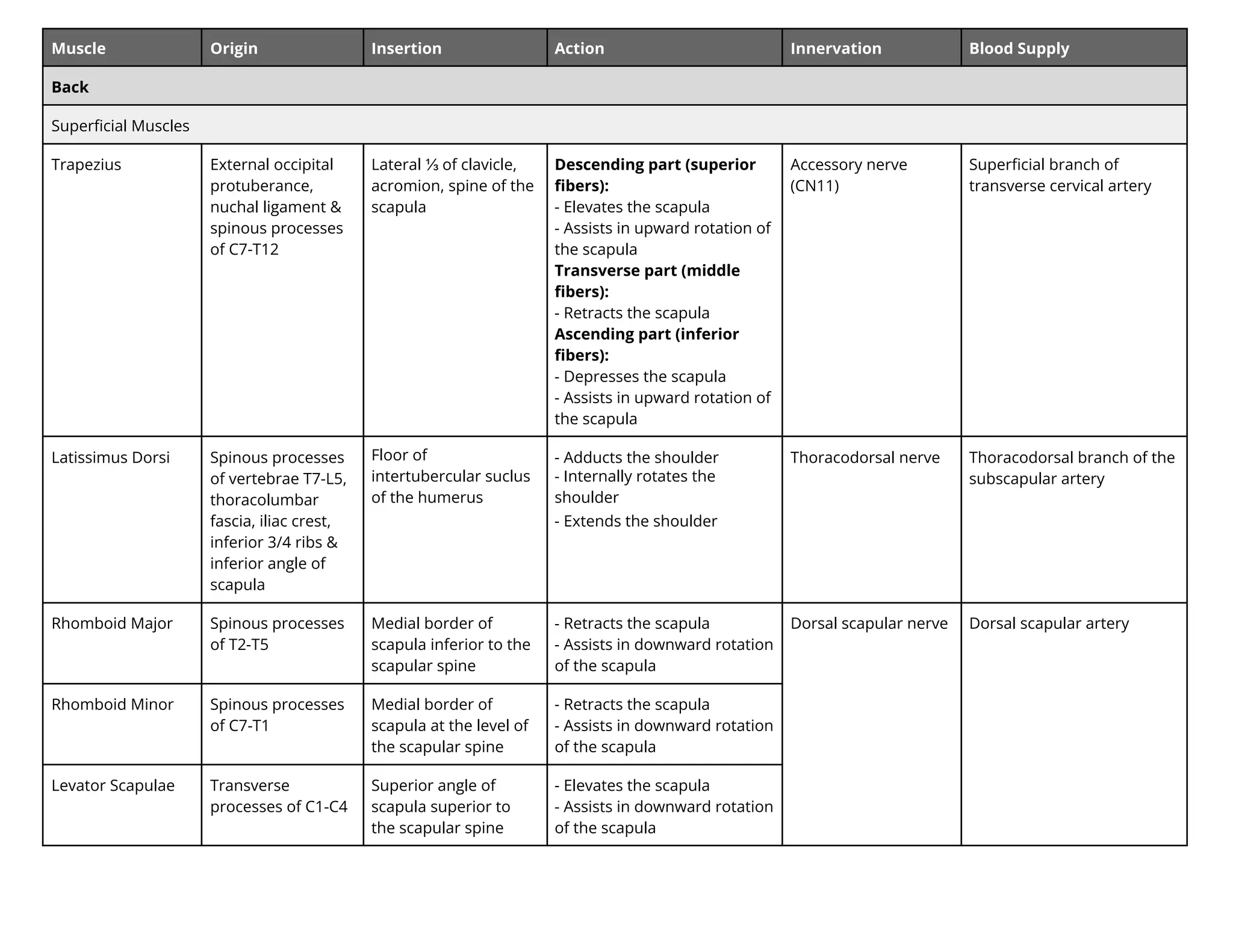
Muscle Origin InsertionAction Innervation Blood Supply
Back
Superficial Muscles
Trapezius External occipital
protuberance,
nuchal ligament &
spinous processes
of C7-T12
Lateral ⅓ of clavicle,
acromion, spine of the
scapula
Descending part (superior
fibers):
- Elevates the scapula
- Assists in upward rotation of
the scapula
Transverse part (middle
fibers):
- Retracts the scapula
Ascending part (inferior
fibers):
- Depresses the scapula
- Assists in upward rotation of
the scapula
Accessory nerve
(CN11)
Superficial branch of
transverse cervical artery
Latissimus Dorsi Spinous processes
of vertebrae T7-L5,
thoracolumbar
fascia, iliac crest,
inferior 3/4 ribs &
inferior angle of
scapula
Floor of
intertubercular suclus
of the humerus
- Adducts the shoulder
- nternally rotates the
shoulder
- Extends the shoulder
Thoracodorsal nerve Thoracodorsal branch of the
subscapular artery
Rhomboid Major Spinous processes
of T2-T5
Medial border of
scapula inferior to the
scapular spine
- Retracts the scapula
- Assists in downward rotation
of the scapula
Dorsal scapular nerve Dorsal scapular artery
Rhomboid Minor Spinous processes
of C7-T1
Medial border of
scapula at the level of
the scapular spine
- Retracts the scapula
- Assists in downward rotation
of the scapula
Levator Scapulae Transverse
processes of C1-C4
Superior angle of
scapula superior to
the scapular spine
- Elevates the scapula
- Assists in downward rotation
of the scapula
12.
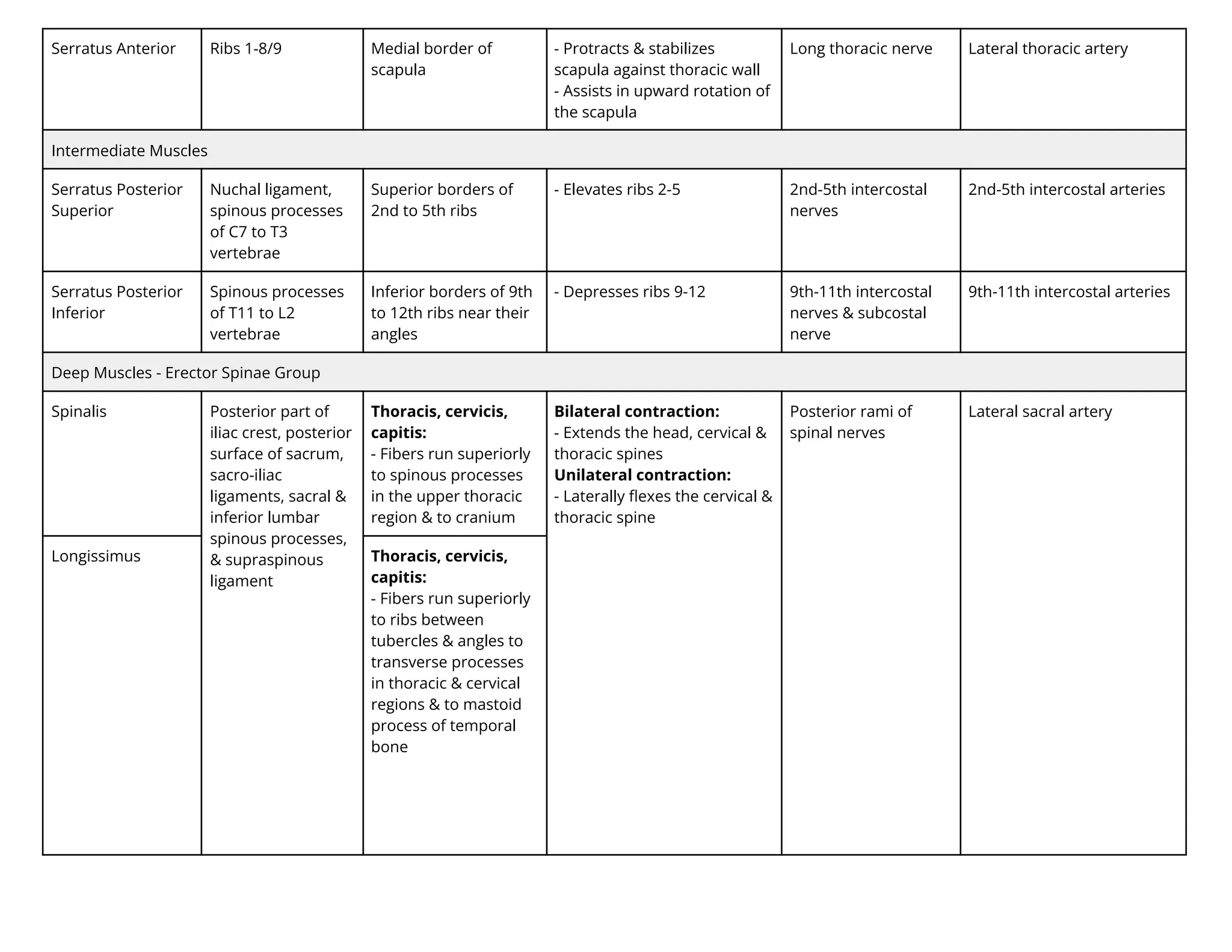
Serratus Anterior Ribs1-8/9 Medial border of
scapula
- Protracts & stabilizes
scapula against thoracic wall
- Assists in upward rotation of
the scapula
Long thoracic nerve Lateral thoracic artery
Intermediate Muscles
Serratus Posterior
Superior
Nuchal ligament,
spinous processes
of C7 to T3
vertebrae
Superior borders of
2nd to 5th ribs
- Elevates ribs 2-5 2nd-5th intercostal
nerves
2nd-5th intercostal arteries
Serratus Posterior
Inferior
Spinous processes
of T11 to L2
vertebrae
Inferior borders of 9th
to 12th ribs near their
angles
- Depresses ribs 9-12 9th-11th intercostal
nerves & subcostal
nerve
9th-11th intercostal arteries
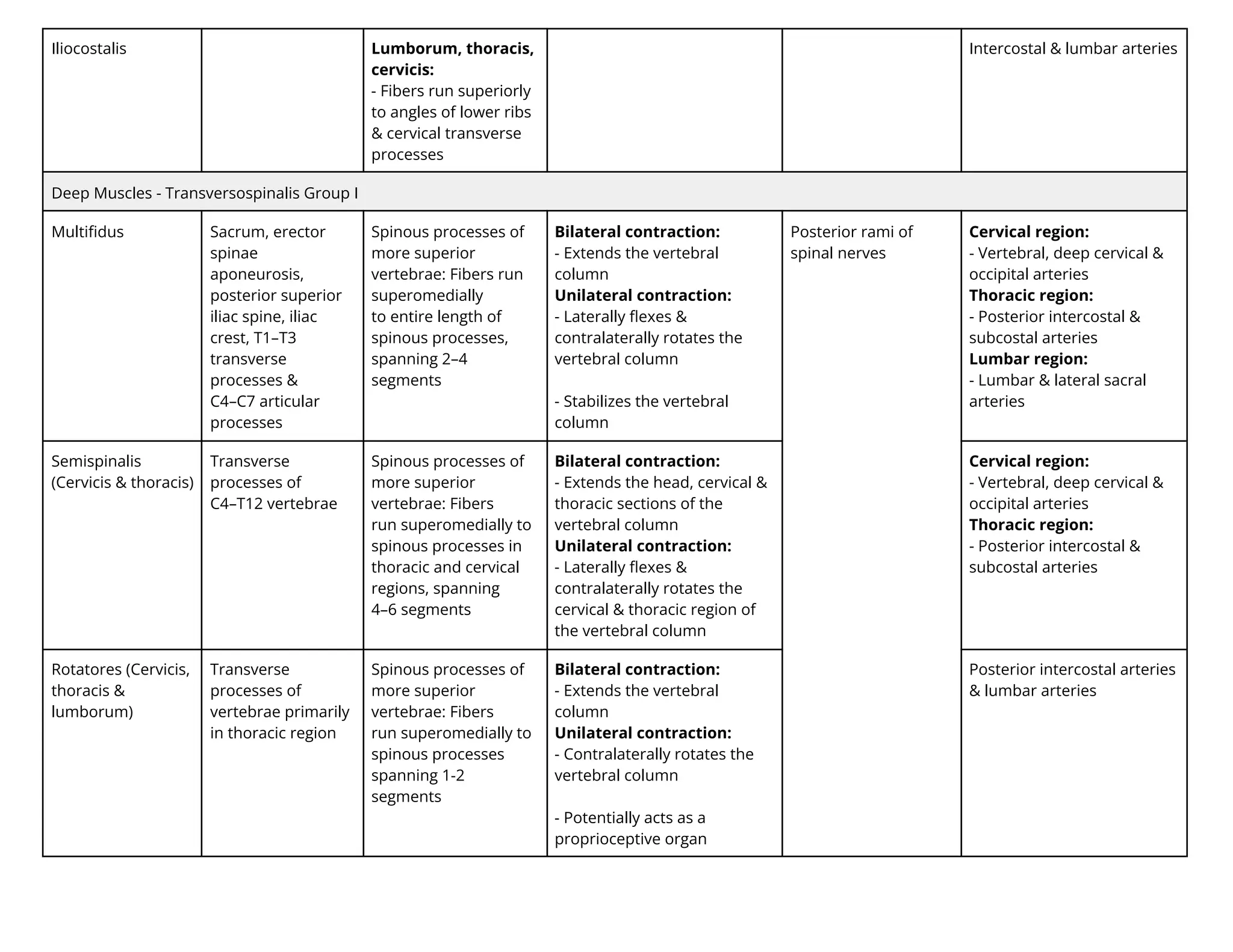
Deep Muscles - Erector Spinae Group
Spinalis Posterior part of
iliac crest, posterior
surface of sacrum,
sacro-iliac
ligaments, sacral &
inferior lumbar
spinous processes,
& supraspinous
ligament
Thoracis, cervicis,
capitis:
- Fibers run superiorly
to spinous processes
in the upper thoracic
region & to cranium
Bilateral contraction:
- Extends the head, cervical &
thoracic spines
Unilateral contraction:
- Laterally flexes the cervical &
thoracic spine
Posterior rami of
spinal nerves
Lateral sacral artery
Longissimus Thoracis, cervicis,
capitis:
- Fibers run superiorly
to ribs between
tubercles & angles to
transverse processes
in thoracic & cervical
regions & to mastoid
process of temporal
bone
13.
Iliocostalis Lumborum, thoracis,
cervicis:
-Fibers run superiorly
to angles of lower ribs
& cervical transverse
processes
Intercostal & lumbar arteries
Deep Muscles - Transversospinalis Group I
Multifidus Sacrum, erector
spinae
aponeurosis,
posterior superior
iliac spine, iliac
crest, T1–T3
transverse
processes &
C4–C7 articular
processes
Spinous processes of
more superior
vertebrae: Fibers run
superomedially
to entire length of
spinous processes,
spanning 2–4
segments
Bilateral contraction:
- Extends the vertebral
column
Unilateral contraction:
- Laterally flexes &
contralaterally rotates the
vertebral column
- Stabilizes the vertebral
column
Posterior rami of
spinal nerves
Cervical region:
- Vertebral, deep cervical &
occipital arteries
Thoracic region:
- Posterior intercostal &
subcostal arteries
Lumbar region:
- Lumbar & lateral sacral
arteries
Semispinalis
(Cervicis & thoracis)
Transverse
processes of
C4–T12 vertebrae
Spinous processes of
more superior
vertebrae: Fibers
run superomedially to
spinous processes in
thoracic and cervical
regions, spanning
4–6 segments
Bilateral contraction:
- Extends the head, cervical &
thoracic sections of the
vertebral column
Unilateral contraction:
- Laterally flexes &
contralaterally rotates the
cervical & thoracic region of
the vertebral column
Cervical region:
- Vertebral, deep cervical &
occipital arteries
Thoracic region:
- Posterior intercostal &
subcostal arteries
Rotatores (Cervicis,
thoracis &
lumborum)
Transverse
processes of
vertebrae primarily
in thoracic region
Spinous processes of
more superior
vertebrae: Fibers
run superomedially to
spinous processes
spanning 1-2
segments
Bilateral contraction:
- Extends the vertebral
column
Unilateral contraction:
- Contralaterally rotates the
vertebral column
- Potentially acts as a
proprioceptive organ
Posterior intercostal arteries
& lumbar arteries
14.
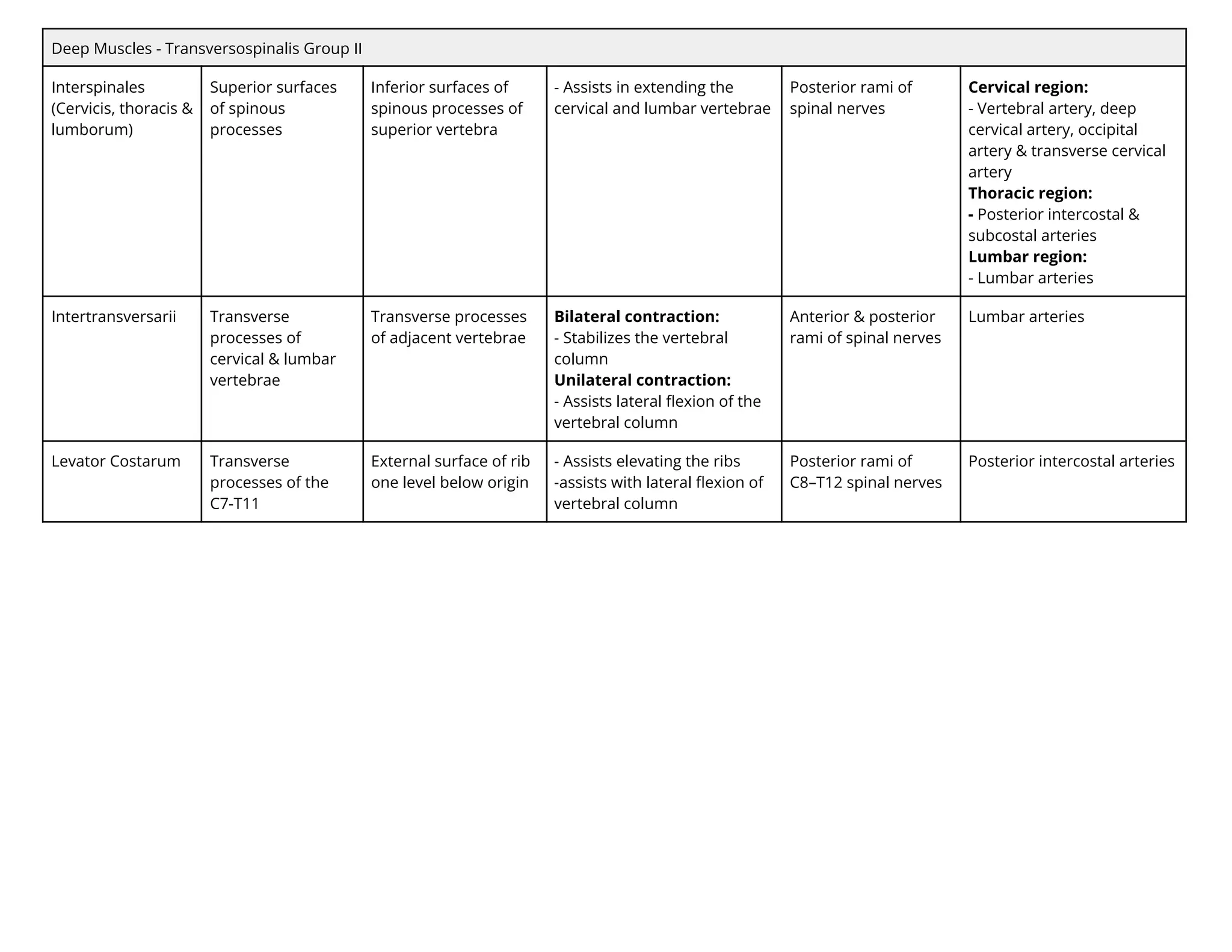
Deep Muscles -Transversospinalis Group II
Interspinales
(Cervicis, thoracis &
lumborum)
Superior surfaces
of spinous
processes
Inferior surfaces of
spinous processes of
superior vertebra
- Assists in extending the
cervical and lumbar vertebrae
Posterior rami of
spinal nerves
Cervical region:
- Vertebral artery, deep
cervical artery, occipital
artery & transverse cervical
artery
Thoracic region:
- Posterior intercostal &
subcostal arteries
Lumbar region:
- Lumbar arteries
Intertransversarii Transverse
processes of
cervical & lumbar
vertebrae
Transverse processes
of adjacent vertebrae
Bilateral contraction:
- Stabilizes the vertebral
column
Unilateral contraction:
- Assists lateral flexion of the
vertebral column
Anterior & posterior
rami of spinal nerves
Lumbar arteries
Levator Costarum Transverse
processes of the
C7-T11
External surface of rib
one level below origin
- Assists elevating the ribs
-assists with lateral flexion of
vertebral column
Posterior rami of
C8–T12 spinal nerves
Posterior intercostal arteries
15.
Muscle Origin InsertionAction Innervation Blood Supply
Anterior & Medial Thigh, Knee
Anterior Compartment
Iliopsoas Sides of T12-L5
vertebrae,
transverse
processes L1-L5
vertebrae & the
ilium (crest & fossa)
Lesser trochanter of
femur
- Flexes the hip Anterior rami of
lumbar nerves
- External iliac artery
- Femoral artery
- Iliolumbar artery
- Obturator artery
Pectineus Superior ramus of
pubis
Pectineal line of femur
just inferior to lesser
trochanter
- Adducts the hip
- Flexes the hip
- Assists in internal &
e ternal rotation of the hip
Femoral nerve Medial femoral circumflex
artery & obturator artery
Sartorius Anterior superior
iliac spine
Superior part of
medial surface of tibia
- Flexes the hip
- Abducts the hip
- ternally rotates the
hip an lexes the knee
Femoral artery
Tensor Fascia Latae Iliotibial tract - Abducts the hip
- nternally rotates the hip
Superior gluteal nerve Lateral circumflex femoral a.
& superior gluteal a.
Rectus Femoris Anterior inferior
iliac spine
Quadriceps tendon,
indirectly via patellar
ligament to tibial
tuberosity
- Extends the knee
- Flexes the hip
- Steadies the hip joint
Femoral nerve - Femoral artery
- Deep femoral artery
- Lateral circumflex femoral
artery
Vastus Lateralis Greater trochanter
& lateral lip of linea
aspera
- Extends the knee Lateral circumflex femoral
artery & deep femoral artery
Vastus Intermedius Anterior & lateral
surfaces of shaft of
femur
Deep femoral artery
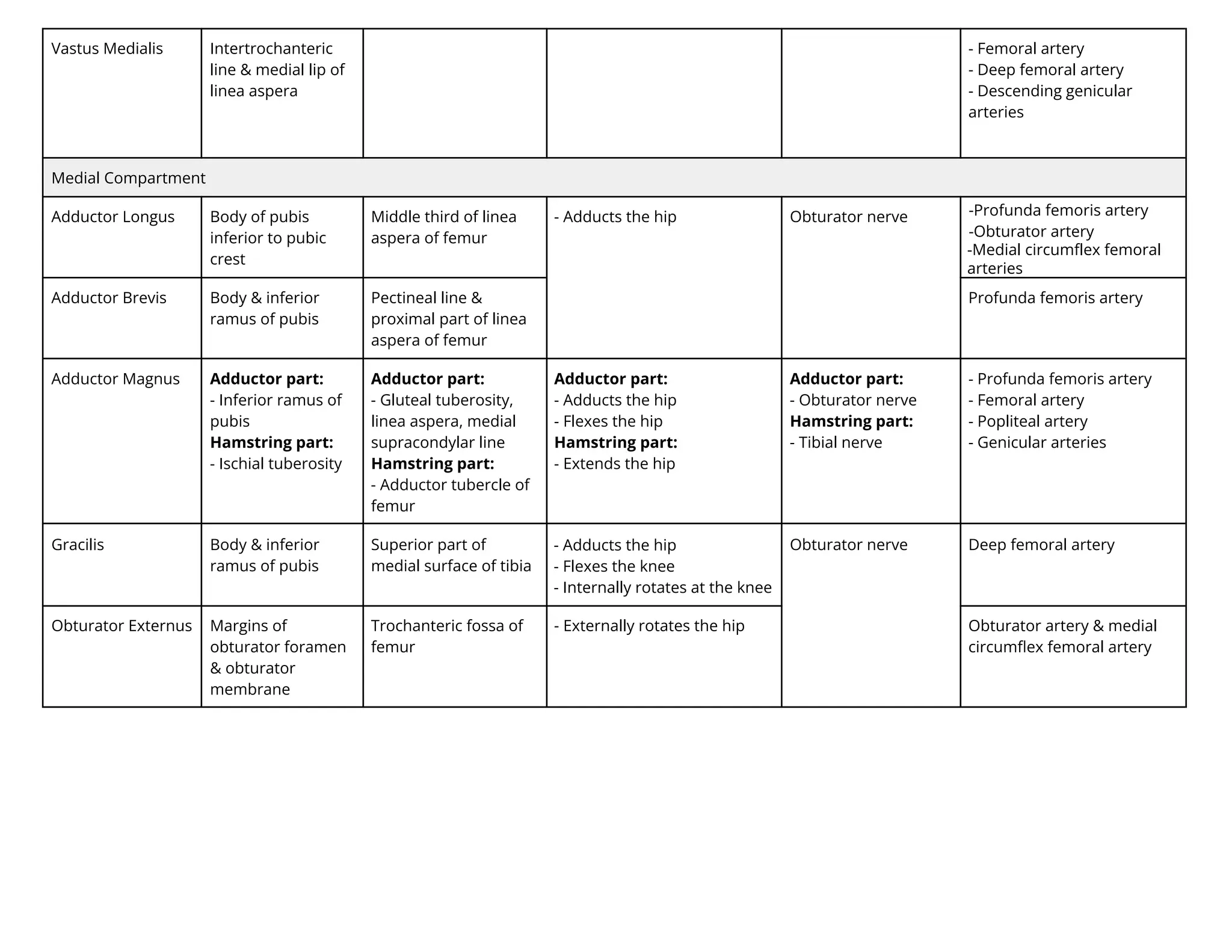
16.
Vastus Medialis Intertrochanteric
line& medial lip of
linea aspera
- Femoral artery
- Deep femoral artery
- Descending genicular
arteries
Medial Compartment
Adductor Longus Body of pubis
inferior to pubic
crest
Middle third of linea
aspera of femur
- Adducts the hip Obturator nerve r n a e ri artery
t rat r artery
Adductor Brevis Body & inferior
ramus of pubis
Pectineal line &
proximal part of linea
aspera of femur
Profunda femoris artery
Adductor Magnus Adductor part:
- Inferior ramus of
pubis
Hamstring part:
- Ischial tuberosity
Adductor part:
- Gluteal tuberosity,
linea aspera, medial
supracondylar line
Hamstring part:
- Adductor tubercle of
femur
Adductor part:
- Adducts the hip
- Flexes the hip
Hamstring part:
- Extends the hip
Adductor part:
- Obturator nerve
Hamstring part:
- Tibial nerve
- Profunda femoris artery
- Femoral artery
- Popliteal artery
- Genicular arteries
Gracilis Body & inferior
ramus of pubis
Superior part of
medial surface of tibia
- Adducts the hip
- Flexes the knee
- nternally rotates at the knee
Obturator nerve Deep femoral artery
Obturator Externus Margins of
obturator foramen
& obturator
membrane
Trochanteric fossa of
femur
- ternally rotates the hip Obturator artery & medial
circumflex femoral artery
e ial ir le e ral
arterie
17.
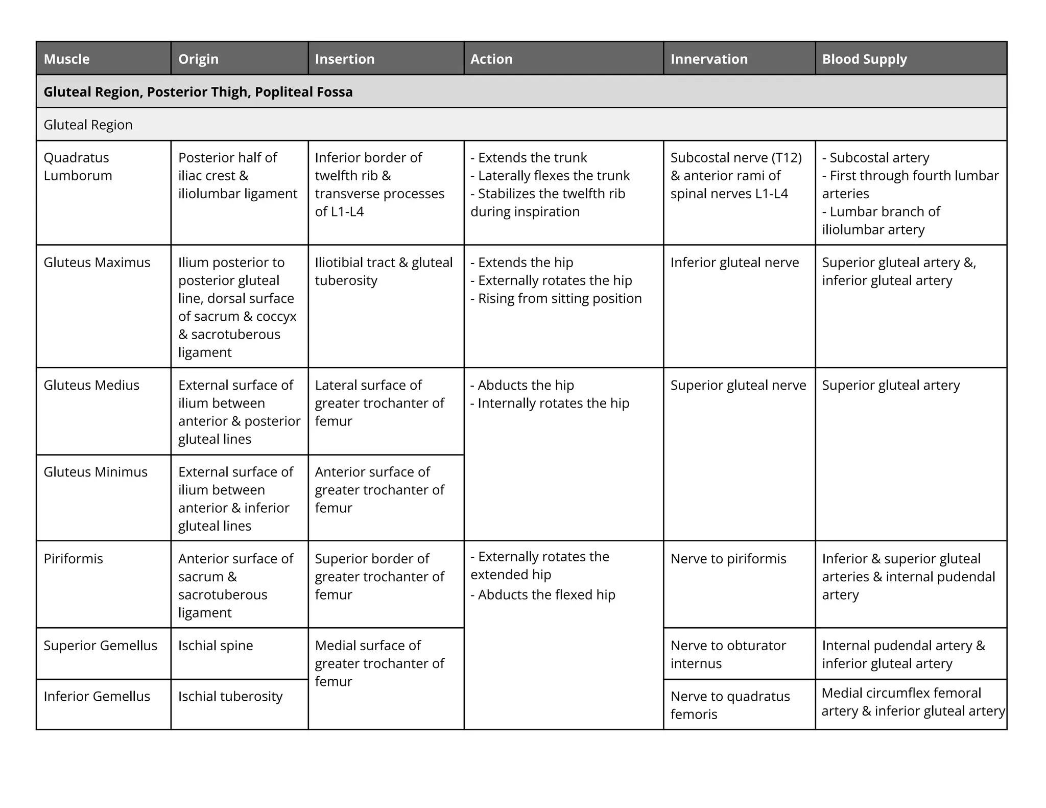
Muscle Origin InsertionAction Innervation Blood Supply
Gluteal Region, Posterior Thigh, Popliteal Fossa
Gluteal Region
Quadratus
Lumborum
Posterior half of
iliac crest &
iliolumbar ligament
Inferior border of
twelfth rib &
transverse processes
of L1-L4
- Extends the trunk
- Laterally flexes the trunk
- Stabilizes the twelfth rib
during inspiration
Subcostal nerve (T12)
& anterior rami of
spinal nerves L1-L4
- Subcostal artery
- First through fourth lumbar
arteries
- Lumbar branch of
iliolumbar artery
Gluteus Maximus Ilium posterior to
posterior gluteal
line, dorsal surface
of sacrum & coccyx
& sacrotuberous
ligament
Iliotibial tract & gluteal
tuberosity
- Extends the hip
- ternally rotates the hip
- Rising from sitting position
Inferior gluteal nerve Superior gluteal artery &,
inferior gluteal artery
Gluteus Medius External surface of
ilium between
anterior & posterior
gluteal lines
Lateral surface of
greater trochanter of
femur
- Abducts the hip
- nternally rotates the hip
Superior gluteal nerve Superior gluteal artery
Gluteus Minimus External surface of
ilium between
anterior & inferior
gluteal lines
Anterior surface of
greater trochanter of
femur
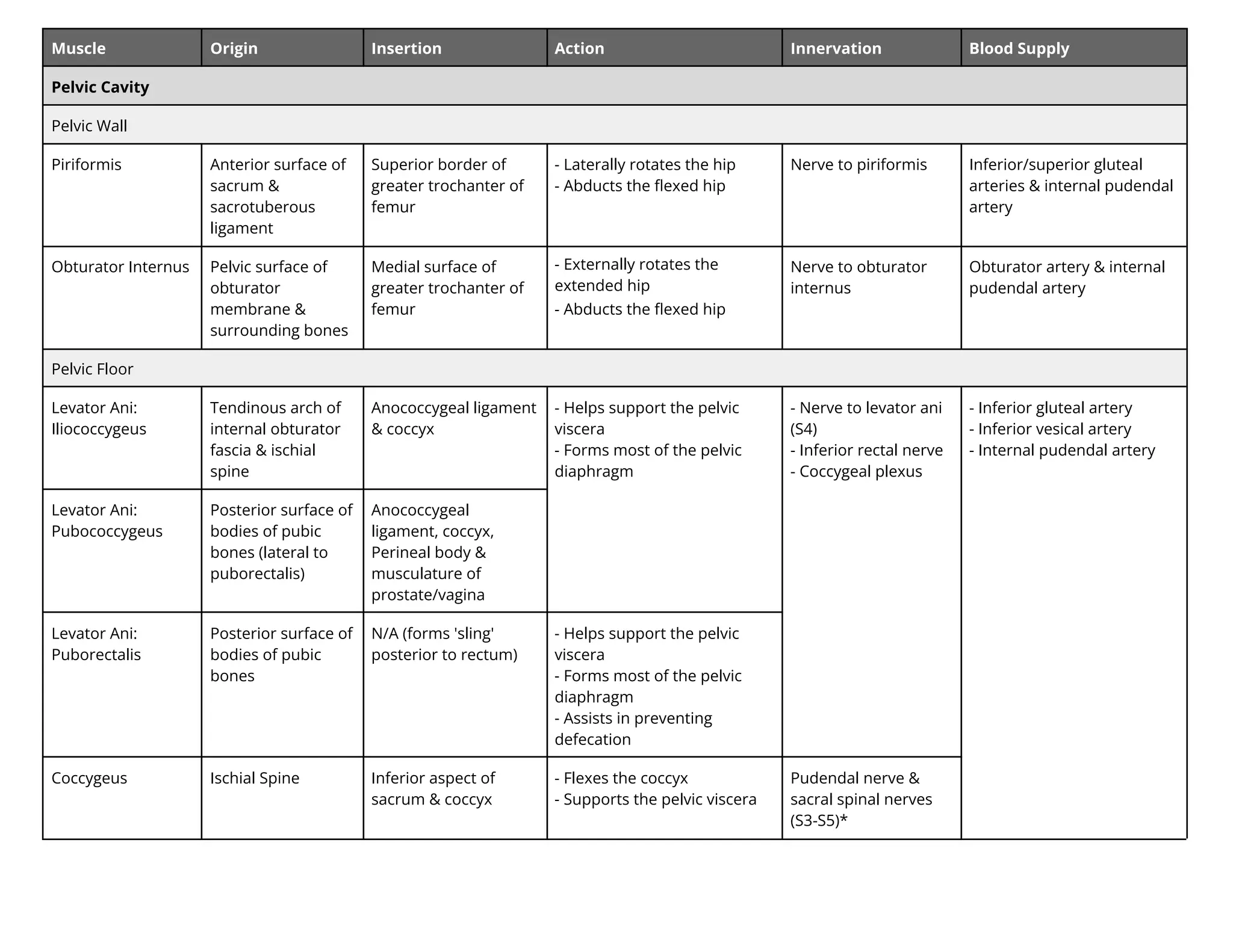
Piriformis Anterior surface of
sacrum &
sacrotuberous
ligament
Superior border of
greater trochanter of
femur
- ternally rotates the
extended hip
- Abducts the flexed hip
Nerve to piriformis Inferior & superior gluteal
arteries & internal pudendal
artery
Superior Gemellus Ischial spine Medial surface of
greater trochanter of
femur
Nerve to obturator
internus
Internal pudendal artery &
inferior gluteal artery
Inferior Gemellus Ischial tuberosity Nerve to quadratus
femoris
Medial circumflex femoral
artery in eri r l teal artery
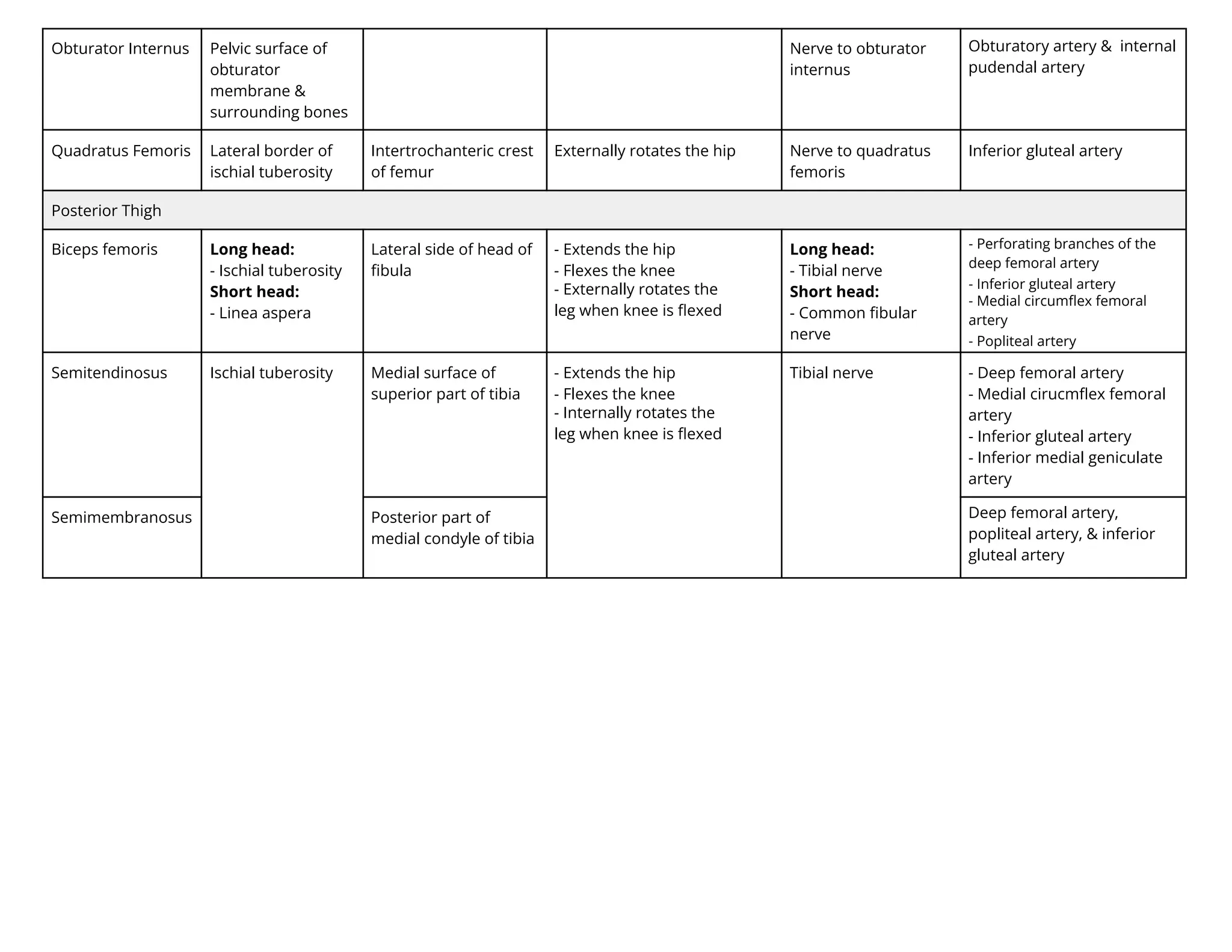
18.
Obturator Internus Pelvicsurface of
obturator
membrane &
surrounding bones
Nerve to obturator
internus
Obturatory artery & internal
pudendal artery
Quadratus Femoris Lateral border of
ischial tuberosity
Intertrochanteric crest
of femur
ternally rotates the hip Nerve to quadratus
femoris
Inferior gluteal artery
Posterior Thigh
Biceps femoris Long head:
- Ischial tuberosity
Short head:
- Linea aspera
Lateral side of head of
fibula
- Extends the hip
- Flexes the knee
- ternally rotates the
leg when knee is flexed
Long head:
- Tibial nerve
Short head:
- Common fibular
nerve
er ratin ran e t e
ee e ral artery
Inferior gluteal artery
e ial ir le e ral
artery
opliteal artery
Semitendinosus Ischial tuberosity Medial surface of
superior part of tibia
- Extends the hip
- Flexes the knee
- nternally rotates the
leg when knee is flexed
Tibial nerve - Deep femoral artery
- Medial cirucmflex femoral
artery
- Inferior gluteal artery
- Inferior medial geniculate
artery
Semimembranosus Posterior part of
medial condyle of tibia
Deep femoral artery,
popliteal artery, & inferior
gluteal artery
19.
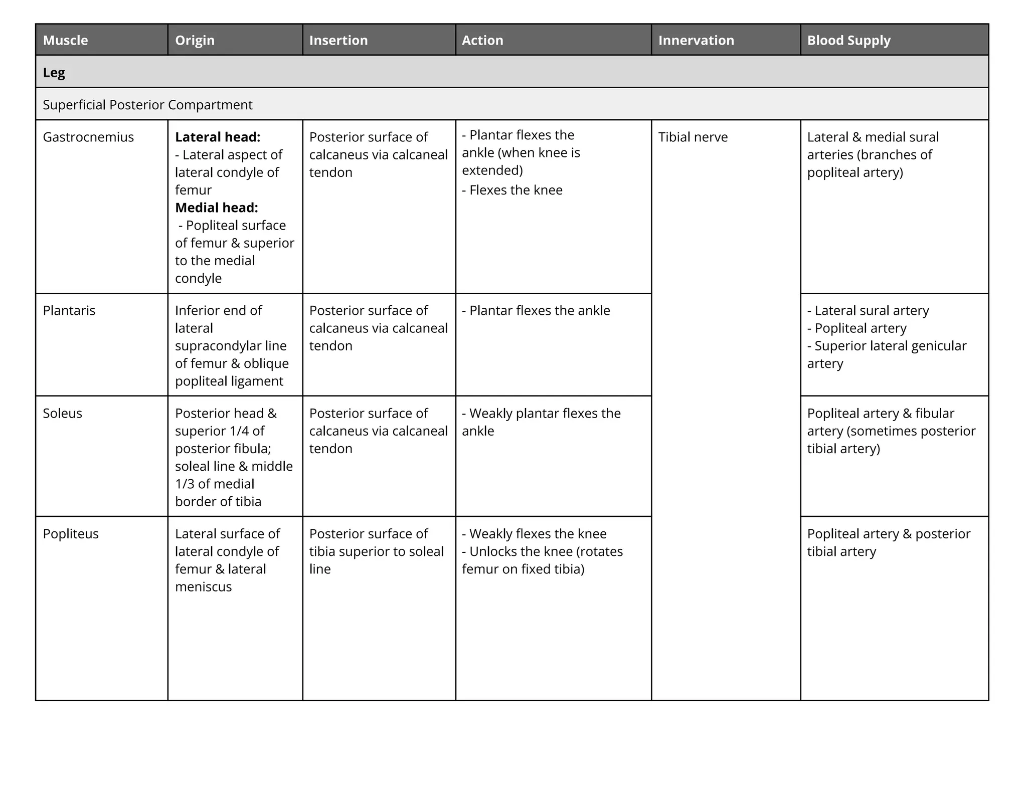
Muscle Origin InsertionAction Innervation Blood Supply
Leg
Superficial Posterior Compartment
Gastrocnemius Lateral head:
- Lateral aspect of
lateral condyle of
femur
Medial head:
- Popliteal surface
of femur & superior
to the medial
condyle
Posterior surface of
calcaneus via calcaneal
tendon
- Plantar flexes the
ankle (when knee is
extended)
- Flexes the knee
Tibial nerve Lateral & medial sural
arteries (branches of
popliteal artery)
Plantaris Inferior end of
lateral
supracondylar line
of femur & oblique
popliteal ligament
Posterior surface of
calcaneus via calcaneal
tendon
- Plantar flexes the ankle - Lateral sural artery
- Popliteal artery
- Superior lateral genicular
artery
Soleus Posterior head &
superior 1/4 of
posterior fibula;
soleal line & middle
1/3 of medial
border of tibia
Posterior surface of
calcaneus via calcaneal
tendon
- Weakly plantar flexes the
ankle
Popliteal artery & fibular
artery (sometimes posterior
tibial artery)
Popliteus Lateral surface of
lateral condyle of
femur & lateral
meniscus
Posterior surface of
tibia superior to soleal
line
- Weakly flexes the knee
- Unlocks the knee (rotates
femur on fixed tibia)
Popliteal artery & posterior
tibial artery
20.
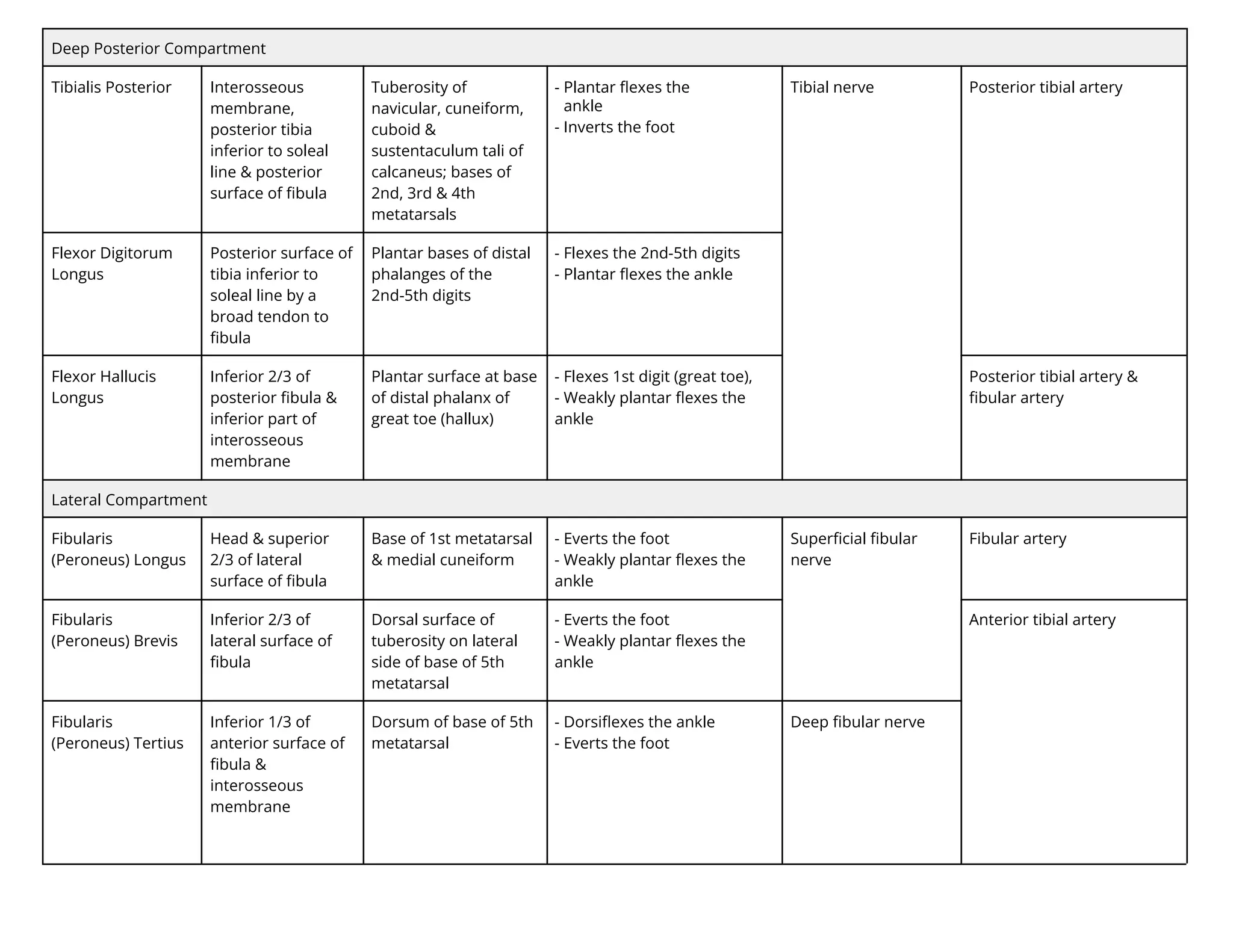
Deep Posterior Compartment
TibialisPosterior Interosseous
membrane,
posterior tibia
inferior to soleal
line & posterior
surface of fibula
Tuberosity of
navicular, cuneiform,
cuboid &
sustentaculum tali of
calcaneus; bases of
2nd, 3rd & 4th
metatarsals
- Plantar flexes the
ankle
- Inverts the foot
Tibial nerve Posterior tibial artery
Flexor Digitorum
Longus
Posterior surface of
tibia inferior to
soleal line by a
broad tendon to
fibula
Plantar bases of distal
phalanges of the
2nd-5th digits
- Flexes the 2nd-5th digits
- Plantar flexes the ankle
Flexor Hallucis
Longus
Inferior 2/3 of
posterior fibula &
inferior part of
interosseous
membrane
Plantar surface at base
of distal phalanx of
great toe (hallux)
- Flexes 1st digit (great toe),
- Weakly plantar flexes the
ankle
Posterior tibial artery &
fibular artery
Lateral Compartment
Fibularis
(Peroneus) Longus
Head & superior
2/3 of lateral
surface of fibula
Base of 1st metatarsal
& medial cuneiform
- Everts the foot
- Weakly plantar flexes the
ankle
Superficial fibular
nerve
Fibular artery
Fibularis
(Peroneus) Brevis
Inferior 2/3 of
lateral surface of
fibula
Dorsal surface of
tuberosity on lateral
side of base of 5th
metatarsal
- Everts the foot
- Weakly plantar flexes the
ankle
Anterior tibial artery
Fibularis
(Peroneus) Tertius
Inferior 1/3 of
anterior surface of
fibula &
interosseous
membrane
Dorsum of base of 5th
metatarsal
- Dorsiflexes the ankle
- Everts the foot
Deep fibular nerve
21.
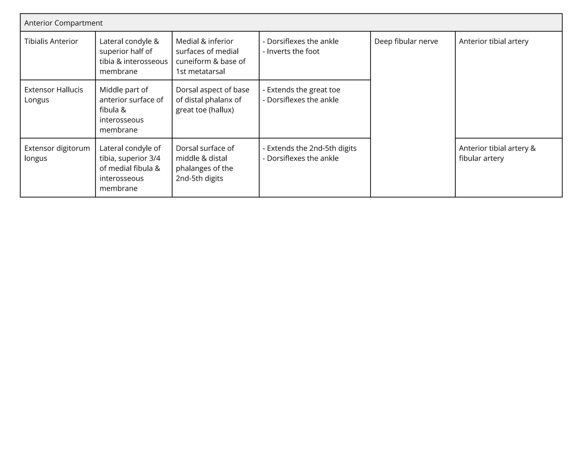
Anterior Compartment
Tibialis AnteriorLateral condyle &
superior half of
tibia & interosseous
membrane
Medial & inferior
surfaces of medial
cuneiform & base of
1st metatarsal
- Dorsiflexes the ankle
- Inverts the foot
Deep fibular nerve Anterior tibial artery
Extensor Hallucis
Longus
Middle part of
anterior surface of
fibula &
interosseous
membrane
Dorsal aspect of base
of distal phalanx of
great toe (hallux)
- Extends the great toe
- Dorsiflexes the ankle
Extensor digitorum
longus
Lateral condyle of
tibia, superior 3/4
of medial fibula &
interosseous
membrane
Dorsal surface of
middle & distal
phalanges of the
2nd-5th digits
- Extends the 2nd-5th digits
- Dorsiflexes the ankle
Anterior tibial artery &
fibular artery
22.
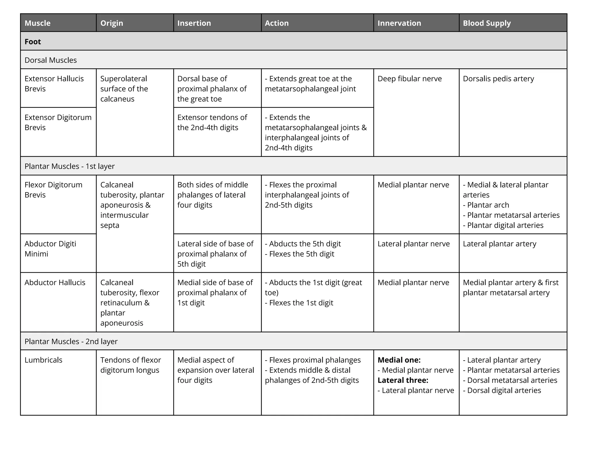
Muscle Origin InsertionAction Innervation Blood Supply
Foot
Dorsal Muscles
Extensor Hallucis
Brevis
Superolateral
surface of the
calcaneus
Dorsal base of
proximal phalanx of
the great toe
- Extends great toe at the
metatarsophalangeal joint
Deep fibular nerve Dorsalis pedis artery
Extensor Digitorum
Brevis
Extensor tendons of
the 2nd-4th digits
- Extends the
metatarsophalangeal joints &
interphalangeal joints of
2nd-4th digits
Plantar Muscles - 1st layer
Flexor Digitorum
Brevis
Calcaneal
tuberosity, plantar
aponeurosis &
intermuscular
septa
Both sides of middle
phalanges of lateral
four digits
- Flexes the proximal
interphalangeal joints of
2nd-5th digits
Medial plantar nerve - Medial & lateral plantar
arteries
- Plantar arch
- Plantar metatarsal arteries
- Plantar digital arteries
Abductor Digiti
Minimi
Lateral side of base of
proximal phalanx of
5th digit
- Abducts the 5th digit
- Flexes the 5th digit
Lateral plantar nerve Lateral plantar artery
Abductor Hallucis Calcaneal
tuberosity, flexor
retinaculum &
plantar
aponeurosis
Medial side of base of
proximal phalanx of
1st digit
- Abducts the 1st digit (great
toe)
- Flexes the 1st digit
Medial plantar nerve Medial plantar artery & first
plantar metatarsal artery
Plantar Muscles - 2nd layer
Lumbricals Tendons of flexor
digitorum longus
Medial aspect of
expansion over lateral
four digits
- Flexes proximal phalanges
- Extends middle & distal
phalanges of 2nd-5th digits
Medial one:
- Medial plantar nerve
Lateral three:
- Lateral plantar nerve
- Lateral plantar artery
- Plantar metatarsal arteries
- Dorsal metatarsal arteries
- Dorsal digital arteries
23.
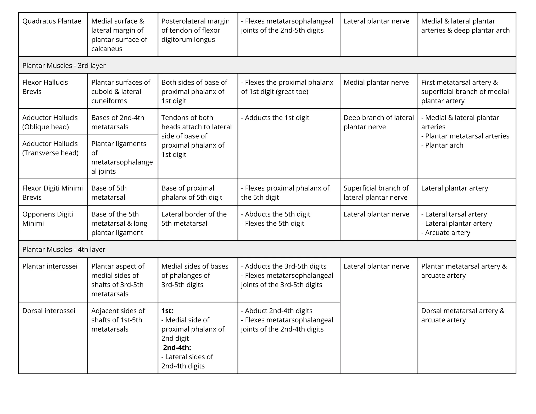
Quadratus Plantae Medialsurface &
lateral margin of
plantar surface of
calcaneus
Posterolateral margin
of tendon of flexor
digitorum longus
- Flexes metatarsophalangeal
joints of the 2nd-5th digits
Lateral plantar nerve Medial & lateral plantar
arteries & deep plantar arch
Plantar Muscles - 3rd layer
Flexor Hallucis
Brevis
Plantar surfaces of
cuboid & lateral
cuneiforms
Both sides of base of
proximal phalanx of
1st digit
- Flexes the proximal phalanx
of 1st digit (great toe)
Medial plantar nerve First metatarsal artery &
superficial branch of medial
plantar artery
Adductor Hallucis
(Oblique head)
Bases of 2nd-4th
metatarsals
Tendons of both
heads attach to lateral
side of base of
proximal phalanx of
1st digit
- Adducts the 1st digit Deep branch of lateral
plantar nerve
- Medial & lateral plantar
arteries
- Plantar metatarsal arteries
- Plantar arch
Adductor Hallucis
(Transverse head)
Plantar ligaments
of
metatarsophalange
al joints
Flexor Digiti Minimi
Brevis
Base of 5th
metatarsal
Base of proximal
phalanx of 5th digit
- Flexes proximal phalanx of
the 5th digit
Superficial branch of
lateral plantar nerve
Lateral plantar artery
Opponens Digiti
Minimi
Base of the 5th
metatarsal & long
plantar ligament
Lateral border of the
5th metatarsal
- Abducts the 5th digit
- Flexes the 5th digit
Lateral plantar nerve - Lateral tarsal artery
- Lateral plantar artery
- Arcuate artery
Plantar Muscles - 4th layer
Plantar interossei Plantar aspect of
medial sides of
shafts of 3rd-5th
metatarsals
Medial sides of bases
of phalanges of
3rd-5th digits
- Adducts the 3rd-5th digits
- Flexes metatarsophalangeal
joints of the 3rd-5th digits
Lateral plantar nerve Plantar metatarsal artery &
arcuate artery
Dorsal interossei Adjacent sides of
shafts of 1st-5th
metatarsals
1st:
- Medial side of
proximal phalanx of
2nd digit
2nd-4th:
- Lateral sides of
2nd-4th digits
- Abduct 2nd-4th digits
- Flexes metatarsophalangeal
joints of the 2nd-4th digits
Dorsal metatarsal artery &
arcuate artery
24.
Muscle Origin InsertionAction Innervation Blood Supply
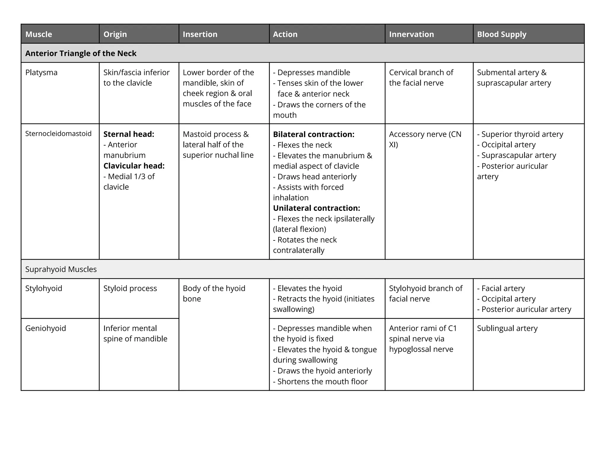
Anterior Triangle of the Neck
Platysma Skin/fascia inferior
to the clavicle
Lower border of the
mandible, skin of
cheek region & oral
muscles of the face
- Depresses mandible
- Tenses skin of the lower
face & anterior neck
- Draws the corners of the
mouth
Cervical branch of
the facial nerve
Submental artery &
suprascapular artery
Sternocleidomastoid Sternal head:
- Anterior
manubrium
Clavicular head:
- Medial 1/3 of
clavicle
Mastoid process &
lateral half of the
superior nuchal line
Bilateral contraction:
- Flexes the neck
- Elevates the manubrium &
medial aspect of clavicle
- Draws head anteriorly
- Assists with forced
inhalation
Unilateral contraction:
- Flexes the neck ipsilaterally
(lateral flexion)
- Rotates the neck
contralaterally
Accessory nerve (CN
XI)
- Superior thyroid artery
- Occipital artery
- Suprascapular artery
- Posterior auricular
artery
Suprahyoid Muscles
Stylohyoid Styloid process Body of the hyoid
bone
- Elevates the hyoid
- Retracts the hyoid (initiates
swallowing)
Stylohyoid branch of
facial nerve
- Facial artery
- Occipital artery
- Posterior auricular artery
Geniohyoid Inferior mental
spine of mandible
- Depresses mandible when
the hyoid is fixed
- Elevates the hyoid & tongue
during swallowing
- Draws the hyoid anteriorly
- Shortens the mouth floor
Anterior rami of C1
spinal nerve via
hypoglossal nerve
Sublingual artery
25.
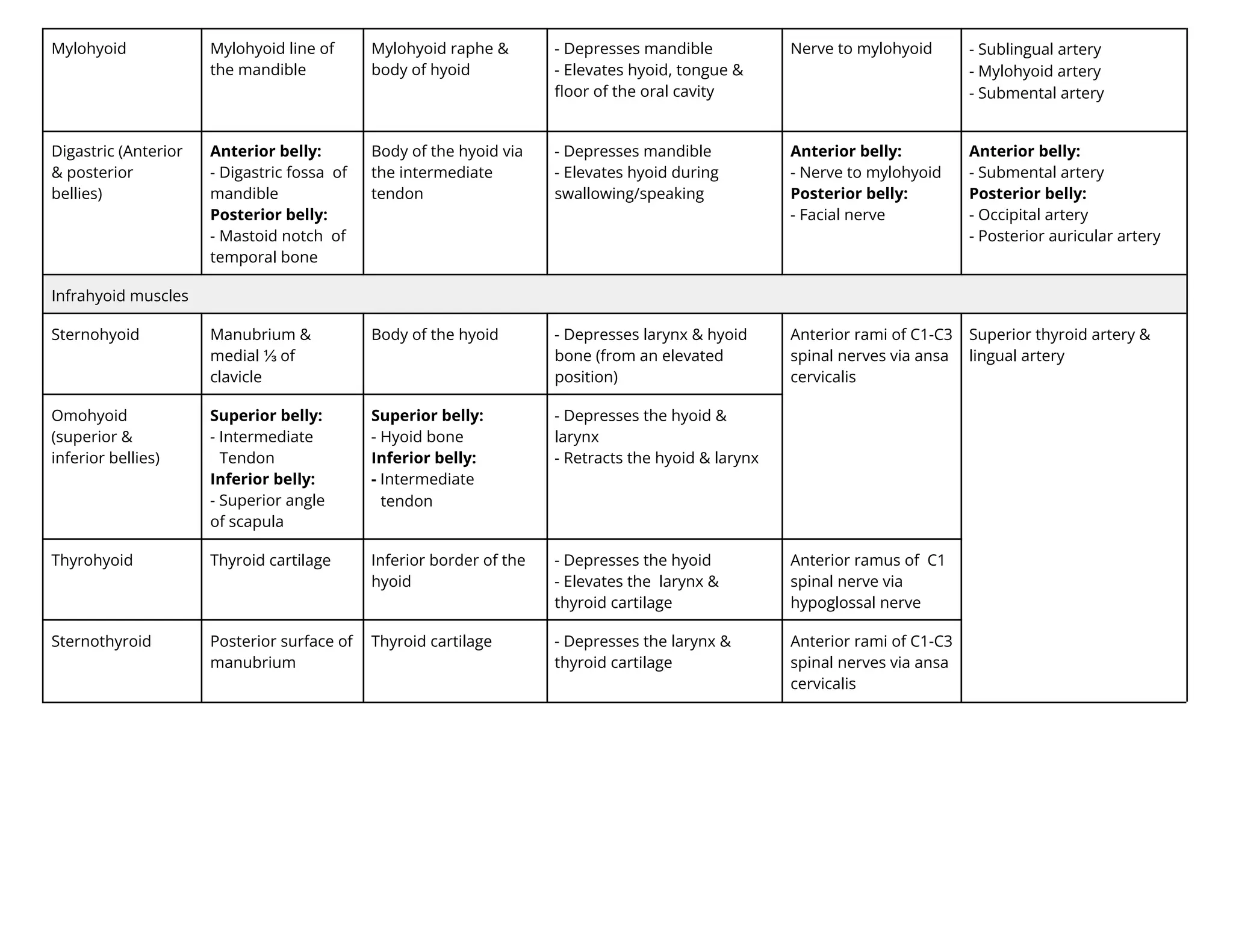
Mylohyoid Mylohyoid lineof
the mandible
Mylohyoid raphe &
body of hyoid
- Depresses mandible
- Elevates hyoid, tongue &
floor of the oral cavity
Nerve to mylohyoid - Sublingual artery
- Mylohyoid artery
- Submental artery
Digastric (Anterior
& posterior
bellies)
Anterior belly:
- Digastric fossa of
mandible
Posterior belly:
- Mastoid notch of
temporal bone
Body of the hyoid via
the intermediate
tendon
- Depresses mandible
- Elevates hyoid during
swallowing/speaking
Anterior belly:
- Nerve to mylohyoid
Posterior belly:
- Facial nerve
Anterior belly:
- Submental artery
Posterior belly:
- Occipital artery
- Posterior auricular artery
Infrahyoid muscles
Sternohyoid Manubrium &
medial ⅓ of
clavicle
Body of the hyoid - Depresses larynx & hyoid
bone (from an elevated
position)
Anterior rami of C1-C3
spinal nerves via ansa
cervicalis
Superior thyroid artery &
lingual artery
Omohyoid
(superior &
inferior bellies)
Superior belly:
- Intermediate
Tendon
Inferior belly:
- Superior angle
of scapula
Superior belly:
- Hyoid bone
Inferior belly:
- Intermediate
tendon
- Depresses the hyoid &
larynx
- Retracts the hyoid & larynx
Thyrohyoid Thyroid cartilage Inferior border of the
hyoid
- Depresses the hyoid
- Elevates the larynx &
thyroid cartilage
Anterior ramus of C1
spinal nerve via
hypoglossal nerve
Sternothyroid Posterior surface of
manubrium
Thyroid cartilage - Depresses the larynx &
thyroid cartilage
Anterior rami of C1-C3
spinal nerves via ansa
cervicalis
26.
Muscle Origin InsertionAction Innervation Blood Supply
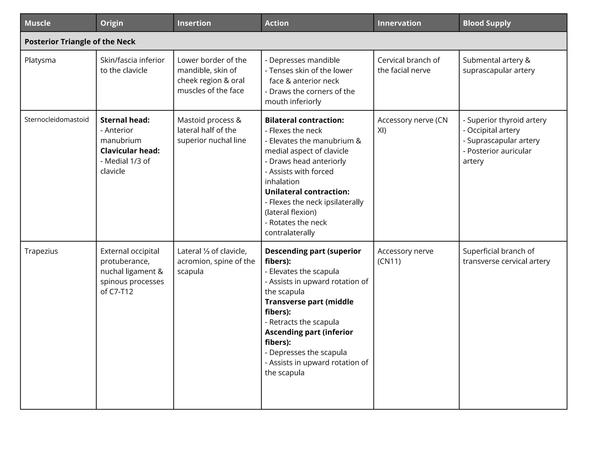
Posterior Triangle of the Neck
Platysma Skin/fascia inferior
to the clavicle
Lower border of the
mandible, skin of
cheek region & oral
muscles of the face
- Depresses mandible
- Tenses skin of the lower
face & anterior neck
- Draws the corners of the
mouth inferiorly
Cervical branch of
the facial nerve
Submental artery &
suprascapular artery
Sternocleidomastoid Sternal head:
- Anterior
manubrium
Clavicular head:
- Medial 1/3 of
clavicle
Mastoid process &
lateral half of the
superior nuchal line
Bilateral contraction:
- Flexes the neck
- Elevates the manubrium &
medial aspect of clavicle
- Draws head anteriorly
- Assists with forced
inhalation
Unilateral contraction:
- Flexes the neck ipsilaterally
(lateral flexion)
- Rotates the neck
contralaterally
Accessory nerve (CN
XI)
- Superior thyroid artery
- Occipital artery
- Suprascapular artery
- Posterior auricular
artery
Trapezius External occipital
protuberance,
nuchal ligament &
spinous processes
of C7-T12
Lateral ⅓ of clavicle,
acromion, spine of the
scapula
Descending part (superior
fibers):
- Elevates the scapula
- Assists in upward rotation of
the scapula
Transverse part (middle
fibers):
- Retracts the scapula
Ascending part (inferior
fibers):
- Depresses the scapula
- Assists in upward rotation of
the scapula
Accessory nerve
(CN11)
Superficial branch of
transverse cervical artery
27.
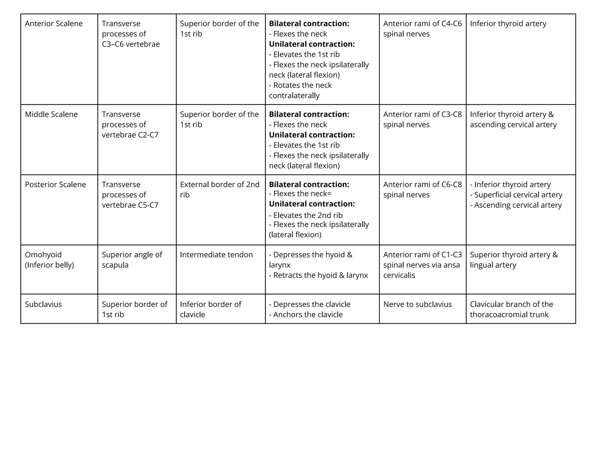
Anterior Scalene Transverse
processesof
C3–C6 vertebrae
Superior border of the
1st rib
Bilateral contraction:
- Flexes the neck
Unilateral contraction:
- Elevates the 1st rib
- Flexes the neck ipsilaterally
neck (lateral flexion)
- Rotates the neck
contralaterally
Anterior rami of C4-C6
spinal nerves
Inferior thyroid artery
Middle Scalene Transverse
processes of
vertebrae C2-C7
Superior border of the
1st rib
Bilateral contraction:
- Flexes the neck
Unilateral contraction:
- Elevates the 1st rib
- Flexes the neck ipsilaterally
neck (lateral flexion)
Anterior rami of C3-C8
spinal nerves
Inferior thyroid artery &
ascending cervical artery
Posterior Scalene Transverse
processes of
vertebrae C5-C7
External border of 2nd
rib
Bilateral contraction:
- Flexes the neck=
Unilateral contraction:
- Elevates the 2nd rib
- Flexes the neck ipsilaterally
(lateral flexion)
Anterior rami of C6-C8
spinal nerves
- Inferior thyroid artery
- Superficial cervical artery
- Ascending cervical artery
Omohyoid
(Inferior belly)
Superior angle of
scapula
Intermediate tendon - Depresses the hyoid &
larynx
- Retracts the hyoid & larynx
Anterior rami of C1-C3
spinal nerves via ansa
cervicalis
Superior thyroid artery &
lingual artery
Subclavius Superior border of
1st rib
Inferior border of
clavicle
- Depresses the clavicle
- Anchors the clavicle
Nerve to subclavius Clavicular branch of the
thoracoacromial trunk
28.
Muscle Origin InsertionAction Innervation Blood Supply
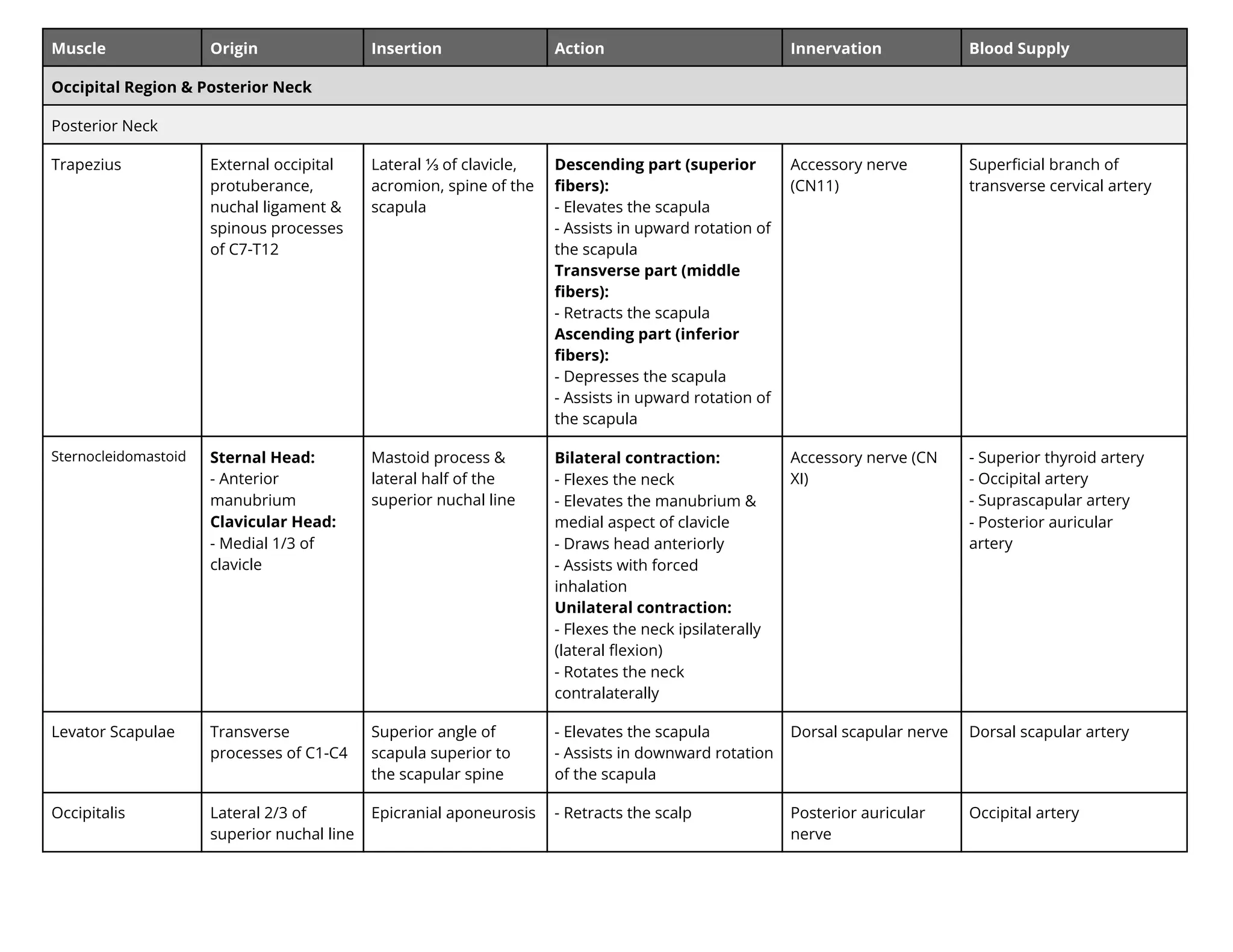
Occipital Region & Posterior Neck
Posterior Neck
Trapezius External occipital
protuberance,
nuchal ligament &
spinous processes
of C7-T12
Lateral ⅓ of clavicle,
acromion, spine of the
scapula
Descending part (superior
fibers):
- Elevates the scapula
- Assists in upward rotation of
the scapula
Transverse part (middle
fibers):
- Retracts the scapula
Ascending part (inferior
fibers):
- Depresses the scapula
- Assists in upward rotation of
the scapula
Accessory nerve
(CN11)
Superficial branch of
transverse cervical artery
Sternocleidomastoid Sternal Head:
- Anterior
manubrium
Clavicular Head:
- Medial 1/3 of
clavicle
Mastoid process &
lateral half of the
superior nuchal line
Bilateral contraction:
- Flexes the neck
- Elevates the manubrium &
medial aspect of clavicle
- Draws head anteriorly
- Assists with forced
inhalation
Unilateral contraction:
- Flexes the neck ipsilaterally
(lateral flexion)
- Rotates the neck
contralaterally
Accessory nerve (CN
XI)
- Superior thyroid artery
- Occipital artery
- Suprascapular artery
- Posterior auricular
artery
Levator Scapulae Transverse
processes of C1-C4
Superior angle of
scapula superior to
the scapular spine
- Elevates the scapula
- Assists in downward rotation
of the scapula
Dorsal scapular nerve Dorsal scapular artery
Occipitalis Lateral 2/3 of
superior nuchal line
Epicranial aponeurosis - Retracts the scalp Posterior auricular
nerve
Occipital artery
29.
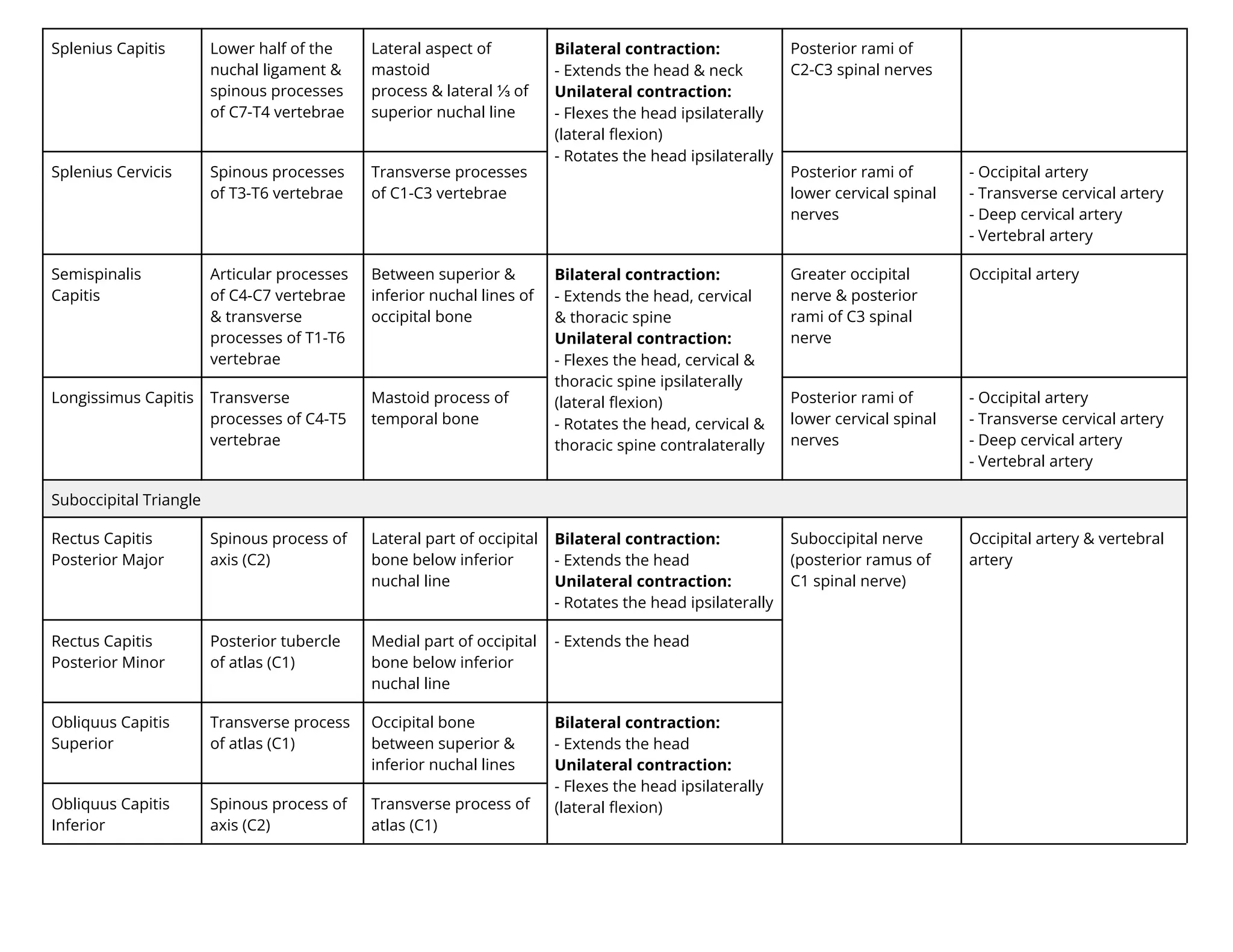
Splenius Capitis Lowerhalf of the
nuchal ligament &
spinous processes
of C7-T4 vertebrae
Lateral aspect of
mastoid
process & lateral ⅓ of
superior nuchal line
Bilateral contraction:
- Extends the head & neck
Unilateral contraction:
- Flexes the head ipsilaterally
(lateral flexion)
- Rotates the head ipsilaterally
Posterior rami of
C2-C3 spinal nerves
Splenius Cervicis Spinous processes
of T3-T6 vertebrae
Transverse processes
of C1-C3 vertebrae
Posterior rami of
lower cervical spinal
nerves
- Occipital artery
- Transverse cervical artery
- Deep cervical artery
- Vertebral artery
Semispinalis
Capitis
Articular processes
of C4-C7 vertebrae
& transverse
processes of T1-T6
vertebrae
Between superior &
inferior nuchal lines of
occipital bone
Bilateral contraction:
- Extends the head, cervical
& thoracic spine
Unilateral contraction:
- Flexes the head, cervical &
thoracic spine ipsilaterally
(lateral flexion)
- Rotates the head, cervical &
thoracic spine contralaterally
Greater occipital
nerve & posterior
rami of C3 spinal
nerve
Occipital artery
Longissimus Capitis Transverse
processes of C4-T5
vertebrae
Mastoid process of
temporal bone
Posterior rami of
lower cervical spinal
nerves
- Occipital artery
- Transverse cervical artery
- Deep cervical artery
- Vertebral artery
Suboccipital Triangle
Rectus Capitis
Posterior Major
Spinous process of
axis (C2)
Lateral part of occipital
bone below inferior
nuchal line
Bilateral contraction:
- Extends the head
Unilateral contraction:
- Rotates the head ipsilaterally
Suboccipital nerve
(posterior ramus of
C1 spinal nerve)
Occipital artery & vertebral
artery
Rectus Capitis
Posterior Minor
Posterior tubercle
of atlas (C1)
Medial part of occipital
bone below inferior
nuchal line
- Extends the head
Obliquus Capitis
Superior
Transverse process
of atlas (C1)
Occipital bone
between superior &
inferior nuchal lines
Bilateral contraction:
- Extends the head
Unilateral contraction:
- Flexes the head ipsilaterally
(lateral flexion)
Obliquus Capitis
Inferior
Spinous process of
axis (C2)
Transverse process of
atlas (C1)
30.
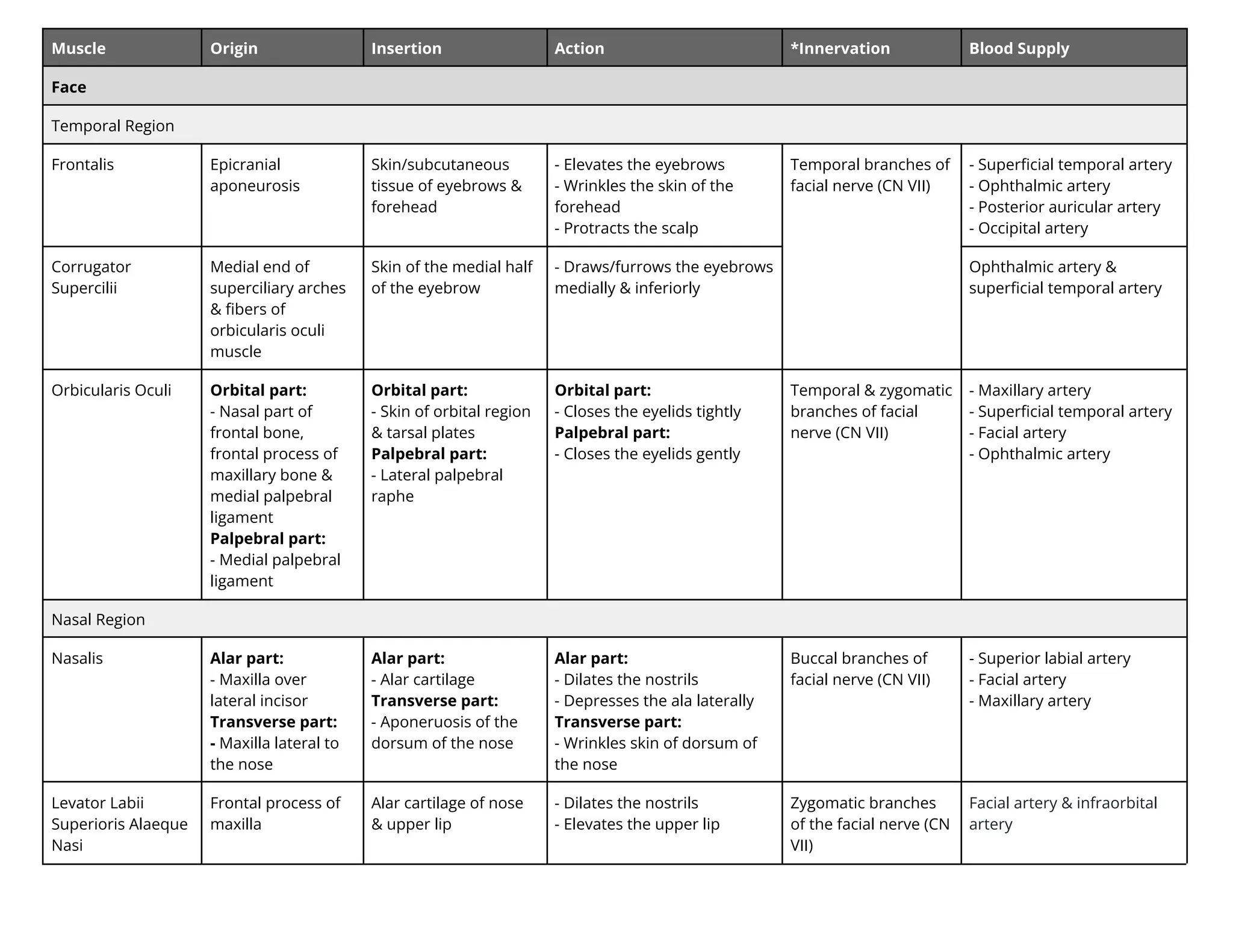
Muscle Origin InsertionAction *Innervation Blood Supply
Face
Temporal Region
Frontalis Epicranial
aponeurosis
Skin/subcutaneous
tissue of eyebrows &
forehead
- Elevates the eyebrows
- Wrinkles the skin of the
forehead
- Protracts the scalp
Temporal branches of
facial nerve (CN VII)
- Superficial temporal artery
- Ophthalmic artery
- Posterior auricular artery
- Occipital artery
Corrugator
Supercilii
Medial end of
superciliary arches
& fibers of
orbicularis oculi
muscle
Skin of the medial half
of the eyebrow
- Draws/furrows the eyebrows
medially & inferiorly
Ophthalmic artery &
superficial temporal artery
Orbicularis Oculi Orbital part:
- Nasal part of
frontal bone,
frontal process of
maxillary bone &
medial palpebral
ligament
Palpebral part:
- Medial palpebral
ligament
Orbital part:
- Skin of orbital region
& tarsal plates
Palpebral part:
- Lateral palpebral
raphe
Orbital part:
- Closes the eyelids tightly
Palpebral part:
- Closes the eyelids gently
Temporal & zygomatic
branches of facial
nerve (CN VII)
- Maxillary artery
- Superficial temporal artery
- Facial artery
- Ophthalmic artery
Nasal Region
Nasalis Alar part:
- Maxilla over
lateral incisor
Transverse part:
- Maxilla lateral to
the nose
Alar part:
- Alar cartilage
Transverse part:
- Aponeruosis of the
dorsum of the nose
Alar part:
- Dilates the nostrils
- Depresses the ala laterally
Transverse part:
- Wrinkles skin of dorsum of
the nose
Buccal branches of
facial nerve (CN VII)
- Superior labial artery
- Facial artery
- Maxillary artery
Levator Labii
Superioris Alaeque
Nasi
Frontal process of
maxilla
Alar cartilage of nose
& upper lip
- Dilates the nostrils
- Elevates the upper lip
Zygomatic branches
of the facial nerve (CN
VII)
Facial artery & infraorbital
artery
31.
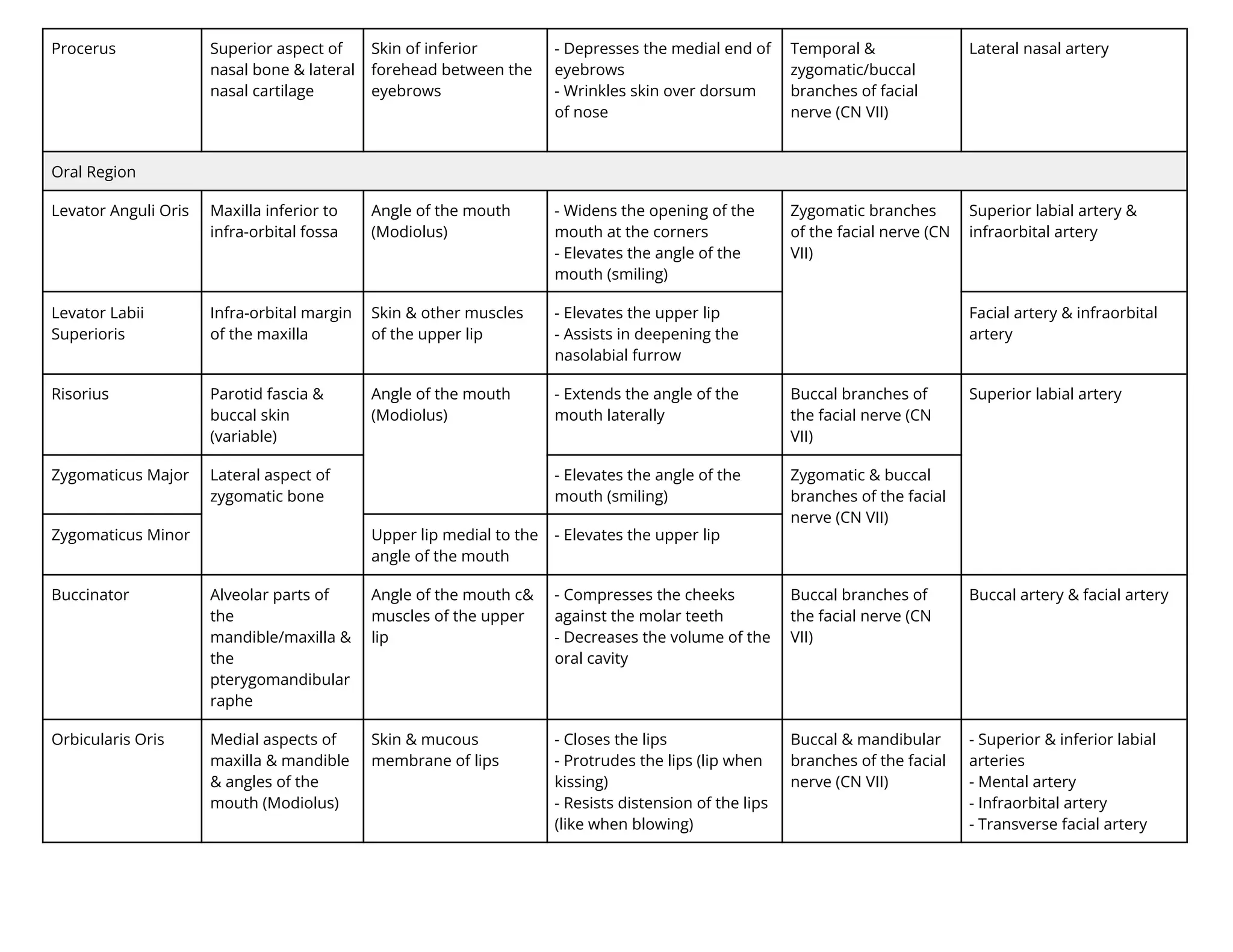
Procerus Superior aspectof
nasal bone & lateral
nasal cartilage
Skin of inferior
forehead between the
eyebrows
- Depresses the medial end of
eyebrows
- Wrinkles skin over dorsum
of nose
Temporal &
zygomatic/buccal
branches of facial
nerve (CN VII)
Lateral nasal artery
Oral Region
Levator Anguli Oris Maxilla inferior to
infra-orbital fossa
Angle of the mouth
(Modiolus)
- Widens the opening of the
mouth at the corners
- Elevates the angle of the
mouth (smiling)
Zygomatic branches
of the facial nerve (CN
VII)
Superior labial artery &
infraorbital artery
Levator Labii
Superioris
Infra-orbital margin
of the maxilla
Skin & other muscles
of the upper lip
- Elevates the upper lip
- Assists in deepening the
nasolabial furrow
Facial artery & infraorbital
artery
Risorius Parotid fascia &
buccal skin
(variable)
Angle of the mouth
(Modiolus)
- Extends the angle of the
mouth laterally
Buccal branches of
the facial nerve (CN
VII)
Superior labial artery
Zygomaticus Major Lateral aspect of
zygomatic bone
- Elevates the angle of the
mouth (smiling)
Zygomatic & buccal
branches of the facial
nerve (CN VII)
Zygomaticus Minor Upper lip medial to the
angle of the mouth
- Elevates the upper lip
Buccinator Alveolar parts of
the
mandible/maxilla &
the
pterygomandibular
raphe
Angle of the mouth c&
muscles of the upper
lip
- Compresses the cheeks
against the molar teeth
- Decreases the volume of the
oral cavity
Buccal branches of
the facial nerve (CN
VII)
Buccal artery & facial artery
Orbicularis Oris Medial aspects of
maxilla & mandible
& angles of the
mouth (Modiolus)
Skin & mucous
membrane of lips
- Closes the lips
- Protrudes the lips (lip when
kissing)
- Resists distension of the lips
(like when blowing)
Buccal & mandibular
branches of the facial
nerve (CN VII)
- Superior & inferior labial
arteries
- Mental artery
- Infraorbital artery
- Transverse facial artery
32.
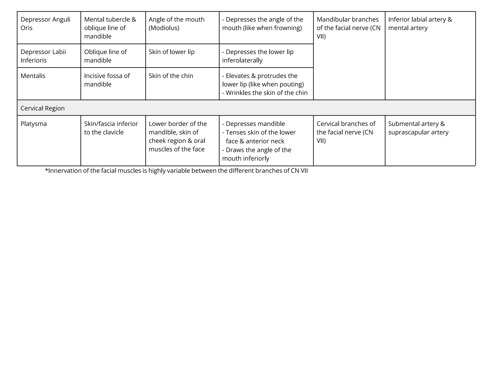
Depressor Anguli
Oris
Mental tubercle&
oblique line of
mandible
Angle of the mouth
(Modiolus)
- Depresses the angle of the
mouth (like when frowning)
Mandibular branches
of the facial nerve (CN
VII)
Inferior labial artery &
mental artery
Depressor Labii
Inferioris
Oblique line of
mandible
Skin of lower lip - Depresses the lower lip
inferolaterally
Mentalis Incisive fossa of
mandible
Skin of the chin - Elevates & protrudes the
lower lip (like when pouting)
- Wrinkles the skin of the chin
Cervical Region
Platysma Skin/fascia inferior
to the clavicle
Lower border of the
mandible, skin of
cheek region & oral
muscles of the face
- Depresses mandible
- Tenses skin of the lower
face & anterior neck
- Draws the angle of the
mouth inferiorly
Cervical branches of
the facial nerve (CN
VII)
Submental artery &
suprascapular artery
*Innervation of the facial muscles is highly variable between the different branches of CN VII
33.
Muscle Origin InsertionAction Innervation Blood Supply
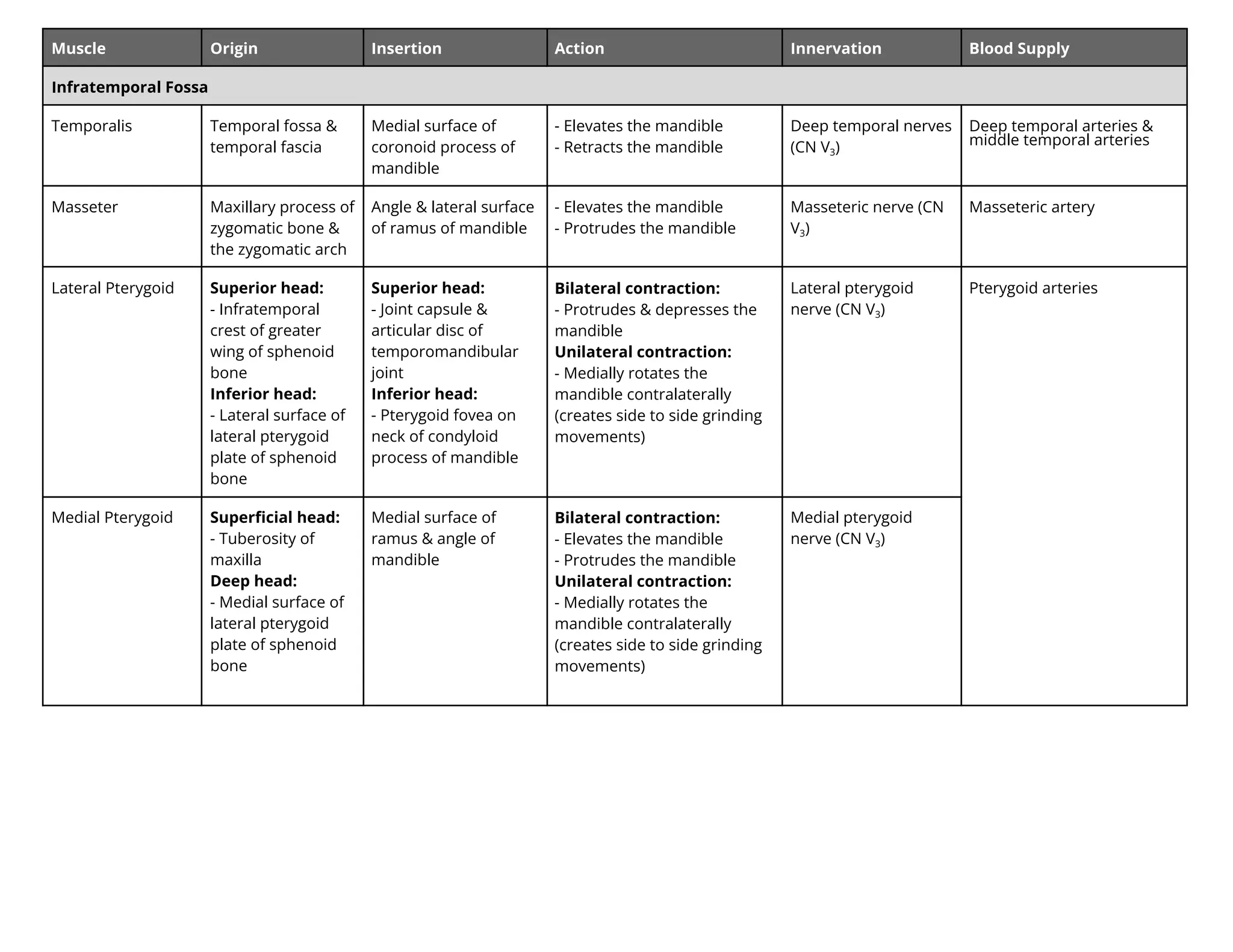
Infratemporal Fossa
Temporalis Temporal fossa &
temporal fascia
Medial surface of
coronoid process of
mandible
- Elevates the mandible
- Retracts the mandible
Deep temporal nerves
(CN V3)
Deep temporal arteries
i le te ral arterie
Masseter Maxillary process of
zygomatic bone &
the zygomatic arch
Angle & lateral surface
of ramus of mandible
- Elevates the mandible
- Protrudes the mandible
Masseteric nerve (CN
V3)
Masseteric artery
Lateral Pterygoid Superior head:
- Infratemporal
crest of greater
wing of sphenoid
bone
Inferior head:
- Lateral surface of
lateral pterygoid
plate of sphenoid
bone
Superior head:
- Joint capsule &
articular disc of
temporomandibular
joint
Inferior head:
- Pterygoid fovea on
neck of condyloid
process of mandible
Bilateral contraction:
- Protrudes & depresses the
mandible
Unilateral contraction:
- Medially rotates the
mandible contralaterally
(creates side to side grinding
movements)
Lateral pterygoid
nerve (CN V3)
Pterygoid arteries
Medial Pterygoid Superficial head:
- Tuberosity of
maxilla
Deep head:
- Medial surface of
lateral pterygoid
plate of sphenoid
bone
Medial surface of
ramus & angle of
mandible
Bilateral contraction:
- Elevates the mandible
- Protrudes the mandible
Unilateral contraction:
- Medially rotates the
mandible contralaterally
(creates side to side grinding
movements)
Medial pterygoid
nerve (CN V3)
34.
Muscle Origin InsertionAction Innervation Blood Supply
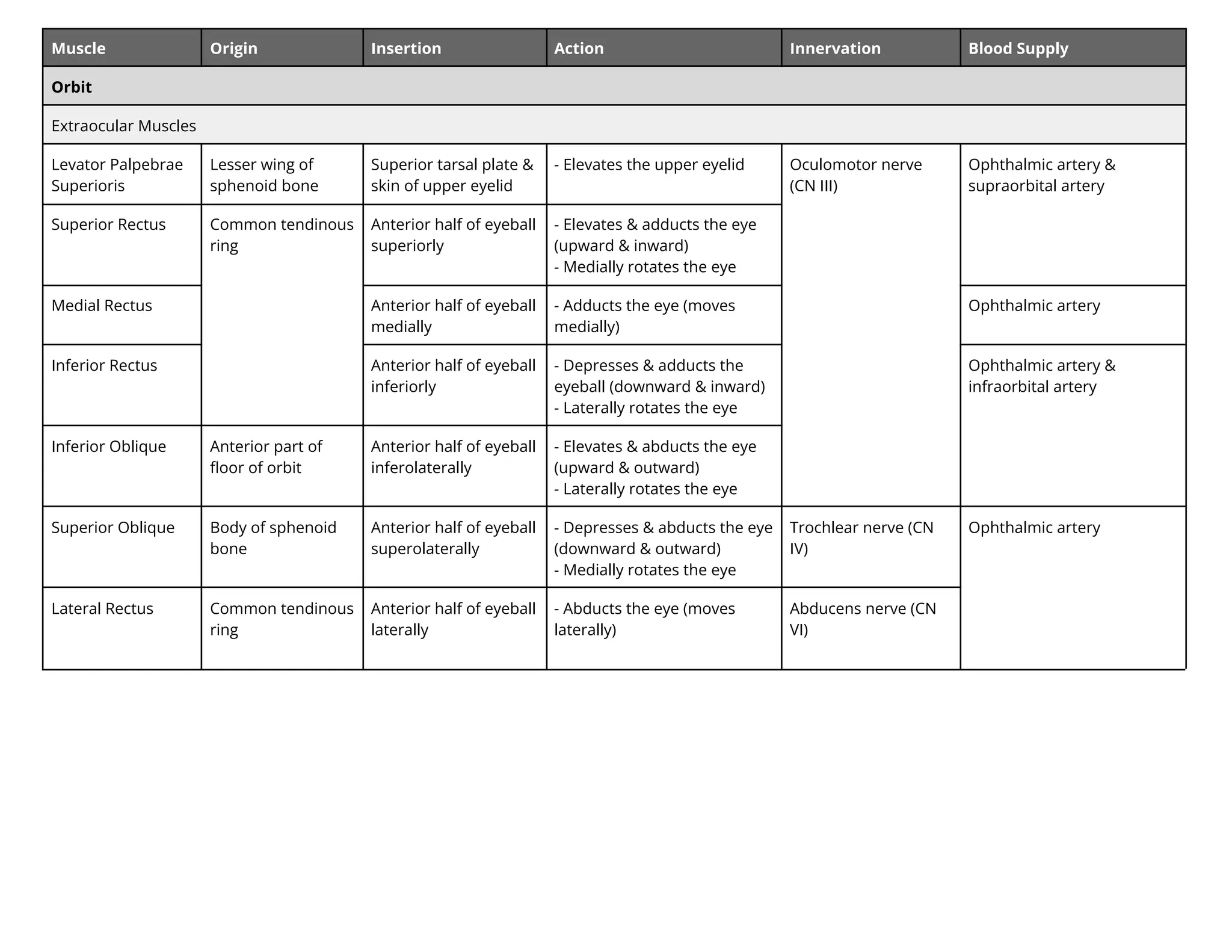
Orbit
Extraocular Muscles
Levator Palpebrae
Superioris
Lesser wing of
sphenoid bone
Superior tarsal plate &
skin of upper eyelid
- Elevates the upper eyelid Oculomotor nerve
(CN III)
Ophthalmic artery &
supraorbital artery
Superior Rectus Common tendinous
ring
Anterior half of eyeball
superiorly
- Elevates & adducts the eye
(upward & inward)
- Medially rotates the eye
Medial Rectus Anterior half of eyeball
medially
- Adducts the eye (moves
medially)
Ophthalmic artery
Inferior Rectus Anterior half of eyeball
inferiorly
- Depresses & adducts the
eyeball (downward & inward)
- Laterally rotates the eye
Ophthalmic artery &
infraorbital artery
Inferior Oblique Anterior part of
floor of orbit
Anterior half of eyeball
inferolaterally
- Elevates & abducts the eye
(upward & outward)
- Laterally rotates the eye
Superior Oblique Body of sphenoid
bone
Anterior half of eyeball
superolaterally
- Depresses & abducts the eye
(downward & outward)
- Medially rotates the eye
Trochlear nerve (CN
IV)
Ophthalmic artery
Lateral Rectus Common tendinous
ring
Anterior half of eyeball
laterally
- Abducts the eye (moves
laterally)
Abducens nerve (CN
VI)
35.
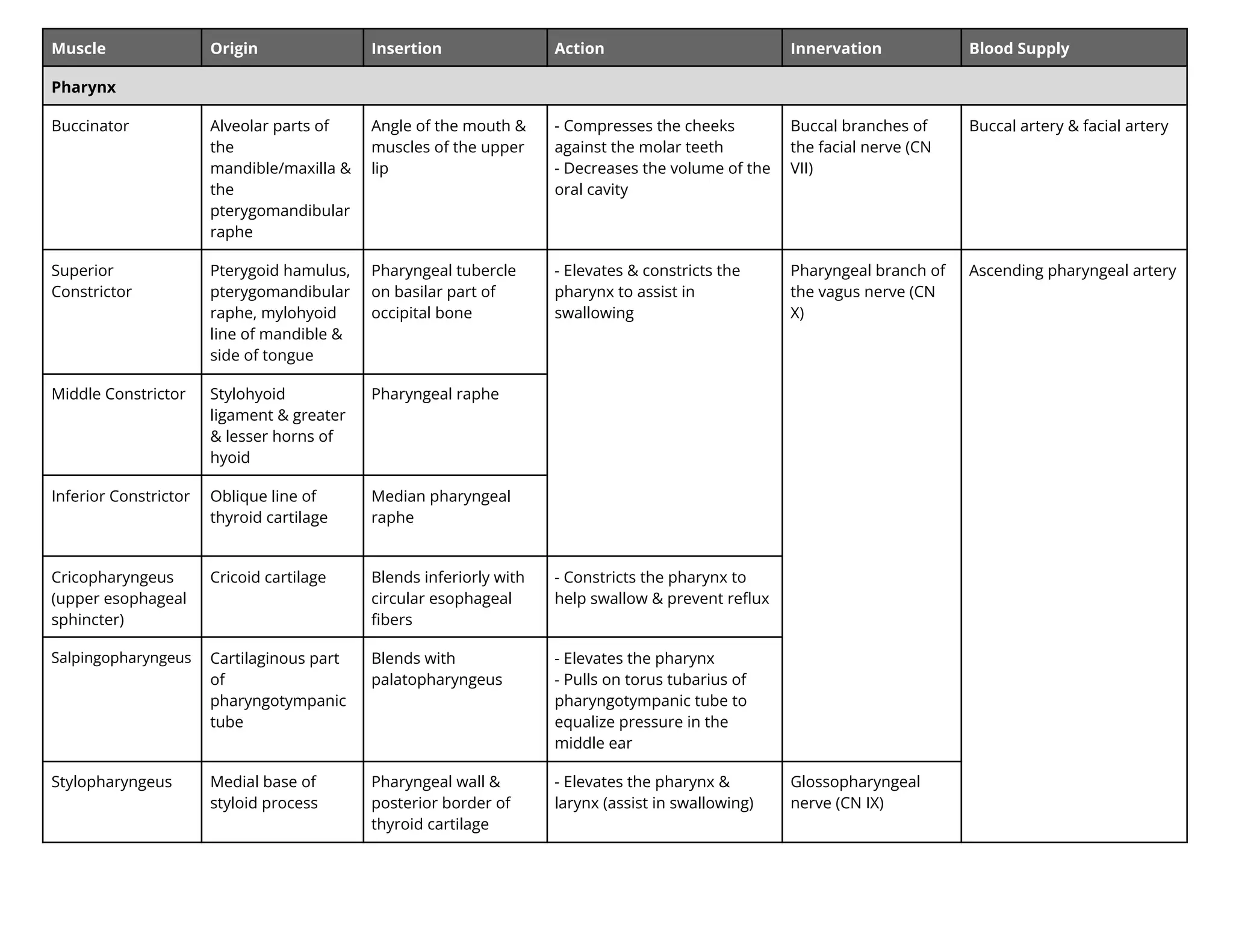
Muscle Origin InsertionAction Innervation Blood Supply
Pharynx
Buccinator Alveolar parts of
the
mandible/maxilla &
the
pterygomandibular
raphe
Angle of the mouth &
muscles of the upper
lip
- Compresses the cheeks
against the molar teeth
- Decreases the volume of the
oral cavity
Buccal branches of
the facial nerve (CN
VII)
Buccal artery & facial artery
Superior
Constrictor
Pterygoid hamulus,
pterygomandibular
raphe, mylohyoid
line of mandible &
side of tongue
Pharyngeal tubercle
on basilar part of
occipital bone
- Elevates & constricts the
pharynx to assist in
swallowing
Pharyngeal branch of
the vagus nerve (CN
X)
Ascending pharyngeal artery
Middle Constrictor Stylohyoid
ligament & greater
& lesser horns of
hyoid
Pharyngeal raphe
Inferior Constrictor Oblique line of
thyroid cartilage
Median pharyngeal
raphe
Cricopharyngeus
(upper esophageal
sphincter)
Cricoid cartilage Blends inferiorly with
circular esophageal
fibers
- Constricts the pharynx to
help swallow & prevent reflux
Salpingopharyngeus Cartilaginous part
of
pharyngotympanic
tube
Blends with
palatopharyngeus
- Elevates the pharynx
- Pulls on torus tubarius of
pharyngotympanic tube to
equalize pressure in the
middle ear
Stylopharyngeus Medial base of
styloid process
Pharyngeal wall &
posterior border of
thyroid cartilage
- Elevates the pharynx &
larynx (assist in swallowing)
Glossopharyngeal
nerve (CN IX)
36.
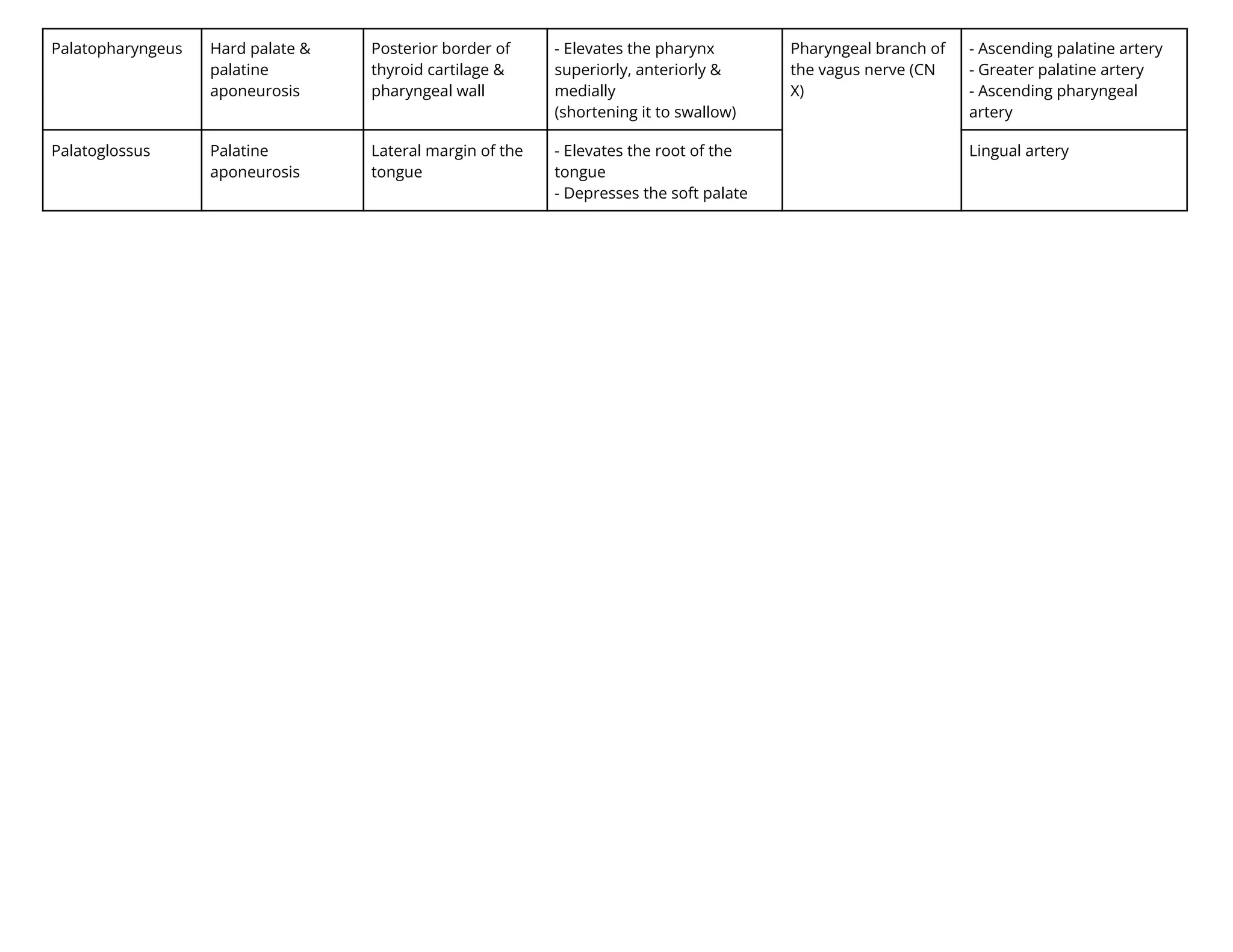
Palatopharyngeus Hard palate&
palatine
aponeurosis
Posterior border of
thyroid cartilage &
pharyngeal wall
- Elevates the pharynx
superiorly, anteriorly &
medially
(shortening it to swallow)
Pharyngeal branch of
the vagus nerve (CN
X)
- Ascending palatine artery
- Greater palatine artery
- Ascending pharyngeal
artery
Palatoglossus Palatine
aponeurosis
Lateral margin of the
tongue
- Elevates the root of the
tongue
- Depresses the soft palate
Lingual artery
37.
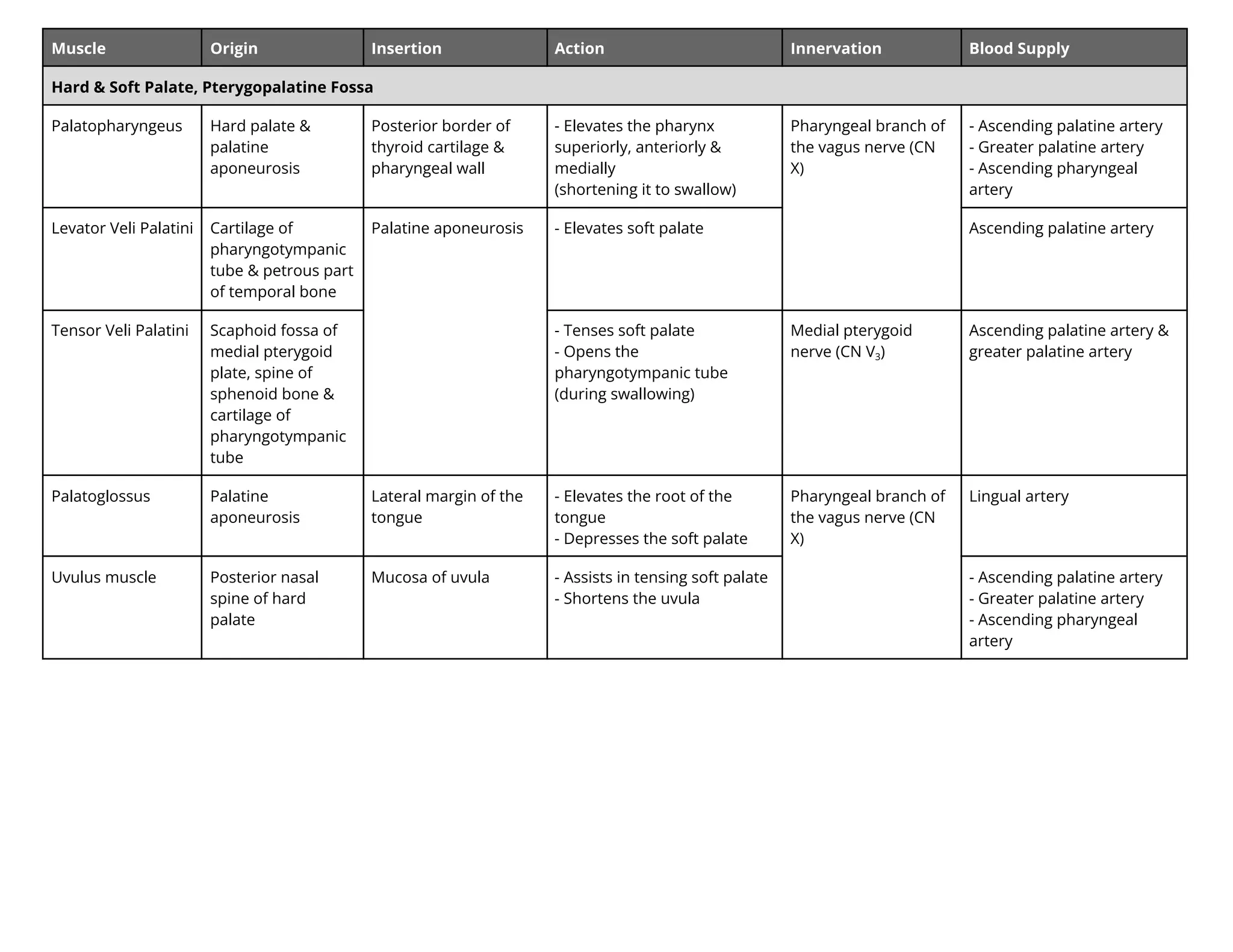
Muscle Origin InsertionAction Innervation Blood Supply
Hard & Soft Palate, Pterygopalatine Fossa
Palatopharyngeus Hard palate &
palatine
aponeurosis
Posterior border of
thyroid cartilage &
pharyngeal wall
- Elevates the pharynx
superiorly, anteriorly &
medially
(shortening it to swallow)
Pharyngeal branch of
the vagus nerve (CN
X)
- Ascending palatine artery
- Greater palatine artery
- Ascending pharyngeal
artery
Levator Veli Palatini Cartilage of
pharyngotympanic
tube & petrous part
of temporal bone
Palatine aponeurosis - Elevates soft palate Ascending palatine artery
Tensor Veli Palatini Scaphoid fossa of
medial pterygoid
plate, spine of
sphenoid bone &
cartilage of
pharyngotympanic
tube
- Tenses soft palate
- Opens the
pharyngotympanic tube
(during swallowing)
Medial pterygoid
nerve (CN V3)
Ascending palatine artery &
greater palatine artery
Palatoglossus Palatine
aponeurosis
Lateral margin of the
tongue
- Elevates the root of the
tongue
- Depresses the soft palate
Pharyngeal branch of
the vagus nerve (CN
X)
Lingual artery
Uvulus muscle Posterior nasal
spine of hard
palate
Mucosa of uvula - Assists in tensing soft palate
- Shortens the uvula
- Ascending palatine artery
- Greater palatine artery
- Ascending pharyngeal
artery
38.
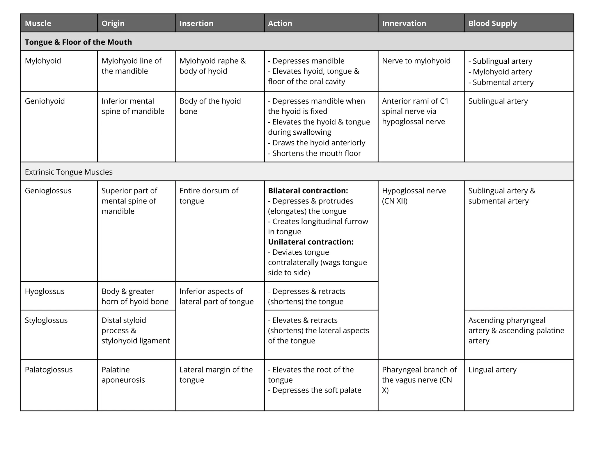
Muscle Origin InsertionAction Innervation Blood Supply
Tongue & Floor of the Mouth
Mylohyoid Mylohyoid line of
the mandible
Mylohyoid raphe &
body of hyoid
- Depresses mandible
- Elevates hyoid, tongue &
floor of the oral cavity
Nerve to mylohyoid - Sublingual artery
- Mylohyoid artery
- Submental artery
Geniohyoid Inferior mental
spine of mandible
Body of the hyoid
bone
- Depresses mandible when
the hyoid is fixed
- Elevates the hyoid & tongue
during swallowing
- Draws the hyoid anteriorly
- Shortens the mouth floor
Anterior rami of C1
spinal nerve via
hypoglossal nerve
Sublingual artery
Extrinsic Tongue Muscles
Genioglossus Superior part of
mental spine of
mandible
Entire dorsum of
tongue
Bilateral contraction:
- Depresses & protrudes
(elongates) the tongue
- Creates longitudinal furrow
in tongue
Unilateral contraction:
- Deviates tongue
contralaterally (wags tongue
side to side)
Hypoglossal nerve
(CN XII)
Sublingual artery &
submental artery
Hyoglossus Body & greater
horn of hyoid bone
Inferior aspects of
lateral part of tongue
- Depresses & retracts
(shortens) the tongue
Styloglossus Distal styloid
process &
stylohyoid ligament
- Elevates & retracts
(shortens) the lateral aspects
of the tongue
Ascending pharyngeal
artery & ascending palatine
artery
Palatoglossus Palatine
aponeurosis
Lateral margin of the
tongue
- Elevates the root of the
tongue
- Depresses the soft palate
Pharyngeal branch of
the vagus nerve (CN
X)
Lingual artery
39.
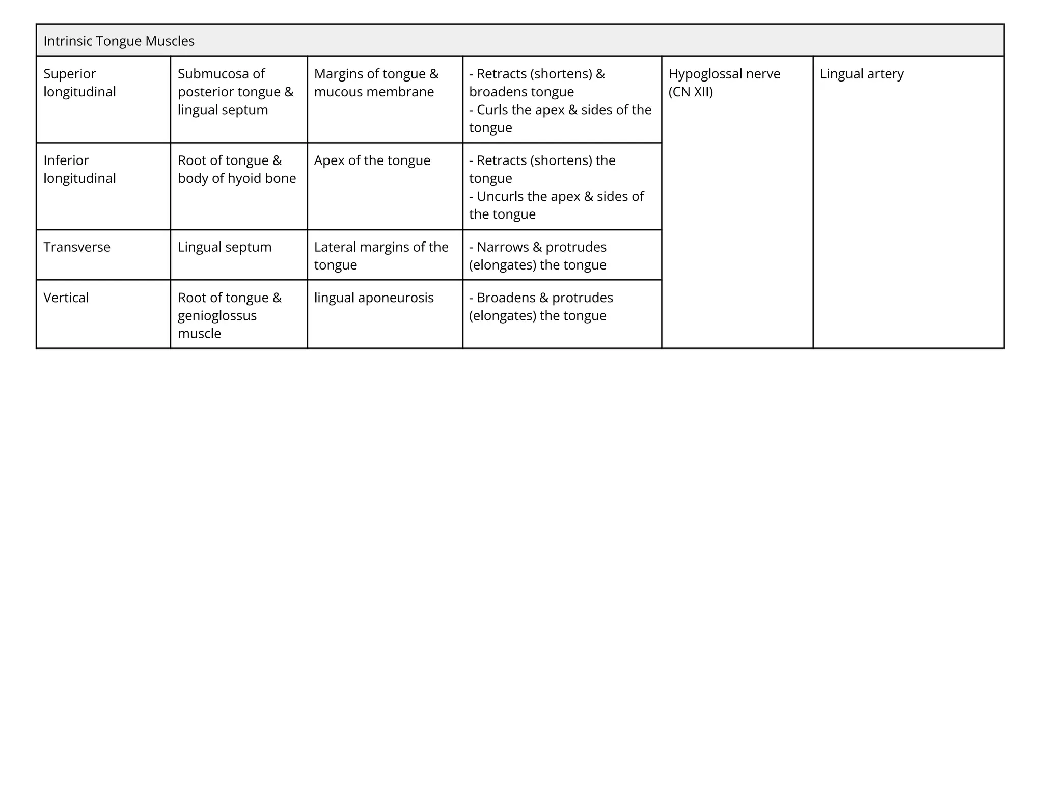
Intrinsic Tongue Muscles
Superior
longitudinal
Submucosaof
posterior tongue &
lingual septum
Margins of tongue &
mucous membrane
- Retracts (shortens) &
broadens tongue
- Curls the apex & sides of the
tongue
Hypoglossal nerve
(CN XII)
Lingual artery
Inferior
longitudinal
Root of tongue &
body of hyoid bone
Apex of the tongue - Retracts (shortens) the
tongue
- Uncurls the apex & sides of
the tongue
Transverse Lingual septum Lateral margins of the
tongue
- Narrows & protrudes
(elongates) the tongue
Vertical Root of tongue &
genioglossus
muscle
lingual aponeurosis - Broadens & protrudes
(elongates) the tongue
40.
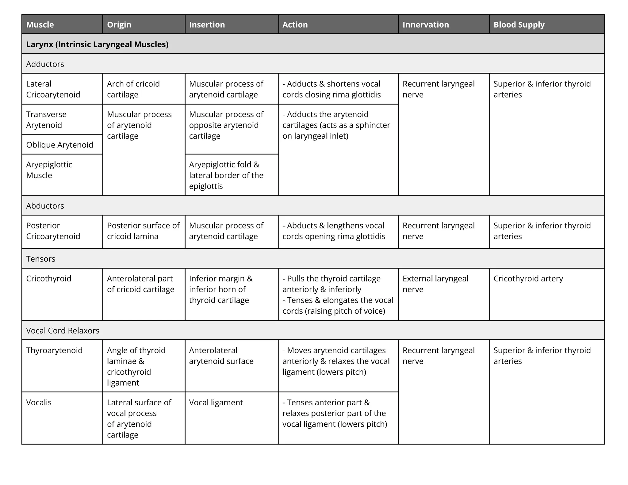
Muscle Origin InsertionAction Innervation Blood Supply
Larynx (Intrinsic Laryngeal Muscles)
Adductors
Lateral
Cricoarytenoid
Arch of cricoid
cartilage
Muscular process of
arytenoid cartilage
- Adducts & shortens vocal
cords closing rima glottidis
Recurrent laryngeal
nerve
Superior & inferior thyroid
arteries
Transverse
Arytenoid
Muscular process
of arytenoid
cartilage
Muscular process of
opposite arytenoid
cartilage
- Adducts the arytenoid
cartilages (acts as a sphincter
on laryngeal inlet)
Oblique Arytenoid
Aryepiglottic
Muscle
Aryepiglottic fold &
lateral border of the
epiglottis
Abductors
Posterior
Cricoarytenoid
Posterior surface of
cricoid lamina
Muscular process of
arytenoid cartilage
- Abducts & lengthens vocal
cords opening rima glottidis
Recurrent laryngeal
nerve
Superior & inferior thyroid
arteries
Tensors
Cricothyroid Anterolateral part
of cricoid cartilage
Inferior margin &
inferior horn of
thyroid cartilage
- Pulls the thyroid cartilage
anteriorly & inferiorly
- Tenses & elongates the vocal
cords (raising pitch of voice)
External laryngeal
nerve
Cricothyroid artery
Vocal Cord Relaxors
Thyroarytenoid Angle of thyroid
laminae &
cricothyroid
ligament
Anterolateral
arytenoid surface
- Moves arytenoid cartilages
anteriorly & relaxes the vocal
ligament (lowers pitch)
Recurrent laryngeal
nerve
Superior & inferior thyroid
arteries
Vocalis Lateral surface of
vocal process
of arytenoid
cartilage
Vocal ligament - Tenses anterior part &
relaxes posterior part of the
vocal ligament (lowers pitch)
41.
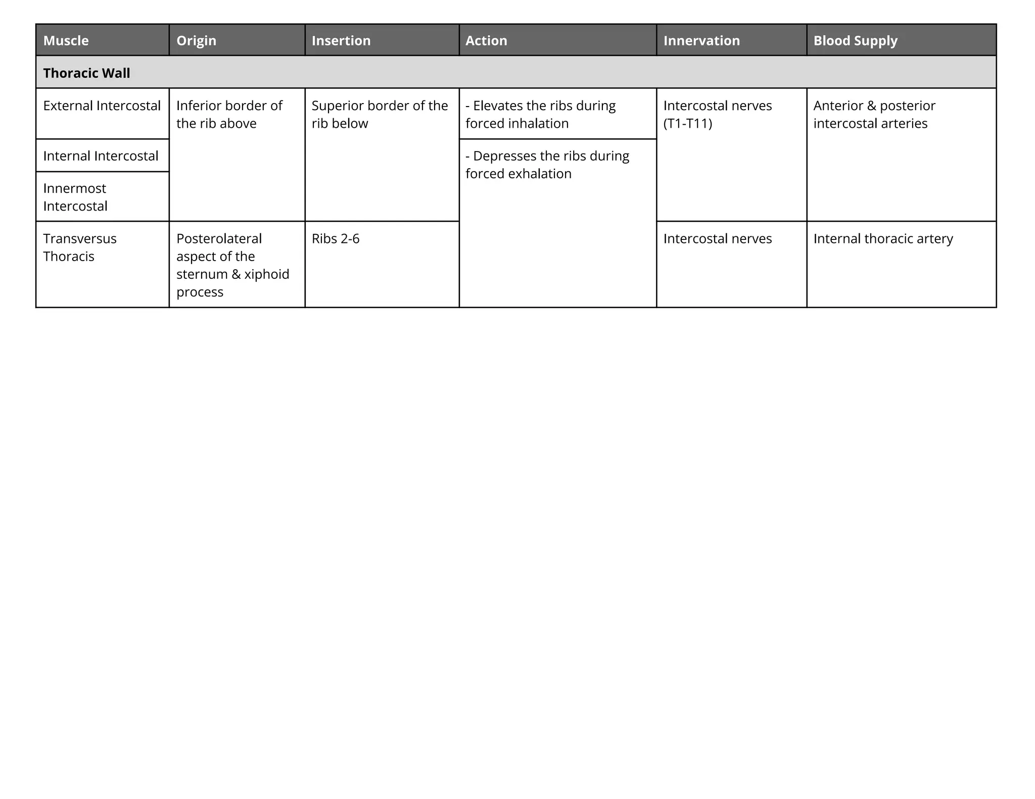
Muscle Origin InsertionAction Innervation Blood Supply
Thoracic Wall
External Intercostal Inferior border of
the rib above
Superior border of the
rib below
- Elevates the ribs during
forced inhalation
Intercostal nerves
(T1-T11)
Anterior & posterior
intercostal arteries
Internal Intercostal - Depresses the ribs during
forced exhalation
Innermost
Intercostal
Transversus
Thoracis
Posterolateral
aspect of the
sternum & xiphoid
process
Ribs 2-6 Intercostal nerves Internal thoracic artery
42.
Muscle Origin InsertionAction Innervation Blood Supply
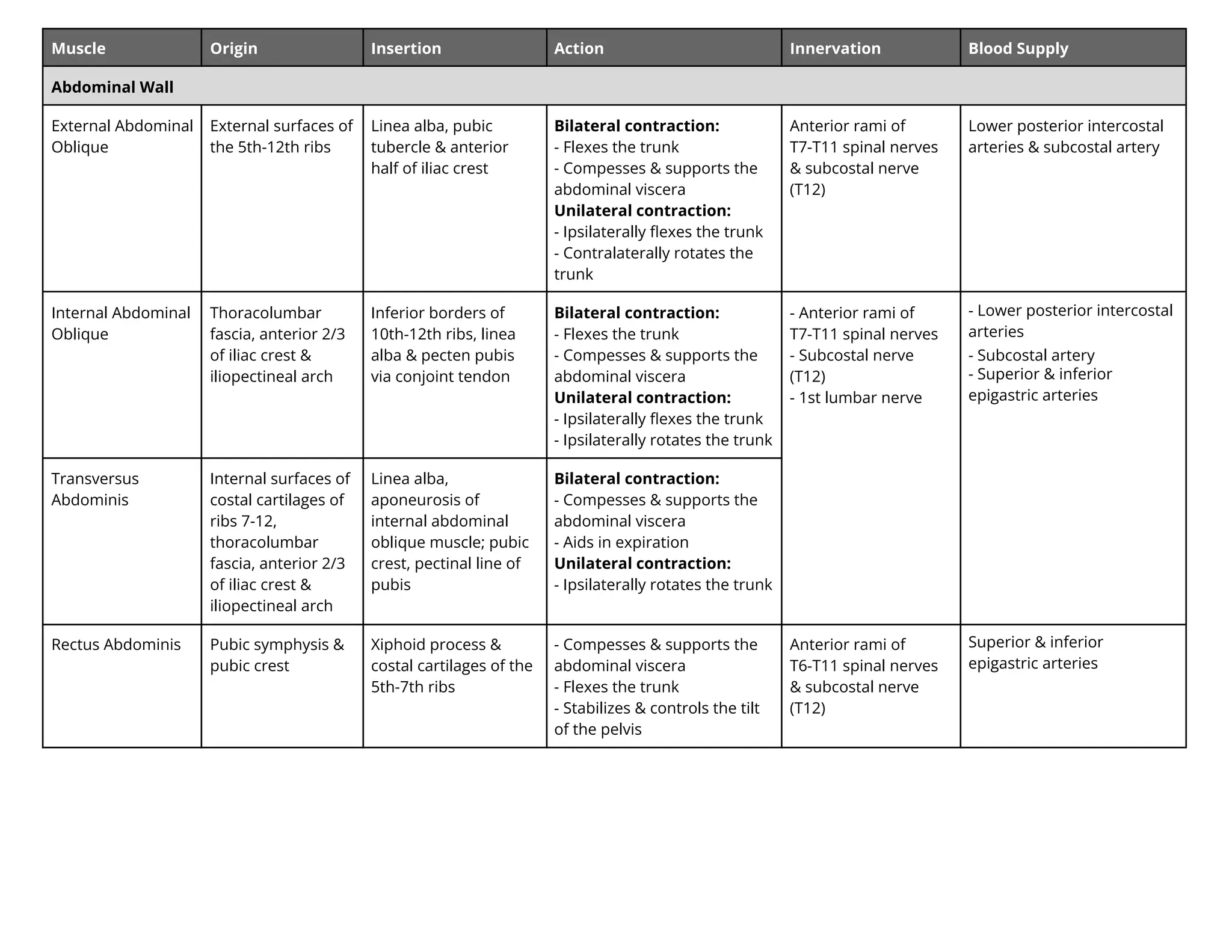
Abdominal Wall
External Abdominal
Oblique
External surfaces of
the 5th-12th ribs
Linea alba, pubic
tubercle & anterior
half of iliac crest
Bilateral contraction:
- Flexes the trunk
- Compesses & supports the
abdominal viscera
Unilateral contraction:
- Ipsilaterally flexes the trunk
- Contralaterally rotates the
trunk
Anterior rami of
T7-T11 spinal nerves
& subcostal nerve
(T12)
Lower posterior intercostal
arteries & subcostal artery
Internal Abdominal
Oblique
Thoracolumbar
fascia, anterior 2/3
of iliac crest &
iliopectineal arch
Inferior borders of
10th-12th ribs, linea
alba & pecten pubis
via conjoint tendon
Bilateral contraction:
- Flexes the trunk
- Compesses & supports the
abdominal viscera
Unilateral contraction:
- Ipsilaterally flexes the trunk
- Ipsilaterally rotates the trunk
- Anterior rami of
T7-T11 spinal nerves
- Subcostal nerve
(T12)
- 1st lumbar nerve
- Lower posterior intercostal
arteries
- Subcostal artery
- Superior & inferior
epigastric arteries
Transversus
Abdominis
Internal surfaces of
costal cartilages of
ribs 7-12,
thoracolumbar
fascia, anterior 2/3
of iliac crest &
iliopectineal arch
Linea alba,
aponeurosis of
internal abdominal
oblique muscle; pubic
crest, pectinal line of
pubis
Bilateral contraction:
- Compesses & supports the
abdominal viscera
- Aids in expiration
Unilateral contraction:
- Ipsilaterally rotates the trunk
Rectus Abdominis Pubic symphysis &
pubic crest
Xiphoid process &
costal cartilages of the
5th-7th ribs
- Compesses & supports the
abdominal viscera
- Flexes the trunk
- Stabilizes & controls the tilt
of the pelvis
Anterior rami of
T6-T11 spinal nerves
& subcostal nerve
(T12)
Superior & inferior
epigastric arteries
43.
Muscle Origin InsertionAction Innervation Blood Supply
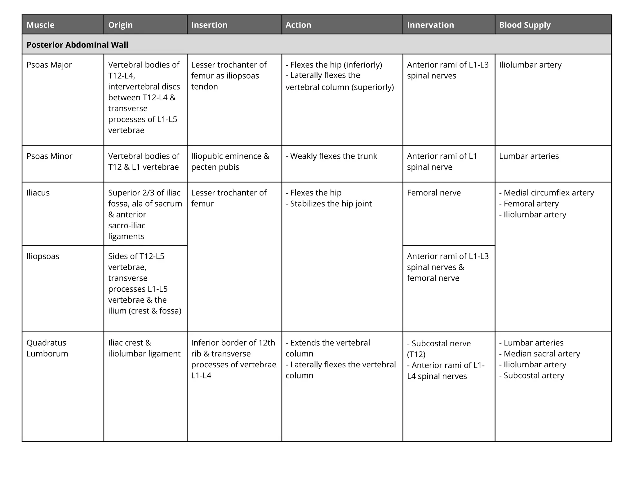
Posterior Abdominal Wall
Psoas Major Vertebral bodies of
T12-L4,
intervertebral discs
between T12-L4 &
transverse
processes of L1-L5
vertebrae
Lesser trochanter of
femur as iliopsoas
tendon
- Flexes the hip (inferiorly)
- Laterally flexes the
vertebral column (superiorly)
Anterior rami of L1-L3
spinal nerves
Iliolumbar artery
Psoas Minor Vertebral bodies of
T12 & L1 vertebrae
Iliopubic eminence &
pecten pubis
- Weakly flexes the trunk Anterior rami of L1
spinal nerve
Lumbar arteries
Iliacus Superior 2/3 of iliac
fossa, ala of sacrum
& anterior
sacro-iliac
ligaments
Lesser trochanter of
femur
- Flexes the hip
- Stabilizes the hip joint
Femoral nerve - Medial circumflex artery
- Femoral artery
- Iliolumbar artery
Iliopsoas Sides of T12-L5
vertebrae,
transverse
processes L1-L5
vertebrae & the
ilium (crest & fossa)
Anterior rami of L1-L3
spinal nerves &
femoral nerve
Quadratus
Lumborum
Iliac crest &
iliolumbar ligament
Inferior border of 12th
rib & transverse
processes of vertebrae
L1-L4
- Extends the vertebral
column
- Laterally flexes the vertebral
column
- Subcostal nerve
(T12)
- Anterior rami of L1-
L4 spinal nerves
- Lumbar arteries
- Median sacral artery
- Iliolumbar artery
- Subcostal artery
44.
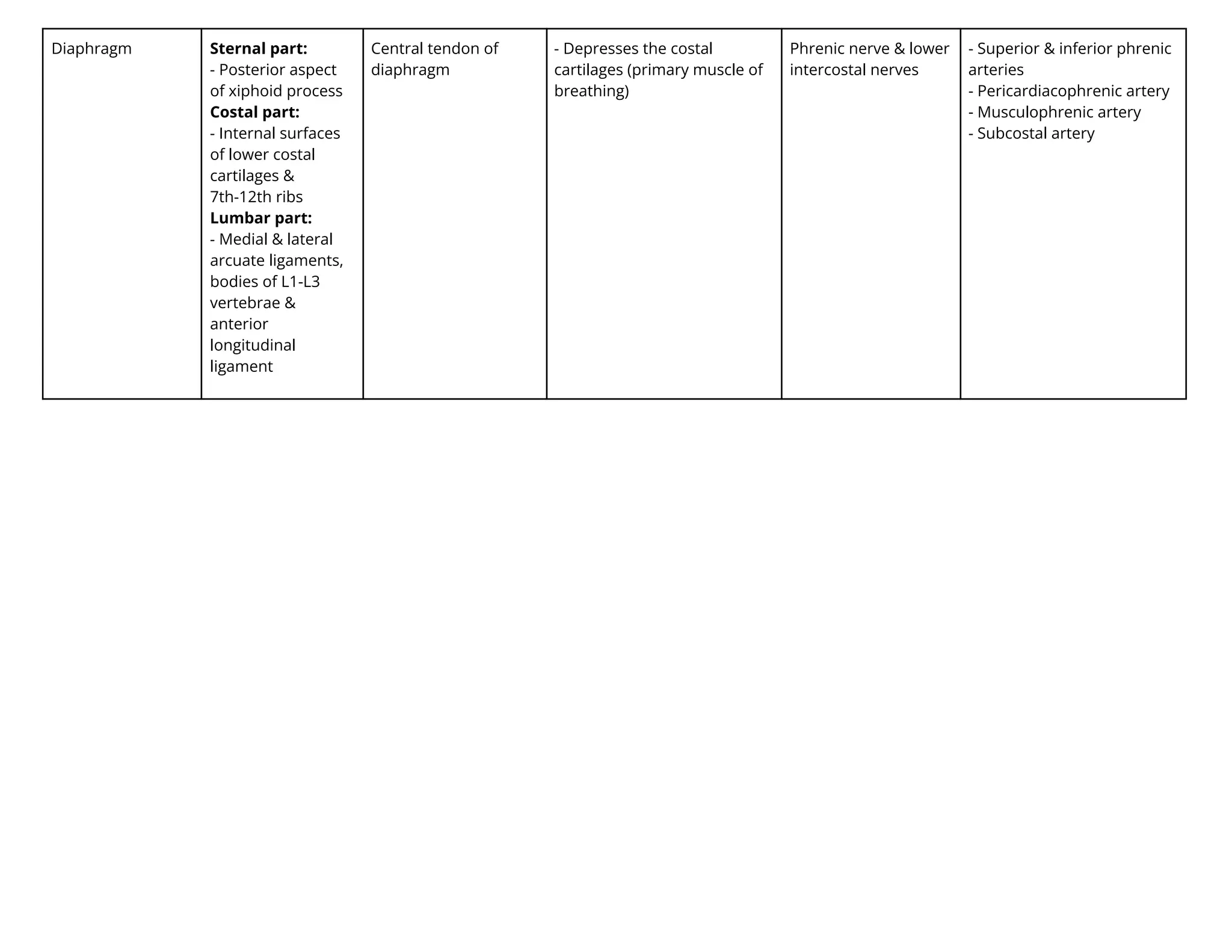
Diaphragm Sternal part:
-Posterior aspect
of xiphoid process
Costal part:
- Internal surfaces
of lower costal
cartilages &
7th-12th ribs
Lumbar part:
- Medial & lateral
arcuate ligaments,
bodies of L1-L3
vertebrae &
anterior
longitudinal
ligament
Central tendon of
diaphragm
- Depresses the costal
cartilages (primary muscle of
breathing)
Phrenic nerve & lower
intercostal nerves
- Superior & inferior phrenic
arteries
- Pericardiacophrenic artery
- Musculophrenic artery
- Subcostal artery
45.
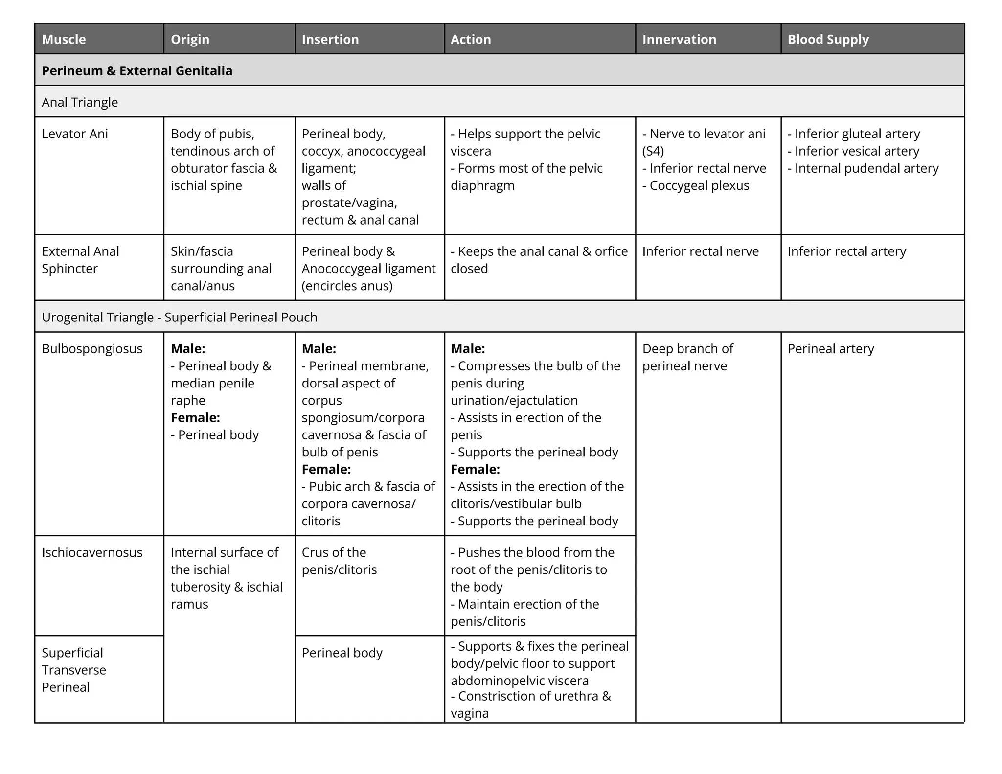
Muscle Origin InsertionAction Innervation Blood Supply
Perineum & External Genitalia
Anal Triangle
Levator Ani Body of pubis,
tendinous arch of
obturator fascia &
ischial spine
Perineal body,
coccyx, anococcygeal
ligament;
walls of
prostate/vagina,
rectum & anal canal
- Helps support the pelvic
viscera
- Forms most of the pelvic
diaphragm
- Nerve to levator ani
(S4)
- Inferior rectal nerve
- Coccygeal plexus
- Inferior gluteal artery
- Inferior vesical artery
- Internal pudendal artery
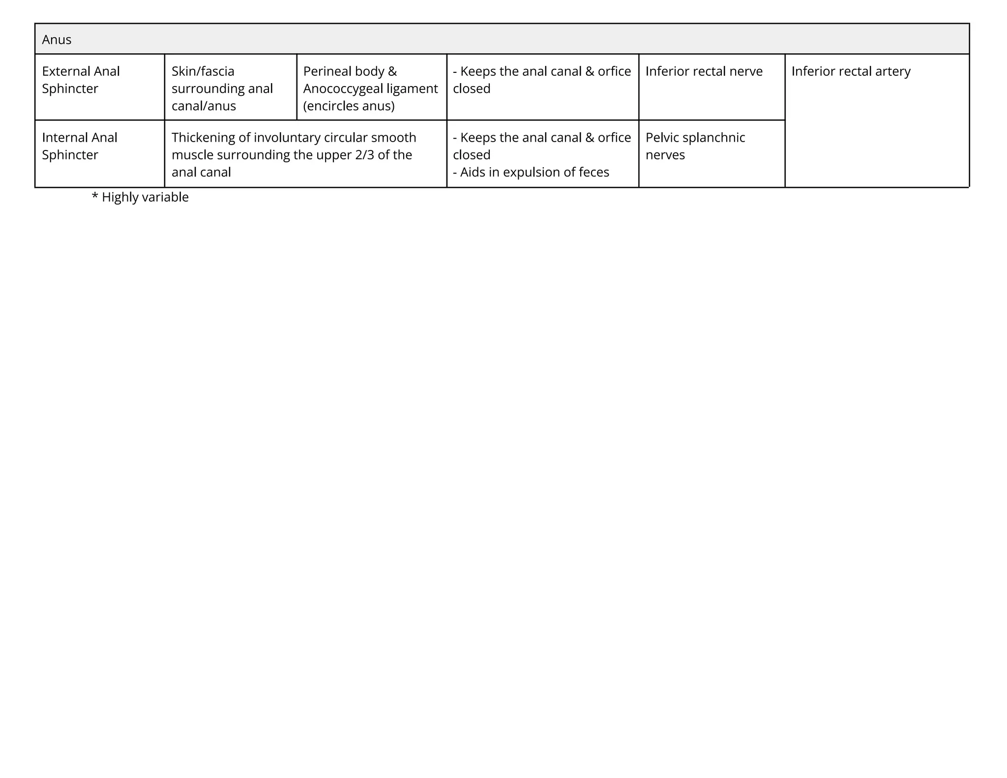
External Anal
Sphincter
Skin/fascia
surrounding anal
canal/anus
Perineal body &
Anococcygeal ligament
(encircles anus)
- Keeps the anal canal & orfice
closed
Inferior rectal nerve Inferior rectal artery
Urogenital Triangle - Superficial Perineal Pouch
Bulbospongiosus Male:
- Perineal body &
median penile
raphe
Female:
- Perineal body
Male:
- Perineal membrane,
dorsal aspect of
corpus
spongiosum/corpora
cavernosa & fascia of
bulb of penis
Female:
- Pubic arch & fascia of
corpora cavernosa/
clitoris
Male:
- Compresses the bulb of the
penis during
urination/ejactulation
- Assists in erection of the
penis
- Supports the perineal body
Female:
- Assists in the erection of the
clitoris/vestibular bulb
- Supports the perineal body
Deep branch of
perineal nerve
Perineal artery
Ischiocavernosus Internal surface of
the ischial
tuberosity & ischial
ramus
Crus of the
penis/clitoris
- Pushes the blood from the
root of the penis/clitoris to
the body
- Maintain erection of the
penis/clitoris
Superficial
Transverse
Perineal
Perineal body
- Supports & fixes the perineal
body/pelvic floor to support
abdominopelvic viscera
- Constrisction of urethra &
vagina
46.
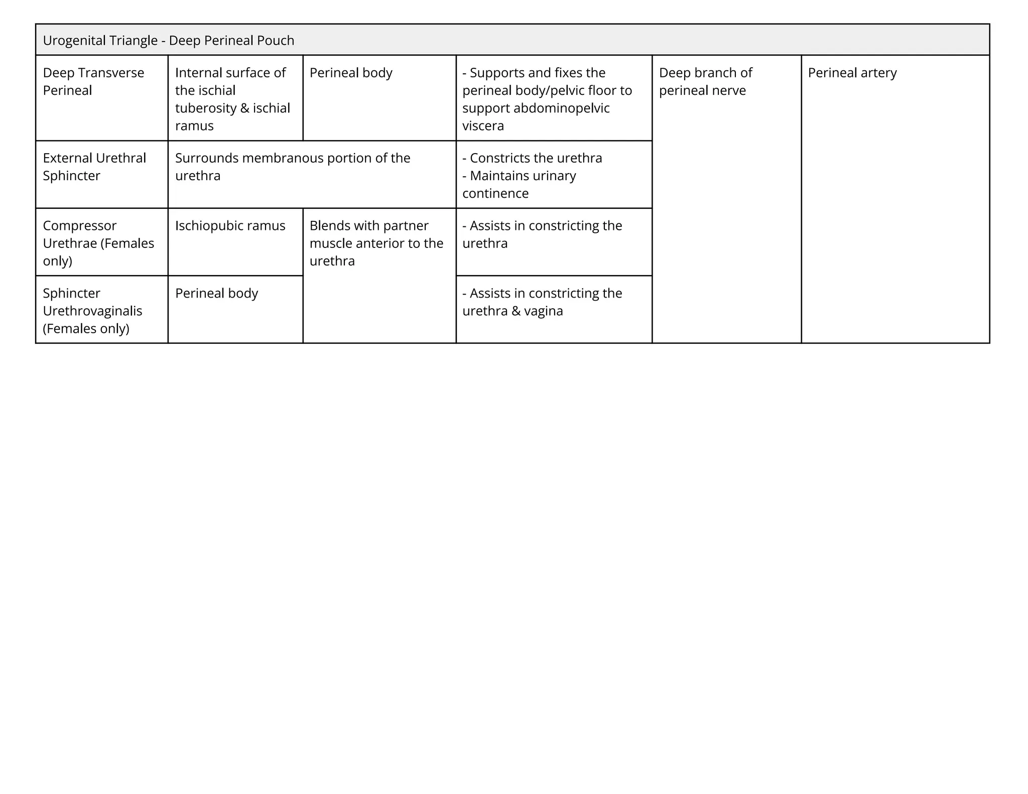
Urogenital Triangle -Deep Perineal Pouch
Deep Transverse
Perineal
Internal surface of
the ischial
tuberosity & ischial
ramus
Perineal body - Supports and fixes the
perineal body/pelvic floor to
support abdominopelvic
viscera
Deep branch of
perineal nerve
Perineal artery
External Urethral
Sphincter
Surrounds membranous portion of the
urethra
- Constricts the urethra
- Maintains urinary
continence
Compressor
Urethrae (Females
only)
Ischiopubic ramus Blends with partner
muscle anterior to the
urethra
- Assists in constricting the
urethra
Sphincter
Urethrovaginalis
(Females only)
Perineal body - Assists in constricting the
urethra & vagina
47.
Muscle Origin InsertionAction Innervation Blood Supply
Pelvic Cavity
Pelvic Wall
Piriformis Anterior surface of
sacrum &
sacrotuberous
ligament
Superior border of
greater trochanter of
femur
- Laterally rotates the hip
- Abducts the flexed hip
Nerve to piriformis Inferior/superior gluteal
arteries & internal pudendal
artery
Obturator Internus Pelvic surface of
obturator
membrane &
surrounding bones
Medial surface of
greater trochanter of
femur
- ternally rotates the
extended hip
- Abducts the flexed hip
Nerve to obturator
internus
Obturator artery & internal
pudendal artery
Pelvic Floor
Levator Ani:
Iliococcygeus
Tendinous arch of
internal obturator
fascia & ischial
spine
Anococcygeal ligament
& coccyx
- Helps support the pelvic
viscera
- Forms most of the pelvic
diaphragm
- Nerve to levator ani
(S4)
- Inferior rectal nerve
- Coccygeal plexus
- Inferior gluteal artery
- Inferior vesical artery
- Internal pudendal artery
Levator Ani:
Pubococcygeus
Posterior surface of
bodies of pubic
bones (lateral to
puborectalis)
Anococcygeal
ligament, coccyx,
Perineal body &
musculature of
prostate/vagina
Levator Ani:
Puborectalis
Posterior surface of
bodies of pubic
bones
N/A (forms 'sling'
posterior to rectum)
- Helps support the pelvic
viscera
- Forms most of the pelvic
diaphragm
- Assists in preventing
defecation
Coccygeus Ischial Spine Inferior aspect of
sacrum & coccyx
- Flexes the coccyx
- Supports the pelvic viscera
Pudendal nerve &
sacral spinal nerves
(S3-S5)*
48.
Anus
External Anal
Sphincter
Skin/fascia
surrounding anal
canal/anus
Perinealbody &
Anococcygeal ligament
(encircles anus)
- Keeps the anal canal & orfice
closed
Inferior rectal nerve Inferior rectal artery
Internal Anal
Sphincter
Thickening of involuntary circular smooth
muscle surrounding the upper 2/3 of the
anal canal
- Keeps the anal canal & orfice
closed
- Aids in expulsion of feces
Pelvic splanchnic
nerves
* Highly variable