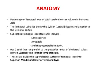
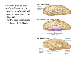
2. ANATOMY
• Percentage of Temporal lobe of total cerebral cortex volume in humans:
22%
• The Temporal Lobe lies below the Sylvian (Lateral) fissure and anterior to
the Occipital cortex.
• Subcortical Temporal lobe structures include –
- Limbic cortex
- Amygdala
- and Hippocampal formation.
• Has 2 sulci that run parallel to the posterior ramus of the lateral sulcus
named Superior and Inferior temporal sulci.
• These sulci divide the superolateral surface of temporal lobe into
Superior, Middle and Inferior Temporal Gyri.
4. Brodmann areas on lateral
suraface of Temporal lobe :
- Auditory area (area 41, 42)
- Auditory association cortex
(area 22)
- Ventral visual stream areas
( area 20, 21, 37 & 38 )
5. The Medial Temporal region (LIMBIC CORTEX) includes :-
- Amygdala and adjacent cortex (Uncus )
- Hippocampus and the surrounding cortex i.e.
- Subiculum
- Entorhinal Cortex (Brodmann area 28 )
- Perirhinal Cortex (Broadmann area 35 & 36 )
6. BLOOD SUPPLY OF TEMPORAL LOBE
• Convexity – inferior branch of MCA
• Medial & inferior aspects (including Hippocampus) – temporal
branch of PCA
7. SUPERIOR TEMPORAL GYRUS
• This is bounded by:
- the lateral sulcus above;
- the superior temporal sulcus below
• It contains several important structures including:
- Brodmann areas 41 and 42, marking the location of the Primary
auditory cortex
- Transverse temporal gyri (also called Heschl's gyri) – PRIMARY
AUDITORY RECEPTIVE AREA
- Wernicke's area - Brodmann 22
8. MIDDLE TEMPORAL LOBE
The region encompasses most of the lateral temporal cortex, a region believed to
play a part in auditory processing and language.
- Language function is left lateralized in most individuals.
- Brodmann area 21.
9. INFERIOR TEMPORAL LOBE
• In humans it corresponds approximately to the inferior temporal
gyrus.
• Brodmann area 20.
• The region encompasses most of the ventral temporal cortex, a
region believed to play a part in high-level visual processing and
recognition memory.
10. AMYGDALA
•The amygdala (Latin,meaning
'almond') are almond-shaped
groups of neurons located
deep within the medial
temporal lobes of the brain, in
close relation to anterior end
of inferior horn of lateral
ventricle, located at anterior
end of hippocampal formation.
11. HIPPOCAMPUS
- The hippocampus is a scrolled structure
located in the medial temporal lobe.
- can be divided into at least 5 different areas.
- Areas CA3 and CA1 are more diffuse; CA2 is
hard to distinguish between them.
- (CA stands for cornu ammonis, from its
ram's horn shape.)
- The subiculum sits at the base of the
hippocampus, and is continuous with
entorhinal cortex, which is part of the
parahippocampal gyrus.
12. DENTATE GYRUS
• Part of the hippocampal formation.
• Contains granule cells, which project to the pyramidal cells
and interneurons of the CA3 subfield of the hippocampus.
• The granule cells are the principal excitatory neurons of the
dentate gyrus.
• The major input to the dentate gyrus (Perforant pathway) is
from the entorhinal cortex, and the dentate gyrus receives no
direct inputs from other cortical structures.
• The dentate gyrus is also one of the few regions of the brain
where neurogenesis takes place. Neurogenesis is thought to
play a role in the formation of new memories.
13. CONNECTIONS OF TEMPORAL LOBES
Five main types:
1. Hierarchical sensory pathway
2. Dorsal auditory pathway
3. Polymodal pathway
4. Medial (mesial) temporal pathway
5. Frontal lobe projection
14. Hierarchical sensory pathway
connections from primary and secondary auditory
and visual cortex
through the lateral temporal cortex
terminate in the temporal pole
• Subserves stimulus recognition
15. Dorsal auditory pathway
• Forms important functional connections from auditory areas
to posterior parietal cortex
• Concerned with directing movements with respect to
auditory information
16. Polymodal Pathway
• A series of parallel projections from the visual and auditory
association areas into the polymodal regions of the superior
temporal sulcus
• Functions in stimulus categorization
17. Medial Temporal Projection
• Projections from auditory and visual areas into the limbic
Regions -
E.g., amygdala and hippocampus
• Also called Perforant pathway
• Serves function in long term memory
18. Frontal lobe Projection
• Auditory and visual information goes to two prefrontal
regions, one on the dorsolateral surface and the other in the
orbital region
• necessary for various aspects of movement control, short-
term memory and affect.
20. Temporal Lobe Functions
• Sensory Processes
– Identification and Categorization of Stimuli
– Cross-Modal Matching
• Process of matching visual and auditory information
• Depends on cortex of the superior temporal sulcus
• Affective Responses
– Emotional response is associated with a particular stimulus
• Spatial Navigation
– Hippocampus – Spatial Memory
21. Superior Temporal Sulcus (STS) and
Biological Motion
• Functional Imaging reveals activation in the STS during perception of
Biological motion.
• Biological Motion
– Movements relevant to a species , Social perceptions
– Allow us to guess others’ intentions
22. Consider an example..
• Imagine that you want to buy a car and now you are riding a
two wheeler. On your ride, you notice many different cars,
and you decide to keep a mental list of the brands and colours
that you encounter so that you can discuss with family and
friends later on.
• As you ride along, you suddenly stop and back up—you have
encountered a traffic policeman on the roadside of the next
square walking with a fineslip towards you . You decide to
change routes and look for cars elsewhere!
• What temporal-lobe functions took part in your experience?
23. • To be aware of specific type, colour and brand- object
recognition by ventral visual pathway
• To categorize quickly as they are in motion-polymodal
pathway in superior temporal sulcus
• Matching acceleration/engine sound to visual input- Cross
modal matching by dorsal auditory pathway
• Policeman walking towards you- “Biological motion” by
superior temporal sulcus
• On seeing fineslip- increased HR and BP and affective
response- by amygdala
• To change route and search elsewhere (spatial orientation)-
hippocampus
• To remember all the cars and discuss- medial temporal lobe
24. TEMPORAL LOBE FUNCTIONS
• SPECIAL SENSES
– Hearing
• FUNCTIONS OF LIMBIC SYSTEM
– Memory
– Emotions & Mood
– Attitudes & Social Behaviour
• LANGUAGE FUNCTIONS
– Semantics
– Naming
• VISUAL & OTHER SENSORY INTEGRATION
25. HEARING
• Auditory cortex lies principally on the supratemporal plane of superior
temporal gyrus (Heschl’s gyrus)
• Primary cortex has tonotopic arrangement:
– high tones terminate in medial portion
– Low tones in lateral portions
• Particular sound frequency causes excitation of particular cortical area =
sound frequency perception
• Also helps in discrimination of sound patterns like combination or
sequence of tones
• Also takes part along with superior olivary nucleus in mechanism for
detecting sound direction
26. HEARING
Nondominant hemisphere- non-verbal aspect, recognition
of harmony & melody
Dominant hemisphere- verbal aspect,naming of musical
scores & all the semantic (writing, reading)
aspects of music
27. LIMBIC SYSTEM
• Coordinate sensory events with bodily and visceral needs
• Regulate innate automatised activities concerned with
feeding, searching, sex, & emotion-provoking situations
• Neurotransmitters:
– For memory- acetyl choline
– Norepinephrine in medial parts of limbic system
– Amygdala, septal nuclei & lateral parts are rich in serotonin
28. • Limbic system regulates emotions and motivations—
particularly those related to survival—such as fear, anger, and
pleasure (sex and eating)
• Feelings of pleasure/reward are very powerful and self-
sustaining. Pleasurable behaviors activate a circuit of
specialized nerve cells in the limbic area that is devoted to
producing and regulating pleasure called the reward system
29
29. Emotion and Motivation
(Individual function)
Rage, aggression Amygdala and hypothalamus.
Fear Amygdala and hypothalamus
Feeding Hypothalamus (satiety centers).
Sexual drive and behavior
Hypothalamus and limbic system
Goal directed behavior
(reward and punishment):
Hypothalamus and frontal cortex
30. MEMORY-
Medial temporal lobes
• The MTL is comprised of multiple structures
including the
Hippocampal formation,
Amygdala,
Entorhinal cortex, and
Surrounding perirhinal and parahippocampal cortices
• This is involved in explicit or declarative memory.
• It has essential role in the formation of new
memories about experienced events
(episodic or autobiographical memory).
31. Memory : Hippocampus
• Critical in laying down declarative memory, but is not necessary
for working memory / procedural memory / memory storage.
• The hippocampal formation plays a role in memory
consolidation
• Damage to the hippocampus usually results in profound
difficulties in forming new memories (anterograde amnesia),
and normally also affects access to memories prior to the
damage (retrograde amnesia) but does not affect procedural
memory
32. Memory : Parahippocampal cortex
•Sensitive to familiarity with stimulus location or the geometry of
surrounding space
•Patients with a lesion limited to parahippocampal cortex lose
the ability to acquire new topographic knowledge
•The Parahippocampal Area responds selectively to visual
stimuli that convey information about the layout of local space.
34. WERNICKE’S AREA
• The sensory area of the dominant hemisphere for interpretation
of language is Wernicke’s area.
• Closely associated with hearing areas
• Provides, at the input stage, an entry point for conversion of
auditory sequences into neural word representations leading to
meaning-appropriate content words.
• Localization of lexicon (vocabulary ) for spoken word recognition
exists in middle part of left superior & middle temporal gyri
• Lexicon for written word recognition exists in posterior part of left
middle temporal gyrus
• A major share of our sensory experience is converted into
language equivalent before stored in memory areas
35. WERNICKE’S AREA
Reception in primary auditory area
of sound signals that encode words
Interpretation of words in
Wernicke’s area
Determination of thoughts & words
to be spoken in Wernicke’s area
Transmission of signals form
wernicke’s area to Broca’s area by
arcuate fasciculus
Activation of skilled motor
programs in Broca’s area for control
of word formation
Transmission of appropriate signals
into motor cortex to control speech
muscles
Primary visual area is the
initial receptive area
Early stage of interpretation
in angular gyrus
Full level of recognition in
Wernicke’s area
Hearing & speaking Reading & speaking
36. VISUAL INTEGRATION
• Middle & inferior temporal gyri (areas 21 & 37) receive massive
contingent fibers form striate cortex (area 17) & parastriate visual
association areas (areas 18,19)
• Subserves visual discriminative functions like spatial orientation,
estimation of depth & distance, stereoscopic vision, & hue perception
• It integrates vision intimately with all forms of exteroception &
proprioception
• Bitemporal lobectomy in animals-‘psychic blindness’
37. SENSORY INTEGRATION
• Temporal lobe is great integrator of ‘sensations, emotions and
behaviour’
• It is the site where sensory modalities are integrated into ultimate
self-awareness
• Similar suprasensory integrative mechanisms are operative in
parietal lobe, but only in the temporal lobe are they brought into
close relationship to one’s instinctive & emotional life
38. • Stream of thinking (internal conversation that is constant in
waking state) requires language & memory functions, both of
which involve temporal lobe
• Temporal lobe throughout the life assembles all fragments of
present & past experiences of all kinds into awareness of
personal integrity which is only interrupted by sleep
• Temporal neocortex functions to bring ultimate awareness
that ‘I am’.
39. OTHER FUNCTIONS
• Vestibular functions:
– Some fibers from vestibular input are relayed in superior temporal
gyrus
– Episodic vertigo may represent the aura or sole manifestation of
temporal CPS
• Time perception:
– Main neural mechanism subserving time perception & integrating it
with other sensory inputs and memory lies in temporal lobe
– With disorder of temporal lobe of either side, there may be
intermittent disturbance of time perception
40. Symptoms of Temporal Lobe Lesions
1. Disturbance of Auditory Sensation and perception
2. Disorders of music perception
3. Disorders of visual perception
4. Disturbance in the selection of visual and auditory input
5. Impaired organization and categorisation of sensory input
6. Inability to use contextual information
7. Impaired long term memory
8. Altered personality and affective behavior
9. Altered sexual behavior
41. Disturbance of Selection of Visual and
Auditory Input
• Selective attention to auditory input is impaired in patients
with temporal lobe damage and can be tested with dichotic
listening
• Damage to the left temporal lobe impairs recall of visual
stimuli in the right visual field
• Damage to the right temporal lobe impairs recall of visual
stimuli in both visual fields
42. Organization and Categorization
• Left temporal lobe lobectomies lead to impairment in the
ability to categorize words or pictures of objects
• Posterior lesions lead to a difficulty in recognizing specific
word categories
43. Using Contextual Information
• Stimuli can be interpreted in different ways depending on the
context
– Example: Fall - the season or a tumble
44. Memory
• Antereograde Amnesia
– Amnesia for events after bilateral removal of the medial
temporal lobes
• Inferotemporal Cortex
– Conscious recall of information
• Left temporal lobe
– Verbal memory
• Right temporal lobe
– Impaired recall of nonverbal material
45. Affect and Personality
• Stimulation of anterior and medial temporal cortex produces feelings of
fear
• Temporal lobe personality
– Personality that overemphasizes trivial and petty details of life
– Pedantic speech (an overly formal speaking style inappropriate to the
conversational setting)
– Egocentricity (preoccupation with one’s own internal world)
– Perseveration
– Paranoia
– Preoccupation with religion
– Proneness to aggression
46. Changes in Sexual Behavior
• Release of sexual behavior seen after bilateral temporal
damage
47. Superior temporal Gyrus
• Auditory radiations run from the medial geniculate body to the Auditory
cortex ( areas 41 and 42) in the superior temporal gyrus.
• Hearing is represented bilaterally in the temporal lobes.
(contralateral predominance)
• Nearby areas - differentiation and interpretation of sounds.
• Electrical stimulaton of auditory area leads to vague auditory hallucination
(tinnitus, sensation of roaring and buzzing ), and adjacent areas causes
vertigo and a sensation of unsteadiness.
• Unilateral destruction of the Auditory cortex lead to difficulty in sound
localisation and a bilateral decrease of auditory acuity.
• Bilateral disease lead to Cortical deafness (may be unaware of their
deficits).
• Audiometric tests may be helpful to reveal mild deficits.
• Involvement of vestibular areas may cause difficulty in equillibrium and
imbalance
48. Asymmetry of Temporal lobe function
• Damage to left temporal lobe – deficits in verbal memory, processing
speech sounds
• Damage to right temporal lobe – deficits in nonverbal memory
processing certain aspects of music
face recognition
56. TEMPORAL LOBE SYNDROMES
Homonymous upper
quadrantanopia
Wernicke’s aphasia
Amusia
Anomia
Visual agnosia
Impairment on tests of
visually presented
material
Homonymous upper
quadrantanopia
Agnosia for sounds &
some qualities of music
Impairment on tests of
visually presented
material
U/L disease of dominant lobe U/L disease of nondominant lobe
57. TEMPORAL LOBE SYNDROMES
Auditory, visual, olfactory
& gustatory hallucinations
Dreamy states
Emotional & behavioural
changes
Delirium
Disturbance of time
perception
Korsakoff amnesic defect
Apathy & placidity
Hypermetamorphopsia,
hyperorality, hypersexulaity,
blunted emotional reactivity
(Kluver-Bucy syndrome)
Either temporal lobe Bilateral disease
58. Wernickes Aphasia
• Also known as Fluent / Recepetive / Sensory / Postrolandic Aphasia
• Due to destruction of posterior superior temporal area of the dominant
hemisphere supplied by inferior division of MCA.
– Fluency is preserved with a normal or even increased word
output (LOGORRHEA)
– Speech although effortless is devoid of meaningful content
– Paraphasias, Neologisms and defective sentence structure
(PARAGRAMMATISM) (JARGON APHASIA)
– Auditory comprehension is impaired, even unaware of his own
speech, and doesnot correct himself.
– Repetition impaired
– Reading impaired
– Naming impaired
– Writing impaired
– Patient often is unaware of the defect
59. Temporal lobe / psychomotor seizures / CPS
• Patients with Temporal lobe lesion may have visual hallucinations and
perversions.
• Visual hallucinations are complex with image distortion (macropsia /
micropsia / too near / too far away)
• May have an auditory component – hallucinated figure may speak .
• Lesions involving medial temporal lobe in the region of Uncus ( k/a
UNCINATE FITS - olfactory hallucination only )
• Seizures may present as : automatisms, illusions , hallucinations and
pilomotor erections.
• Amnesia for the period of event is seen.
60. • DÉJÀ VU [already seen]
• DEJA PENSEE/ DEJA VACU - something new seeming strangely
familiar
• JAMAIS VU - something familiar is strange or new
• EXPERIENTIAL HALLUCINATIONS - Hallucinations based on
remembered experiences
• TORNADO EPILEPSY – vertigo due to involvement of vestibular
cortex in a seizure discharge
61. KLUVER- BUCY SYNDROME
• Due to bilateral ablation of temporal lobes [ anterior ] in monkeys.
• Placidity i.e. extreme calmness , loss of fear & rage reactions
• Lack the ability to visually recognize objects [ psychic blindness or
visual agnosia ]
• Striking tendency to put everything into mouth.
• Hypersexuality
• Increased food intake [bulimia]
• Severe memory loss
• Seen only in partial forms in humans ( placidity and enhanced oral
behavior are the most common presentations)