Gram-Negative cocci. Neisseria. Gonococcal & Meningococcal infections
•Download as DOC, PDF•
9 likes•1,852 views

This document discusses two pathogenic Neisseria species: Neisseria gonorrhoeae, which causes gonorrhea, and Neisseria meningitidis, which causes meningococcal infections. It describes their morphology, cultural characteristics, virulence factors, pathogenicity, epidemiology, and laboratory diagnosis. Diagnosis of gonorrhea involves microscopy and culture of urethral discharge or other specimens, while meningococcal meningitis is diagnosed via microscopy, culture and antigen detection in cerebrospinal fluid and blood cultures. Treatment of both involves antibiotics like penicillin, cephalosporins, or fluoroquinolones.

Recommended
More Related Content
What's hot
What's hot (20)
Similar to Gram-Negative cocci. Neisseria. Gonococcal & Meningococcal infections
Similar to Gram-Negative cocci. Neisseria. Gonococcal & Meningococcal infections (20)
More from Eneutron
More from Eneutron (20)
Recently uploaded
Recently uploaded (20)
Gram-Negative cocci. Neisseria. Gonococcal & Meningococcal infections
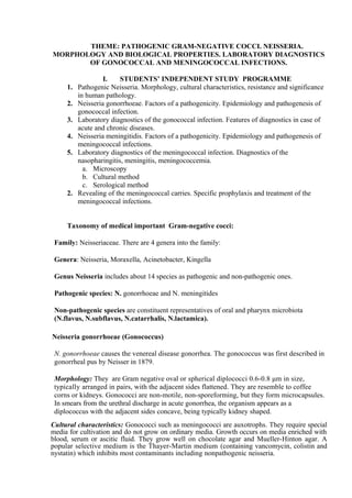
- 1. THEME: PATHOGENIC GRAM-NEGATIVE COCCI. NEISSERIA. MORPHOLOGY AND BIOLOGICAL PROPERTIES. LABORATORY DIAGNOSTICS OF GONOCOCCAL AND MENINGOCOCCAL INFECTIONS. I. STUDENTS’ INDEPENDENT STUDY PROGRAMME 1. Pathogenic Neisseria. Morphology, cultural characteristics, resistance and significance in human pathology. 2. Neisseria gonorrhoeae. Factors of a pathogenicity. Epidemiology and pathogenesis of gonococcal infection. 3. Laboratory diagnostics of the gonococcal infection. Features of diagnostics in case of acute and chronic diseases. 4. Neisseria meningitidis. Factors of a pathogenicity. Epidemiology and pathogenesis of meningococcal infections. 5. Laboratory diagnostics of the meningococcal infection. Diagnostics of the nasopharingitis, meningitis, meningococcemia. a. Microscopy b. Cultural method c. Serological method 2. Revealing of the meningococcal carries. Specific prophylaxis and treatment of the meningococcal infections. Taxonomy of medical important Gram-negative cocci: Family: Neisseriaceae. There are 4 genera into the family: Genera: Neisseria, Moraxella, Acinetobacter, Kingella Genus Neisseria includes about 14 species as pathogenic and non-pathogenic ones. Pathogenic species: N. gonorrhoeae and N. meningitides Non-pathogenic species are constituent representatives of oral and pharynx microbiota (N.flavus, N.subflavus, N.catarrhalis, N.lactamica). Neisseria gonorrhoeae (Gonococcus) N. gonorrhoeae causes the venereal disease gonorrhea. The gonococcus was first described in gonorrheal pus by Neisser in 1879. Morphology: They are Gram negative oval or spherical diplococci 0.6-0.8 μm in size, typically arranged in pairs, with the adjacent sides flattened. They are resemble to coffee corns or kidneys. Gonococci are non-motile, non-sporeforming, but they form microcapsules. In smears from the urethral discharge in acute gonorrhea, the organism appears as a diplococcus with the adjacent sides concave, being typically kidney shaped. Cultural characteristics: Gonococci such as meningococci are auxotrophs. They require special media for cultivation and do not grow on ordinary media. Growth occurs on media enriched with blood, serum or ascitic fluid. They grow well on chocolate agar and Mueller-Hinton agar. A popular selective medium is the Thayer-Martin medium (containing vancomycin, colistin and nystatin) which inhibits most contaminants including nonpathogenic neisseria.
- 2. Gonococci are more difficult to grow than meningococci. They are aerobic but may grow anaerobically also. Growth occurs best at pH 7.2-7.6 and at a temperature of 35-36 °C. It is es- sential to provide 5-10 per cent CO2. Colonies are small, round, translucent, convex or slightly umbonate, with finely granular surface and lobate margins. They are soft and easily emulsifiable. Four types of colonies have been recognised - Tl to T4. Types 1 and 2 form small brown colonies. The cocci are piliated, autoagglutinable and virulent. Types 3 and 4 form larger, granular, nonpigmented colonies. T3 and T4 cocci are nonpiliated, form smooth suspensions and are avirulent. Fresh isolates from acute cases of gonorrhea generally form Tl and T2 colonies. Biochemical reactions: They possess weak enzymatic activity. Unlike to meningococci, gono- cocci ferment only glucose and not maltose. They can liquefy gelatin. Antigenic properties: Gonococci are antigenically heterogeneous. Their superficial proteins have antigenic properties. Nowadays there are about 40 serotypes are recognized into species which have no cross reactive antigens. The trilaminar outer membrane of gonococci contain many different proteins. Protein I is the major constituent and shows antigenic diversity, which helps in typing gonococcal strains. Virulent factors: 1. Pili (adhesins) which are hair-like structures several mi- crometres long, act as virulence factors by promoting attachment to host cells and inhibiting phagocytosis. 2. Proteins I acts as ligands attaching the coccus to the host cells. 3. Protein II is related to the opacity of the gonococcal colonies and so is called the 'opacity associated' (OPA) outer membrane protein. Strains with the OPA protein form opaque colonies and those lacking it transparent colonies. OPA may be responsible for attachment to the host cells and also for the clumping of cocci seen in urethral exudate smears. 4. The outer membrane also contains lipopolysaccharide (endotoxin) which may be responsible for the toxicity in gonococcal infections 5. IgA – protease inhibits protective secretory immunoglobulins and weakens local immunity Resistance: The gonococcus is a very delicate organism, readily killed by heat, drying and antiseptics. It is a strict parasite and dies in 1-2 hours in exudates outside the body. Formerly, it was highly susceptible to sulphonamides, penicillin and many other antibiotics. However, gonococci have developed resistance to one antibiotic after another. Pathogenicity: Gonorrhea is a venereal disease which are transmitted by sexual intercourse. The only source of infection is a human - carrier or less often a patient. The first step in infection is adhesion of gonococci to the urethra or other mucosal surfaces. Pili are involved in this adhesion. The cocci penetrate through the intercellular spaces and reach the subepithelial connective tissue by the third day after infection. The incubation period is 2-8 days. In men, the disease starts as an acute urethritis with a mucopurulent discharge containing gonococci in large numbers. The infection extends along the urethra to the prostate, seminal vesicles and epididymis. Chronic urethritis may lead to stricture formation. In women, the initial infection involves the urethra and cervix uteri. The vaginal mucosa is not usually affected in adults because the stratified squamous epithelium is resistant to infection by the cocci and also because of the acid pH of vaginal secretions. The infection may extend to Bartholin's glands, endometrium and fallopian tubes. Pelvic inflammatory disease and salpingitis may lead to sterility. Rarely, peritonitis may develop with perihepatic inflammation. Clinical disease is as a rule less severe in women, many of whom may carry gonococci in the cervix without developing any clinical symptoms. Asymptomatic carriage of gonococci is rare in men. . Conjunctivitis may occur, usually due to autoinoculation by the patient's fingers. Blood invasion may occur from the primary site of infection and may lead to metastatic lesions such as arthritis, ulcerative endocarditis and very rarely meningitis.
- 3. A nonvenereal infection is gonococcal ophthalmia in the newborn, which results from direct infection during passage through the birth canal. It has been controlled by the practice of instilling 1% silver nitrate solution into the eyes of all newborn babies Gonococcal bacteremia leads to skin lesions, especially hemorrhagic papules and pustules on the hands, forearm, feet and legs, and to tenosynovitis and suppurative arthritis, usually of the knees, ankles and wrists. Laboratory diagnosis: In the acute stage, diagnosis can be established readily but chronic cases sometimes present great difficulties. In acute gonorrhea the urethral discharge contains gonococci in large numbers. That is why microscopy of stained smears is reliable to diagnose acute cases. In chronic infections, there may not be any urethral discharge. The 'morning drop' of secretion may be examined or some exudate may be obtained after prostatic massage. The collected specimens are stained by Gram or by simple staining with methylene blue. The demonstration of intracellular Gram negative diplococci in stained smears provides a presumptive evidence of gonorrhea in men. The use of fluorescent antibody techniques for the identification of gonococci in smears has increased the sensitivity and specificity of diagnosis by microscopy. For culture, specimens should be inoculated on prewarmed plates, immediately on collection. In acute gonorrhea, cultures can be obtained readily on chocolate agar or Mueller- Hinton agar incubated at 35-36 °C under 5-10 per cent CO2. In chronic cases, where mixed infection is usual, however, it is better to use a selective medium such as the Thayer-Martin medium. The growth is identified by morphology and biochemical reactions. It may not be possible to obtain gonococci in culture from some chronic cases or from patients with metastatic lesions such as arthritis. Serological tests: The complement fixation test becomes positive only some weeks after the infection is established and may remain positive for months or years after the disease has been cured. So, serological method is more often used for diagnostics of chronic infections. Therapy and prophylaxis: The Centers for Disease Control and Prevention, USA in 1993 recommended the following schedule for uncomplicated gonorrhea: Ceftriaxone 125 mg single IM dose or Ciprofloxacin 500 mg (or Ofloxacin 400 mg) single oral dose, plus Doxycycline 100 mg twice daily for 7 days or Erythromycin 1 g single oral dose. The regimen is The Centers for Disease Control and Prevention, USA in 1993 recommended the following schedule for uncomplicated gonorrhea: Ceftriaxone 125 mg single IM dose or Ciprofloxacin 500 mg (or Ofloxacin 400 mg) single oral dose, plus Doxycycline 100 mg twice daily for 7 days or Erythromycin 1 g single oral dose. Control of gonorrhea consists of early detection of cases, contact tracing, health education and other general measures. As even clinical disease does not confer any immunity, vaccination has no place in prophylaxis. Neisseria meningitidis (Meningococcus) Meningococcus was first described and isolated in 1887 by Weichselbaum from the spinal fluid of a patient. N. meningitidis causes meningococcal meningitis (formerly also known as cerebrospinal fever) which may occur sporadically, as localised outbreaks or as epidemics, and also septicemia. Morphology: Meningococci are Gram negative oval or spherical cocci 0.6-0.8 μm in size, typically arranged in pairs, with the adjacent sides flattened. In smears from lesions, the cocci are more regular and generally intracellular. They are nonmotile. Most fresh isolates are capsulated. Cultural characteristics: Such as gonococci meningococci have exacting growth requirements and do not grow on ordinary media. They are strict aerobes, no growth occurring anaerobically. The optimum temperature for growth is 35-36 °C. No growth takes place below 30 °C. Optimum pH is 7.4-7.6. Growth is facilitated by 5-10 per cent CO2 and high humidity. On solid media, after incubation for 24 hours, the colonies are small (about 1 mm in diameter) translucent, round, convex, bluish grey, with a smooth glistening surface and with entire edges. Weak hemolysis occurs on blood agar. Growth is poor in liquid media, producing a granular turbidity with little or no surface growth.
- 4. Blood agar, chocolate agar and Mueller-Hinton starch casein hydrolvsate agar are the media commonly used for culturing meningococci. Modified Thayer -Martin (with vancomycin, colistin and nystatin) is a useful selective medium. Biochemical reactions: They are catalase and oxidase positive. The prompt oxidase reaction helps the identification of neisseria (both meningococcus and gonococcus) in mixed cultures. When freshly prepared 1% solution of oxidase reagent (tetramethyl paraphenylene diamine hydrochloride) is poured on the culture media, the neisseria colonies turn deep purple. Glucose and maltose are fermented, but not sucrose or lactose, producing acid but no gas (gonococci ferment glucose but not maltose). Antigenic properties and classification: Meningococci are capsulated, unlike other neisseriae. Based on their capsular polysaccaride antigens, meningococci are classified into at least 13 serogroups, of which Groups A, B and C are the most important. Group A is usually associated with epidemics and Group C mostly with localised outbreaks, while Group B causes both epidemics and outbreaks. Groups 29-E, W-135 and Y also frequently cause meningitis. Resistance: Meningococci are very delicate organisms, being highly susceptible to heat, dessication, alterations in pH and to disinfectants. They were uniformly sensitive to penicillin and other antibiotics, but resistant strains have emerged and become common in many areas. Virulent factors: 1. Endotoxin (LPS)is released by autolysis. The vascular endothelium is particularly sensi- tive to the endotoxin. 2. Pili (adhesins) are responsible to attachment to nasophayngeal epithelium. 3. Capsule protects from phagocytosis. Pathogenicity: Cerebrospinal meningitis and meningococcal septicemia are the two main types of meningococcal disease. Meningococci are strict human parasites inhabiting the nasopharynx. Infection is usually asymptomatic. In some, local inflammation ensues, with rhinitis and pharyngitis. The manner in which the cocci spread from the nasopharynx to the meninges is controversial. On reaching the central nervous system, a suppurative lesion of the meninges is set up, involving the surface of the spinal cord as well as the base and cortex of the brain. The cocci are invariably found in the spinal fluid, both free and within the leucocytes. Case fatality is variable but in untreated cases may be as high as 80 per cent. Meningococcemia presents as acute fever with chills, malaise and prostration. Typically a petechial rash occurs early in the disease. A few develop fulminant meningococcemia (formerly called Waterhouse-Friderichsen syndrome) which is an overwhelming and usually fatal condition, characterised by shock, disseminated intravascular coagulation and multisystem failure. Epidemiology: The human nasopharynx is the only reservoir of the meningococcus. Asymptomatic nasopharyngeal carriers rarely contract the illness but serve to infect their contacts. Transmission is essentially by airborne droplets or less often by fomites. Meningitis is common in children between 3 months and 5 years of old. Epidemics usually occur in semi- closed communities living in crowded conditions, as in jails and ships formerly, and in army camps in recent times. Laboratory diagnosis: In meningococcal meningitis, the cocci are present in large numbers in the spinal fluid and, in the early stage, in the blood as well. Demonstration of meningococci in the nasopharynx helps in the detection of carriers. Examination of CSF: 1. Microscopy. The fluid will be under pressure and turbid, with a large number of pus cells. Under microscopy CSF contains a large number of meningococci inside polymorphs but often extracellularly also. 2. Detection of capsular antigen with serological tests. They may be demonstrated by latex agglutination, precipitation or counterimmunoelectrophoresis using meningococcal antisera. 3. Culture method: CSF is inoculated on blood agar or chocolate agar plates and incubated at 35-36 °C under 5-10 per cent. CO2. Colonies appear after 18-24 hours and may be identified by morphology and biochemical reactions. Blood culture: In meningococcemia and in early cases of meningitis, blood culture is often positive.Cultures should be incubated for 4-7 days. Nasopharyngeal swab: This is useful for the detection of carriers. Collected specimens are cultivated and identified as it is described for CSF culture. Retrospective diagnose of meningococcal infection may be obtained by detection of
- 5. antibodies with complement fixation test. Treatment and prophylaxis. Intravenous penicillin G is the treatment of choice. Chloramphenicol is equally effective. One of the later cephalosporins (Ceftriaxone, Ceftazidime) may be used for the initiation of treatment before the etiology of meningitis is known. Monovalent and polyvalent vaccines are available containing the capsular polysaccharides of groups A, C, W-135 and Y. The vaccines induce good immunity after a single dose in older children and adults but are of little value in infants. The immunity is group specific. There is no Group B vaccine available at present. As attack rates are very high in the households or close contacts of meningococcal patients, they should be provided with chemoprophylaxis. II.Students practical activities: 1. Microscopy the prepared smears from pure culture of N.meningitidis stained by Gram. Estimate the morphology and sketch the image. 2. Microscopy the prepared smears of pus from urethra stained with methylene blue. Sketch the image. 3. Familiarize with specific media for cultivation of pathogenic neisseria. 4. Write down the scheme of laboratory diagnostics of the gonococcal and meningococcal infections.
- 6. antibodies with complement fixation test. Treatment and prophylaxis. Intravenous penicillin G is the treatment of choice. Chloramphenicol is equally effective. One of the later cephalosporins (Ceftriaxone, Ceftazidime) may be used for the initiation of treatment before the etiology of meningitis is known. Monovalent and polyvalent vaccines are available containing the capsular polysaccharides of groups A, C, W-135 and Y. The vaccines induce good immunity after a single dose in older children and adults but are of little value in infants. The immunity is group specific. There is no Group B vaccine available at present. As attack rates are very high in the households or close contacts of meningococcal patients, they should be provided with chemoprophylaxis. II.Students practical activities: 1. Microscopy the prepared smears from pure culture of N.meningitidis stained by Gram. Estimate the morphology and sketch the image. 2. Microscopy the prepared smears of pus from urethra stained with methylene blue. Sketch the image. 3. Familiarize with specific media for cultivation of pathogenic neisseria. 4. Write down the scheme of laboratory diagnostics of the gonococcal and meningococcal infections.