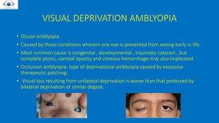
AMBLYOPIA
Presenter : Dr Nikhil Agrawal (1st year resident)
Moderator : Dr Ekta Gupta
DHIR HOSPITAL AND POSTGRADUATE INSTITUTE OF OPHTHALMOLOGY
DEFINITION
• Amblyopia (lazy eye) has been described as unilateral or bilateral reduction in
BCVA caused by vision deprivation , abnormal binocular interaction , without a
visible organic cause .
• Most common cause of preventable uniocular vision loss in children .
• The visual loss is correctable, if appropriate measures are applied at appropriate
time .
Epidemiology
• Onset – Birth to 7 yrs
• Socio-economic factors does not significantly matter .
• Earlier the onset the greater the deficit .
• 4 times more frequent in premature children .
• 6 times more frequent in children with delayed milestones .
• Other risks are Retinopathy of Prematurity, malnutrition, maternal smoking, use of
drugs and/or alcohol , albinism, Down syndrome, neurological problems like
hydrocephaly and cerebral palsy..
Pathophysiology
A Nobel Partnership: Hubel & Wiesel
Significant electrophysiological and anatomical
abnormalities are seen in the striate cortex and
lateral geniculate nucleus (LGN). The
neurophysiology reveals that the deprived eye
shows a marked shrinkage of its input stripes
(ocular dominance columns) and a
corresponding expansion of the non deprived
eye.
Pathophysiology
• Critical period-The period of development of visual acuity (1 month to 3-5 years of
age).
• Sensitive period-The period during which deprivation is effective in causing
amblyopia (a few months to 7 or 8 years of age) .
• Plastic period -The period during which recovery from amblyopia can be obtained
(time of deprivation to the teenage years or even into the adult years)
• Bilateral stimulus deprivation in critical period disrupts the development of fixation
reflex and nystagmus appears as a result of this.
TYPES
Functional Amblyopia Organic Amblyopia
Potentially reversible Irreversible
Refers to obligatory psychical suppression of the
retinal image.
Refers to partial loss of vision caused by undetectable
organic lesions in the eye or in the visual pathway.
Types: Strabismic, Anisometropic, Ametropic,
Meridional, Stimulus, Deprivation
Types: Nutritional, Toxic, d/t retinal diseases,
Idiopathic
Usually seen in childhood Can cause visual acuity defect at any age
CLASSIFICATION
1. Strabismic amblyopia
2. Stimulus deprivation
3. Anisometropic amblyopia
4. Bilateral ametropic amblyopia
5. Meridional amblyopia
6. Amblyopia secondary to nystagmus
STRABISMIC AMBLYOPIA
• Seen in patients with unilateral constant squint who strongly favour one eye for
fixation from birth to 6 years of age.
• Seen far more often in esotropes than the exotropes.
• Types –
Cong. Esotropia
Cong. Exotropia
Accommodative Esotropia
Acquired constant tropia in childhood
Monofixation Syndrome (small angle tropia )
STRABISMIC AMBLYOPIA
• Does not develop in alternating or intermittent strabismus as there are periods of
normal binocular interaction that preserve the integrity of visual system .
• Severity of amblyopia does not correlate with angle of strabismus.
VISUAL DEPRIVATION AMBLYOPIA
• Disuse amblyopia.
• Caused by those conditions wherein one eye is prevented from seeing early in life.
• Most common cause is congenital , developmental , traumatic cataract , but
complete ptosis, corneal opacity and vitreous hemorrhage may also implicated.
• Occlusion amblyopia- type of deprivational amblyopia caused by excessive
therapeutic patching.
• Visual loss resulting from unilateral deprivation is worse than that produced by
bilateral deprivation of similar degree.
ANISOMETROPIC AMBLYOPIA
• Refers to the amblyopia occurring in an eye having higher degree of refractive
error than the fellow eye.
• Most patients have straight eyes and appear normal , so the only way to identify
these patients is through vision screening.
• Amblyopia is more common and is of higher degree in patients with
anisohypermetropia than in those with anisomyopia.
• Potentially Amblyopiogenic Refractive Errors :
ISOMETROPIC AMBLYOPIA
• • Bilateral amblyopia occurring in children with bilateral uncorrected high
refractive error.
• Mechanism – effect of blurred retinal images alone.
• Potentially Amblyopiogenic Refractive Errors :
CLINICAL CHARACTERISTICS
• Decreased visual acuity
• Decreased stereopsis
• Fixation reflex
• Crowding phenomenon
• Effect of neutral density filter
• Contrast sensitivity
• Fixation pattern
• VEP - Reduction in amplitude and slightly prolonged latency
• Afferent pupillary defect may be seen
• Occasionally latent nystagmus
VISUAL ACUITY
• Two line difference between amblyopic and
normal eye
• For B/L amblyopia the VA should be less than
6/12 in each eye
• VA in children-
Infants - Fixation preference preverbal
Children-preferential looking test , OKN test , VEP
2-3 yrs - E charts , pictorial charts
 >3 yrs – Snellen’s charts , HOTV charts
Lea’s padels
OKN TEST
STEREO-ACUITY
Two pencil test Titmus Fly test
FIXATION REFLEX
• Useful tool to assess VA in
children <5yrs of age
• Central steady and maintained
(CSM) fixation implies good VA .
CROWDING PHENOMENON
• Amblyopia patients exhibit better VA for single optotypes than for letters placed in
a row .
• Vision testing with single optotypes is likely to over estimate VA in pts with
amblyopia
• More accurate assessment of mono ocular VA is obtained with the presentation of
line of optotypes or single optotype with crowding bars that surround the
optotype being identified .
NEUTRAL DENSITY FILTER
• A neutral density filter reduces overall luminance without inducing a color change.
• Decreased luminance of the visual target results in diminished central acuity in
normal eyes.
• Decreased illumination of visual targets has less of an effect on amblyopic eyes
because they are not using central acuity
• It was found that neutral filters profoundly reduce vision in eyes with organic
amblyopia whereas vision of eyes with functional amblyopia was not reduced and
occasionally even slightly improved.
Amblyopia is a Diagnosis of Exclusion.
Following three factors are essential to diagnose amblyopia.
1. Evidence of reduced visual acuity - usually unilateral but may be bilateral.
2. Presence of amblyopgenic factors .
3. No cause for visual loss on examination.
Specific emphasis should be given on Bruckner’s test – Studies suggest an accuracy
of 84% in amblyopia detection in patients with strabismus or prescription difference
of 3D or more .
CLINICAL EVALUATION & DIAGNOSIS
• Thorough clinical history
• Evaluation of visual acuity and fixation
pattern
• Stereo-acuity testing
• Binocular alingnment and ocular
motility
• Thorough ocular examination including
fundus examination.
• Cycloplegic retinoscopy/Refraction
• Bruckner’s test
• Neutral density filter and testing for
crowding phenomenon.
Hirschberg corneal reflex test
Grading of Severity
• On the basis of visual acuity :
Mild amblyopia -6/9 to 6/12
Moderate amblyopia – 6/12 to 6/36
Severe amblyopia – worse than 6/36
TREATMENT
• Eliminate any obstacles to vision , such as cataract.
• Correct any significant refractive error.
• Increase use of the poorer eye by limiting use of the better eye.
TREATMENT
• Refractive correction
• Occlusion therapy
• Penalisation
• Drug therapy
• Pleoptics
• Cam stimulator
• Surgery to treat the cause of amblyopia
Cataract Sx
• As early as possible in unilateral cases .
• In bilateral cases , interval between 1st and 2nd eye sx should not be more than 1-2
weeks .
• Proper correction should be given in the form of contact lens after cataract sx .
• Near correction is more important in children .
REFRACTIVE ERROR CORRECTION
• Cycloplegic refraction followed by adequate optical correction
• ATS-5 concluded that amblyopia improved with optical correction in 77% and
resolved in 27%
• ATS-7Treatment of binocular refractive amblyopia with glasses improves binocular
VA.
• Chen et al (AJO 2007) concluded that penalisation and occlusion is required only if
the child doesn’t improve with glasses for four months .
• LASIK in children has been shown to improve VA but regression can occur .
• LASIK is alternate surgical modality for adult anisometropic amblyopia .
OCCLUSION THERAPY
• Occlusion of the sound eye is the most effective treatment for amblyopia
treatment
• When fixation is eccentric, <7yrs central fixation will be recovered.
• Older the child harder to regain central fixation.
• MOA- prevents fixating eye taking part in vision and removes inhibitory stimulus
that arises from stimulation from fixating eye.
OCCLUSION THERAPY
Total Occlusion Partial occlusion
All light is prevented from entering eye. Does not cut off the total light entering eye
Employed in amblyopic eyes with acuity less than
6/24
Degrades the vision of normal eye such that
amblyopic eye gets better vision and preference
Occlusion using elastoplast, gauze pad, tape, doynes
rubber occlude.
Occlusion using cellophane, or a higher plus lens
• There is poor binocular outcome from patching as it primarily involves
monocular mechanism .
Types of Occluders
• Adhesive skin patches made of micropore
• Commercially available opticlude
• Spectacle occluder
• Contact lens occlude
Amblyopia Treatment Study
Pediatric eye disease investigator group(PEDIG) has conducted several amblyopia
treatment trials (amblyopia treatment study or ATS) . Results have shown that
• A Randomized Trial of Atropine Versus Patching for Treatment of Moderate Amblyopia in
Children - ATS 1 :There was substantial improvement in amblyopic eye visual acuity with
both treatments .Improvement was more rapid in the patching group.
• A Randomized Trial Comparing Part-time Versus Full-time Patching for Severe Amblyopia
- ATS 2A: In patients aged 3-7 years with severe amblyopia (VA 6/30 to 6/120) full time
patching produced similar effect to that of six hours patching per day .
• A Randomized Trial Comparing Part-time Versus Minimal-time Patching for Moderate
Amblyopia - ATS 2B :In patients aged 3-7 years with moderate amblyopia (VA better than
6/30) 2 hours patching produced similar effect to that of six hours patching per day .
Amblyopia Treatment Study
• Prospective Study of Recidivism After Cessation of Amblyopia Treatment - ATS 2C :
Most recurrences occur within 3 months – early follow-up is critical . It suggests
weaning is beneficial .
• An Evaluation of Treatment of Amblyopia in Children 7-<18 Years Old – ATS3
:Amblyopic vision improves in 7 to < 13 year old children but not 13 to < 18 year
olds who have been treated previously. All amblyopic eyes have remaining visual
deficit.
• A Randomized Trial of Atropine Regimens for Treatment of Moderate Amblyopia in
Children – ATS4 :Weekend atropine appears to be as effective as daily atropine in
treating moderate amblyopia in children 3 to < 7 years of age . The magnitude of
the visual acuity improvement was similar to that seen for 2-hour and 6-hour
prescribed patching regimens for moderate amblyopia.
OCCLUSION THERAPY
• Active vision exercises by amblyopic eye like dotting O’s and encircling E’s in a
newspaper, joining dots,reading comics and story books.
• In case of vision improvement, occlusion is continued till amblyopic eye has not
only developed equal vision but also equal preference of fixation.
• Maintenance treatment is continued atleast upto 9 yrs of age with part time
occlusion and exercises .
• When VA is stable , patching may be decreased slowly because amblyopia recurred
in large no. of patients .
• If no improvement in vision for 3 months , patching is discontinued .
Full Time Patching
• Recommended for :
-Severe Amblyopia
-Older age group
-Failure of part time occlusion
-Non compliant patient
PENALISATION
• Therapeutic technique performed by optically defocussing the eye with better
vision by using cycloplegia or altering the eye glass lens.
• INDICATIONS
No compliance for occlusion.
Mild degrees of amblyopia.
Maintainence after occlusion.
Anisometropic amblyopia
Methods of Penalization
1. Near penalization: fixing eye is atropinized & fully corrected for distance,
amblyopic eye is overcorrected with +2 to +3D .
2. Distance penalization : fixing eye is atropinized & overcorrected, amblyopic eye
is fully corrected.
3. Total : fixing eye is atropinized & undercorrected by 4 to 5 D, amblyopic eye is
fully corrected.
PLEOPTICS
• Involves active stimulation of fovea to
overcome eccentric fixation.
• The peripheral retina including the
eccentrically fixing area around the
fovea is dazzled.
• After lights are turned off, fovea
functions better because the
surrounding retinal area is in a state
of hypofunction .
CAM STIMULATOR
• Slowly rotating high contrast square wave grating of different spatial frequencies
• Principle – rotating gratings provide specific stimulation for cortical neurons
• The visual improvement was found to be better in emmetropes and
hypermetropes .
• Not used these days.
PHARMACOLOGICAL THERAPY
• • LEVADOPA is the only most extensively studied drug in western and Indian
population
• Precursor of dopamine known to influence visual system at retina and cortical
level
• Advantages - Augments conventional occlusion , Speeds up recovery of visual
functions , Improves compliance , Reduces cost and duration of treatment.
• Catecholamine based medical treatment citicholine has been demonstrated to
improve VA in amblyopic eyes.
SURGERY
• Anisometropia refractive surgeries .
• Surgical therapy for strabismus generally should occur after amblyopia is reversed.
• Disadvantages of prior surgery
Difficulty in telling if amblyopia is present because there is no longer a strabismus
to assess fixation preference
Higher potential to being lost to follow-up.
The improved cosmesis gives the parents a false sense of security about the vision
improving.
Newer Modalities
• Aim at binocular development along with
VA improvement .
• Involves :
- Perceptual Learning
-Dichoptic Training
RECURRENCE
• When amblyopia treatment is discontinued after fully or partially successful
completion, approximately half of patients show some dgree of recurrence,
• Maintenance therapy:
• Patching for 1-3 hours per day
• Optical penalization with spectacles
• Pharmacologic penalization with atropine 1 or 2 day per week.
• This may require periodic monitoring until age 8-10.
REFERENCE
• https://braintour.harvard.edu/archives/portfolio-items/hubel-and-
wiesel
• https://www.seevividly.com/info/Lazy_Eye_Treatments/Dichoptic_Trai
ning
• https://www.djo.org.in/articles/26/4/amblyopia-management.html
• Binocular vision and ocular motility , Noorden
• Clinical ophthalmology , Kanski’s
• Google Images
AMBLYOPIA

AMBLYOPIA

  • 1.
    AMBLYOPIA Presenter : DrNikhil Agrawal (1st year resident) Moderator : Dr Ekta Gupta DHIR HOSPITAL AND POSTGRADUATE INSTITUTE OF OPHTHALMOLOGY
  • 2.
    DEFINITION • Amblyopia (lazyeye) has been described as unilateral or bilateral reduction in BCVA caused by vision deprivation , abnormal binocular interaction , without a visible organic cause . • Most common cause of preventable uniocular vision loss in children . • The visual loss is correctable, if appropriate measures are applied at appropriate time .
  • 4.
    Epidemiology • Onset –Birth to 7 yrs • Socio-economic factors does not significantly matter . • Earlier the onset the greater the deficit . • 4 times more frequent in premature children . • 6 times more frequent in children with delayed milestones . • Other risks are Retinopathy of Prematurity, malnutrition, maternal smoking, use of drugs and/or alcohol , albinism, Down syndrome, neurological problems like hydrocephaly and cerebral palsy..
  • 5.
  • 6.
    Significant electrophysiological andanatomical abnormalities are seen in the striate cortex and lateral geniculate nucleus (LGN). The neurophysiology reveals that the deprived eye shows a marked shrinkage of its input stripes (ocular dominance columns) and a corresponding expansion of the non deprived eye.
  • 7.
    Pathophysiology • Critical period-Theperiod of development of visual acuity (1 month to 3-5 years of age). • Sensitive period-The period during which deprivation is effective in causing amblyopia (a few months to 7 or 8 years of age) . • Plastic period -The period during which recovery from amblyopia can be obtained (time of deprivation to the teenage years or even into the adult years) • Bilateral stimulus deprivation in critical period disrupts the development of fixation reflex and nystagmus appears as a result of this.
  • 8.
    TYPES Functional Amblyopia OrganicAmblyopia Potentially reversible Irreversible Refers to obligatory psychical suppression of the retinal image. Refers to partial loss of vision caused by undetectable organic lesions in the eye or in the visual pathway. Types: Strabismic, Anisometropic, Ametropic, Meridional, Stimulus, Deprivation Types: Nutritional, Toxic, d/t retinal diseases, Idiopathic Usually seen in childhood Can cause visual acuity defect at any age
  • 9.
    CLASSIFICATION 1. Strabismic amblyopia 2.Stimulus deprivation 3. Anisometropic amblyopia 4. Bilateral ametropic amblyopia 5. Meridional amblyopia 6. Amblyopia secondary to nystagmus
  • 10.
    STRABISMIC AMBLYOPIA • Seenin patients with unilateral constant squint who strongly favour one eye for fixation from birth to 6 years of age. • Seen far more often in esotropes than the exotropes. • Types – Cong. Esotropia Cong. Exotropia Accommodative Esotropia Acquired constant tropia in childhood Monofixation Syndrome (small angle tropia )
  • 11.
    STRABISMIC AMBLYOPIA • Doesnot develop in alternating or intermittent strabismus as there are periods of normal binocular interaction that preserve the integrity of visual system . • Severity of amblyopia does not correlate with angle of strabismus.
  • 12.
    VISUAL DEPRIVATION AMBLYOPIA •Disuse amblyopia. • Caused by those conditions wherein one eye is prevented from seeing early in life. • Most common cause is congenital , developmental , traumatic cataract , but complete ptosis, corneal opacity and vitreous hemorrhage may also implicated. • Occlusion amblyopia- type of deprivational amblyopia caused by excessive therapeutic patching. • Visual loss resulting from unilateral deprivation is worse than that produced by bilateral deprivation of similar degree.
  • 13.
    ANISOMETROPIC AMBLYOPIA • Refersto the amblyopia occurring in an eye having higher degree of refractive error than the fellow eye. • Most patients have straight eyes and appear normal , so the only way to identify these patients is through vision screening. • Amblyopia is more common and is of higher degree in patients with anisohypermetropia than in those with anisomyopia. • Potentially Amblyopiogenic Refractive Errors :
  • 14.
    ISOMETROPIC AMBLYOPIA • •Bilateral amblyopia occurring in children with bilateral uncorrected high refractive error. • Mechanism – effect of blurred retinal images alone. • Potentially Amblyopiogenic Refractive Errors :
  • 15.
    CLINICAL CHARACTERISTICS • Decreasedvisual acuity • Decreased stereopsis • Fixation reflex • Crowding phenomenon • Effect of neutral density filter • Contrast sensitivity • Fixation pattern • VEP - Reduction in amplitude and slightly prolonged latency • Afferent pupillary defect may be seen • Occasionally latent nystagmus
  • 16.
    VISUAL ACUITY • Twoline difference between amblyopic and normal eye • For B/L amblyopia the VA should be less than 6/12 in each eye • VA in children- Infants - Fixation preference preverbal Children-preferential looking test , OKN test , VEP 2-3 yrs - E charts , pictorial charts  >3 yrs – Snellen’s charts , HOTV charts Lea’s padels OKN TEST
  • 17.
  • 18.
    FIXATION REFLEX • Usefultool to assess VA in children <5yrs of age • Central steady and maintained (CSM) fixation implies good VA .
  • 19.
    CROWDING PHENOMENON • Amblyopiapatients exhibit better VA for single optotypes than for letters placed in a row . • Vision testing with single optotypes is likely to over estimate VA in pts with amblyopia • More accurate assessment of mono ocular VA is obtained with the presentation of line of optotypes or single optotype with crowding bars that surround the optotype being identified .
  • 20.
    NEUTRAL DENSITY FILTER •A neutral density filter reduces overall luminance without inducing a color change. • Decreased luminance of the visual target results in diminished central acuity in normal eyes. • Decreased illumination of visual targets has less of an effect on amblyopic eyes because they are not using central acuity • It was found that neutral filters profoundly reduce vision in eyes with organic amblyopia whereas vision of eyes with functional amblyopia was not reduced and occasionally even slightly improved.
  • 21.
    Amblyopia is aDiagnosis of Exclusion. Following three factors are essential to diagnose amblyopia. 1. Evidence of reduced visual acuity - usually unilateral but may be bilateral. 2. Presence of amblyopgenic factors . 3. No cause for visual loss on examination. Specific emphasis should be given on Bruckner’s test – Studies suggest an accuracy of 84% in amblyopia detection in patients with strabismus or prescription difference of 3D or more .
  • 22.
    CLINICAL EVALUATION &DIAGNOSIS • Thorough clinical history • Evaluation of visual acuity and fixation pattern • Stereo-acuity testing • Binocular alingnment and ocular motility • Thorough ocular examination including fundus examination. • Cycloplegic retinoscopy/Refraction • Bruckner’s test • Neutral density filter and testing for crowding phenomenon. Hirschberg corneal reflex test
  • 23.
    Grading of Severity •On the basis of visual acuity : Mild amblyopia -6/9 to 6/12 Moderate amblyopia – 6/12 to 6/36 Severe amblyopia – worse than 6/36
  • 24.
    TREATMENT • Eliminate anyobstacles to vision , such as cataract. • Correct any significant refractive error. • Increase use of the poorer eye by limiting use of the better eye.
  • 25.
    TREATMENT • Refractive correction •Occlusion therapy • Penalisation • Drug therapy • Pleoptics • Cam stimulator • Surgery to treat the cause of amblyopia
  • 26.
    Cataract Sx • Asearly as possible in unilateral cases . • In bilateral cases , interval between 1st and 2nd eye sx should not be more than 1-2 weeks . • Proper correction should be given in the form of contact lens after cataract sx . • Near correction is more important in children .
  • 27.
    REFRACTIVE ERROR CORRECTION •Cycloplegic refraction followed by adequate optical correction • ATS-5 concluded that amblyopia improved with optical correction in 77% and resolved in 27% • ATS-7Treatment of binocular refractive amblyopia with glasses improves binocular VA. • Chen et al (AJO 2007) concluded that penalisation and occlusion is required only if the child doesn’t improve with glasses for four months . • LASIK in children has been shown to improve VA but regression can occur . • LASIK is alternate surgical modality for adult anisometropic amblyopia .
  • 29.
    OCCLUSION THERAPY • Occlusionof the sound eye is the most effective treatment for amblyopia treatment • When fixation is eccentric, <7yrs central fixation will be recovered. • Older the child harder to regain central fixation. • MOA- prevents fixating eye taking part in vision and removes inhibitory stimulus that arises from stimulation from fixating eye.
  • 30.
    OCCLUSION THERAPY Total OcclusionPartial occlusion All light is prevented from entering eye. Does not cut off the total light entering eye Employed in amblyopic eyes with acuity less than 6/24 Degrades the vision of normal eye such that amblyopic eye gets better vision and preference Occlusion using elastoplast, gauze pad, tape, doynes rubber occlude. Occlusion using cellophane, or a higher plus lens • There is poor binocular outcome from patching as it primarily involves monocular mechanism .
  • 31.
    Types of Occluders •Adhesive skin patches made of micropore • Commercially available opticlude • Spectacle occluder • Contact lens occlude
  • 32.
    Amblyopia Treatment Study Pediatriceye disease investigator group(PEDIG) has conducted several amblyopia treatment trials (amblyopia treatment study or ATS) . Results have shown that • A Randomized Trial of Atropine Versus Patching for Treatment of Moderate Amblyopia in Children - ATS 1 :There was substantial improvement in amblyopic eye visual acuity with both treatments .Improvement was more rapid in the patching group. • A Randomized Trial Comparing Part-time Versus Full-time Patching for Severe Amblyopia - ATS 2A: In patients aged 3-7 years with severe amblyopia (VA 6/30 to 6/120) full time patching produced similar effect to that of six hours patching per day . • A Randomized Trial Comparing Part-time Versus Minimal-time Patching for Moderate Amblyopia - ATS 2B :In patients aged 3-7 years with moderate amblyopia (VA better than 6/30) 2 hours patching produced similar effect to that of six hours patching per day .
  • 33.
    Amblyopia Treatment Study •Prospective Study of Recidivism After Cessation of Amblyopia Treatment - ATS 2C : Most recurrences occur within 3 months – early follow-up is critical . It suggests weaning is beneficial . • An Evaluation of Treatment of Amblyopia in Children 7-<18 Years Old – ATS3 :Amblyopic vision improves in 7 to < 13 year old children but not 13 to < 18 year olds who have been treated previously. All amblyopic eyes have remaining visual deficit. • A Randomized Trial of Atropine Regimens for Treatment of Moderate Amblyopia in Children – ATS4 :Weekend atropine appears to be as effective as daily atropine in treating moderate amblyopia in children 3 to < 7 years of age . The magnitude of the visual acuity improvement was similar to that seen for 2-hour and 6-hour prescribed patching regimens for moderate amblyopia.
  • 34.
    OCCLUSION THERAPY • Activevision exercises by amblyopic eye like dotting O’s and encircling E’s in a newspaper, joining dots,reading comics and story books. • In case of vision improvement, occlusion is continued till amblyopic eye has not only developed equal vision but also equal preference of fixation. • Maintenance treatment is continued atleast upto 9 yrs of age with part time occlusion and exercises . • When VA is stable , patching may be decreased slowly because amblyopia recurred in large no. of patients . • If no improvement in vision for 3 months , patching is discontinued .
  • 35.
    Full Time Patching •Recommended for : -Severe Amblyopia -Older age group -Failure of part time occlusion -Non compliant patient
  • 36.
    PENALISATION • Therapeutic techniqueperformed by optically defocussing the eye with better vision by using cycloplegia or altering the eye glass lens. • INDICATIONS No compliance for occlusion. Mild degrees of amblyopia. Maintainence after occlusion. Anisometropic amblyopia
  • 37.
    Methods of Penalization 1.Near penalization: fixing eye is atropinized & fully corrected for distance, amblyopic eye is overcorrected with +2 to +3D . 2. Distance penalization : fixing eye is atropinized & overcorrected, amblyopic eye is fully corrected. 3. Total : fixing eye is atropinized & undercorrected by 4 to 5 D, amblyopic eye is fully corrected.
  • 38.
    PLEOPTICS • Involves activestimulation of fovea to overcome eccentric fixation. • The peripheral retina including the eccentrically fixing area around the fovea is dazzled. • After lights are turned off, fovea functions better because the surrounding retinal area is in a state of hypofunction .
  • 39.
    CAM STIMULATOR • Slowlyrotating high contrast square wave grating of different spatial frequencies • Principle – rotating gratings provide specific stimulation for cortical neurons • The visual improvement was found to be better in emmetropes and hypermetropes . • Not used these days.
  • 40.
    PHARMACOLOGICAL THERAPY • •LEVADOPA is the only most extensively studied drug in western and Indian population • Precursor of dopamine known to influence visual system at retina and cortical level • Advantages - Augments conventional occlusion , Speeds up recovery of visual functions , Improves compliance , Reduces cost and duration of treatment. • Catecholamine based medical treatment citicholine has been demonstrated to improve VA in amblyopic eyes.
  • 41.
    SURGERY • Anisometropia refractivesurgeries . • Surgical therapy for strabismus generally should occur after amblyopia is reversed. • Disadvantages of prior surgery Difficulty in telling if amblyopia is present because there is no longer a strabismus to assess fixation preference Higher potential to being lost to follow-up. The improved cosmesis gives the parents a false sense of security about the vision improving.
  • 42.
    Newer Modalities • Aimat binocular development along with VA improvement . • Involves : - Perceptual Learning -Dichoptic Training
  • 43.
    RECURRENCE • When amblyopiatreatment is discontinued after fully or partially successful completion, approximately half of patients show some dgree of recurrence, • Maintenance therapy: • Patching for 1-3 hours per day • Optical penalization with spectacles • Pharmacologic penalization with atropine 1 or 2 day per week. • This may require periodic monitoring until age 8-10.
  • 44.
    REFERENCE • https://braintour.harvard.edu/archives/portfolio-items/hubel-and- wiesel • https://www.seevividly.com/info/Lazy_Eye_Treatments/Dichoptic_Trai ning •https://www.djo.org.in/articles/26/4/amblyopia-management.html • Binocular vision and ocular motility , Noorden • Clinical ophthalmology , Kanski’s • Google Images