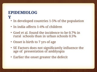
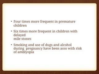
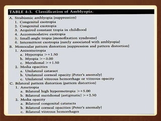
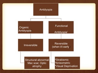
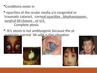
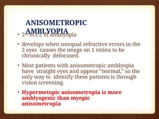
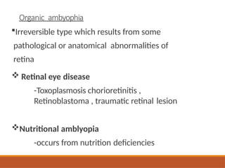
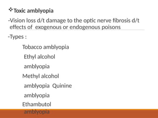
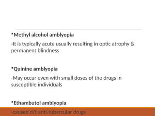
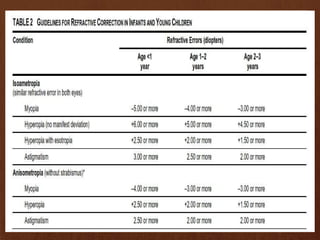
Amblyopia
By ABDUL SALAM
( Amblyos– Dull, ops—vision)
Amblyopia: (greek word means blunt eye) Also
called as: lazy eye is a disorder of sight.
“Defective visual acuity in one or both eyes,
which persists after correction of the
refractive error & removal of any pathological
obstacle to vision”.
Definition
EPIDEMIOLOG
Y
• In developed countries 1-5% of the population
• In india affects 1-4% of children
• Goel et al. found the incidence to be 0.7% in
rural schools than in urban schools 0.5%
• Onset is birth to 7 yrs of age
• SE Factors does not significantly influence the
age of presentation of amblyopia
• Earlier the onset greater the defecit
• Four times more frequent in premature
children
• Six times more frequent in children with
delayed
mile stones
• Smoking and use of dugs and alcohol
during pregnancy have been asso with risk
of amblyopia
PATHOPHYSIOLOG
Y
Amblyogenic factors
Role of retina
Active cortical
inhibition
Amblyogenic
factors
VISUAL
DEPRIVATION
monocula
r
Seen in strabismic ,
anisometropic,
stimulus deprivation
amblyopia
Binocular
Seen in bilateral
cataract, ametropia
and bilateral high
refractive errors
LIGHT DEPRIVATION.
Usually seen in
children with
unilateral or bilateral
complete cataracts.
ABNORMAL
BINOCULAR
INTERACTIO
N
-produces profound
amblyopia due to
competition
amblyopia.
-seen in strabismic,
anisometropic and
unilateral stimulus
deprivation
amblyopia.
CRITICAL PERIODS
DEVELOPMENT OF V/A FROM 6/60 TO
6/6 (From birth to age 3-5 years)
PERIOD OF HIGHEST RISK (From few months to
7-8 years)
PERIOD DURING WHICH RECOVERY FROM
AMBLYOPIA CAN BE OBTAINED
(before 8 years)
Sensitive Period
The capacity of the visual system to develop amblyopia
is limited by its state of maturity
During immaturity of the visual system the retinocortical
connections are not firmly established and may be
modified by the quantity or quality of the visual input
This phase has been described as the sensitive, critical,
or susceptible period
The human is most sensitive to environmental
manipulation during the first 2 years of life
The human critical period is over by approximately 7 to 9
years of age
RETINA IN THE DEVELOPMENT OF
AMBLYOPIA
• Decreased sensitivity of foveal cones in
amblyopia
• The reduced input from rods and cones in
the affected eye causes certain
neurophysiologic changes, transmitted to
the CNS which triggers amblyopia.
ACTIVE CORTICAL
INHIBITION
1)A developmental defect of spatial
visual processing occurring in the
visual pathway.
2)Poor transmission from the fovea, optic nerve
to the Striate Cortex of the affected eye.
3) LGB & Striate cortex develop abnormally.
4)Ganglion cells in foveal area are affected;
Shrinkage of LGB Nucleus & Striate cortical
fibres in the amblyopic eye.
5)Loss of binocularly driven cells in LGB &
Striate Cortex
CLASSIFICATION &
TYPES
• Strabismic amblyopia.
• Stimulus deprivation or amblyopia of
disuse.
• Anisometropic amblyopia.
• Meridional amblyopia.
• Isoametropic amblyopia .
• Amblyopia secondary to nystagmus.
• Idiopathic amblyopia.
• organic amblyopia.
Amblyopia
‘Organic
Amblyopia’
Irreversible
Structural abnormal
Mac scar, Optic
atrophy
‘Functional
Amblyopia’
Reversible
(when t/t early)
•Strabismic
•Anisometric
•Visual Deprivation
STRABISMIC
AMBLYOPIA
• M/C form of amblyopia
• A.K.A amblyopia of arrest
• Seen in unilateral constant squint who
strongly favour one eye for fixation.
• cortical suppression from deviating eye
thought to be due to inhibitory interactions
from neurons carrying non fusable images
which cause visual confusion
• Esotropia more likely to develop
amblyopia as compared to exotropia
• Does not develop in alternating or
intermittent strabismus as there are periods
of normal binocular interaction that preserve
the integrity of visual system
• Severity of amblyopia does not correlate with
angle of strabismus
Strabismic amblyopia
Most common form of amblyopia
Amblyopia is unilateral
Seen in unilateral constant squint who
strongly favour one eye for fixation
Will develop in 100% of pts with constant
untreated acquired esotropia under 3 years
of age resulting in marked decrease in VA
within week(treatment of this type of
amblyopia following acquired esotropia
therefore becomes daytime emergency)
19.7% of congenital cases of esotropia if
untreated
Caused by active inhibition within the retinocortical
pathwayof
visual input originating in the fovea of the deviating eye.
Far more often in esotropes than in exotropes because
exotropia is often intermittent at its onset
Also be related to the nasotemporal asymmetry of the
retinocortical projections. In esotropes the fovea of the
deviating eye has to compete with the strong temporal
hemifield of the fellow eye . In exotropia the fovea
competes with the weaker contralateral nasal hemifield
a/k suppression amblyopia
Contd…
STIMULUS DEPRIVATION
AMBLYOPIA
• Amblyopia ex anopsia disuse amblyopia.
• Least common but most damaging.
• Cause when the visual axis is
obstructed.
• Monocular congenital or traumatic
cataract, complete ptosis, corneal
opacity & prolonged patching of the
normal eye for treatment of amblyopia.
• Less than 6 yrs – severe amblyopia.
• After 6 yrs – less harmful .
• Visual loss resulting from unilateral
deprivation is worse than that produced by
bilateral deprivation of similar degree
Conditions exists in
-opacities of the ocular media s/a congenital or
traumatic cataract, corneal opacities , blepharospasm ,
surgical lid closure , or U/L
Complete ptosis
- B/L ptosis is not amblyogenic because the pt
maintains normal VA with a chin elevation
ANISOMETROPIC
AMBLYOPIA
• 2nd m.c.c of amblyopia
• develops when unequal refractive errors in the
2 eyes causes the image on 1 retina to be
chronically defocused.
• Most patients with anisometropic amblyopia
have straight eyes and appear “normal,” so the
only way to identify these patients is through
vision screening.
• Hypermetopic anisometropia is more
amblyogenic than myopic
anisometropia
• The amount of anisometropia that can induce
amblyopia varies according to the type of
refractive error
• Amount of anisometropia
Hypermetropia > 2D
Myopia > 4D
Astigmatism> 1.25D
However unilateral high hyperopia or myopia
(>6D) causes severe amblyopia
MERIDIONAL
AMBLYOPIA
• Resolution of eye is reduced in selective
meridians as a result of un corrected
astigmatism
• Cylinder >1.5D is comsidered amblyogenic
• Doesn’t develop until first year of age
ISOMETROPIC
AMBLYOPIA
• Bilateral amblyopia occurring in children
with bilateral uncorrected high
refractive error.
Hyperopia > +5
D. Myopia > -10
D. astigmatism
> 2D
Mechanism – effect of blurred
retinal images alone
AMBLYOPIA SECONDARY TO
NYSTAGMUS
• Difficult to establish ascertain whether
nystagmus is the cause or effect of amblyopia
• Bilateral
IDIOPATHIC
AMBLYOPIA
• Occurring in apparently normal patients
with a negative history of strabismus & in
the absence of other amblyogenic factors.
• Mech- foveal suppression of amblyogenic
eye d/t transient amblyogenic factor during
infancy
Organic ambyophia
Irreversible type which results from some
pathological or anatomical abnormalities of
retina
 Retinal eye disease
-Toxoplasmosis chorioretinitis ,
Retinoblastoma , traumatic retinal lesion
Nutritional amblyopia
-occurs from nutrition deficiencies
Toxic amblyopia
-Vision loss d/t damage to the optic nerve fibrosis d/t
effects of exogenous or endogenous poisons
-Types :
Tobacco amblyopia
Ethyl alcohol
amblyopia
Methyl alcohol
amblyopia Quinine
amblyopia
Ethambutol
amblyopia
Tobacco amblyopia
-Typically occurs in men in pipe smokers, heavy
drinkers
Ethyl alcohol amblyopia
-Usually in a/w tobacco amblyopia
-May occur in non-smoker but heavy drinkers
suffering from chronic gastritis
Methyl alcohol amblyopia
-It is typically acute usually resulting in optic atrophy &
permanent blindness
Quinine amblyopia
-May occur even with small doses of the drugs in
susceptible individuals
Ethambutol amblyopia
-caused d/t anti-tubercular drugs
Clinical
characteristics
CLINICAL
CHARACTERISTICS
• Decreased visual acuity
• Decreased stereoacity
• Fixation reflex
• Crowding phenomenon
• Effect of neutral density
filter
• Contrast sensitivity
• Fixation pattern
VISUAL
ACUITY
• Two line difference between amblyopic and normal
eye
• For B/L amblyopia the VA should be less than 20/40 in
each
eye
• But in children there will be difficulty in
assesing VA Infants-fixation preference
preverbal children-preferential looking test,OKN
test,VEP 2-3 yrs- E charts,pictoral charts
>3 yrs-snellens charts,HOTV charts
PREFERENTIAL LOOKING
TEST
STEREOACUIT
Y
• Presence of amblyopia can be detected by
defective performance on various
stereograms
• Two pencil test is a clinically useful test and
can be applied even when VA recording is
unreliable or not possible
• Can also be easured by titmus fly test,random
dot
stereogram
FIXATION
REFLEX
• useful tool to assess VA in children <5yrs of age
• Central steady and maintained (CSM) fixation implies
good
VA
Affixation  <3/60
unsteady fixation  3/60 to 6/60
Central but not maintained 6/60 to 6/18
Central but strong preference for other eye  6/18
to 6/9 Alternate fixation  6/6
CROWDING
PHENOMENON
• Amblyopia pts exhibit better VA for single
optotypes than for letters placed in a row
• Although not specific for amblyopia,it may be
pronounced in amblyopic eye compared to
better eye
• Single line acuity improves more than line
acuity during treatment
• So it is important to record both single letter
and line visual acuity every time as it is
prognostic indicator
• Vision testing with single optotypes is likely to
over estimate VA in pts with amblyopia
• More accurate assesment of mono ocular VA is
obtained with the presentation of line of
optotypes or single optotype with crowding
bars that surround the optotype being
identified
NEUTRAL DENSITY
FILTER
• A neutral density filter reduces overall
luminance without inducing a color change.
• Decreased luminance of the visual target
results in diminished central acuity in
normal eyes.
• Decreased illumination of visual targets has less
of an effect on amblyopic eyes because they are
not using central acuity
• It was found that neutral filters profoundly reduce
vision in eyes with organic amblyopia whereas
vision of eyes with functional amblyopia was not
reduced and occasionally even slightly improved.
• Hence it can be used to differentiate the two.
CONTRAST
SENSITIVITY
• Reduction in contrast
sensitivity more
for higher
frequencies
• Improves during
amblyopia
therapy and
useful to monitor
the progress
• Contrast threshold
becomes normal in
strabismic
amblyopia
when luminance
levels
were reduced,
while the
deficit persists in
anisometropic
pelli robson contrast
sensitivity chart
FIXATION
PATTERN
• Bangerter’s classification of fixation
patterns in amblyopia
I. Central fixation
II. Eccentric fixation (nonfoveolar)-
common type
III. No fixation
• Patients with eccentric fixation appear to be
looking to the side,not directly at the fixation
target. They have poor smooth pursuits,so they do
not accurately follow a moving target.
• Can be tested in old coperative children by
visuoscope
OTHER
FEATURES
• VEP Reduction in amplitude and
slightly prolonged latency
• Afferent pupillary defect may be seen
• Normalisation of VA in dim light
occasionally
• Occasionally latent nystagmus
Visualfield
Monocular VF are usually recorded as normal in
strabismic amblyopia . Although there is obviously
a relative defect in the fovea it is difficult to
demonstrate it on a target screen or goldmann
perimeter
This clearly differentiates strabismic
amblyopia from organic amblyopia in which
a scotoma involving the fovea area can be
plotted
CLINICAL EVALUATION &
DIAGNOSIS
• Thorough clinical history
• Binocular red reflex test
• Binocularity/stereo acuity testig
• Evaluation of visual acuity and fixation pattern
• Binocular alingnment and ocular motility
• External examination
• Pupillary examinaion
• Thorough ocular examination including fundus
examination.
• Cycloplegic retinoscopy/Refraction
• Neutral density filter and testing for crowding
phenomenon.
Pupillary Responses
An afferent pupillary defect of amblyopic eyes has
been reported by several authors(9% to 93%)
(Dole´nek)
On Pupillographic measurements on the eyes of
amblyopic children it was found that on average
the pupil of the amblyopic eye was 0.5 mm larger
than the pupil of the normal eye in the natural
state and
0.3 mm larger in miosis induced by a light stimulus
(Dole’nek &
Kru¨ger).
Prognostic Factors in
Amblyopia
Positive factor Negative factor
functional organic
Central fixation Eccentric fixation
Random dot stereopsis No random dot
stereopsis
Short duration Long duration
Young patient, motivated Older patient, un-
motivated
Degree of prognosis
Strabismic > Anisometropic myopia >
Anisometropic hypermetropia
TREATMENT
MODALITIES
Treatment of amblyopia involves following
steps:-
1)eliminate any obstacles to vision , such as
cataract.
2) correct any significant error.
3)force use of the poorer eye by limiting use
of the better eye.
Strategies to treat amblyopia
Eliminate cause of visual deprivation & provision of
clear retinal image in amblyopic eye.
Correction of ocular dominance
Perceptual training
Recommended treatment
should be based on:-
Patient’s
age
Visual acuity
Compliance with
• Refractive correction
• Occlusion therapy
• Penalisation
• Drug therapy
• Pleoptics
• Cam stimulator
• Surgery to treat the cause of
amblyopia
CATARACT
REMOVAL
• Removal of congenital lens opacity- first 4 -6 week of
life.
• If symmetrical b/l cases- interval b/w 1st & 2nd
eye should not be not more than 1-2 weeks.
• Developing severe traumatic cataract in children
less than
6 yrs removed within few weeks of injury.
REFRACTIVE ERROR
CORRECTION
• Improves VA in 25-33% of patients with
anisometropic amblyopia and also in strabismic
amblyopia
• Cycloplegic refraction followed by adequate
optical correction
• ATS 5 concluded that amblyopia improved with
optical correction in 77% and resolved in 27%
• Chen et al (AJO 2007) concluded that penalisation
and occlusion is required only if the child
doesn’t improve with glasses for four months
• In general eye glasses are well tolerated by
children
especially when there is improvement in visual
function
OCCLUSION
THERAPY
• Occlusion of the sound eye is the most
effective treatment for amblyopia
treatment
• When fixation is central, simple &
effective.
• When fixation is eccentric, <7yrs central
fixation will be recovered.
• Older the child harder to regain central
fixation.
• Success rate 30-92%
• MOAprevent fixating eye taking part in act
of vision and removes inhibitory stimulus
Mode of action
Prevent fixating eye taking part in act of vision and removes
inhibitory stimulus that arises from stimulation from fixating
eye (non-amblyopic eye)
Occlusion goals
• Differential diagnosis
• Improvement of amblyopia
• Elimination of suppression
• Awareness or elimination of diplopia
• Disruption of
anomalous
corresponde
nce.
Types of
occlusion
• Total or partial
• Conventional or inverse
• Full time or part time
• Patching is most commonly
prescribed,however contravercy exists
concerning how much treatment is necessary
• Most data on response according to daily
dosage of patching are retrospective and
uncontrolled
TOTAL VS PARTIAL
OCCLUSION
total partial
•All light is prevented from •Does not cut off the total
entering eye.
•Employed in amblyopic
eyes
light entering eye
•Degrades the vision of
with acuity less than 6/24
•Occlusion using elastoplast,
normal eye such that
amblyopic eye gets better
gauze pad, tape, doynes
rubber occluder.
vision and preference
•Occlusion using cellophane,
transparent nail polish, or a
higher plus lens.
Partial /translucent
occlusion
Total occlusion
Given in
nystagmus
Amblyopia
treatment
CONVENTIONAL VS INVERSE
OCCLUSION
Conventional Inverse
•Occlusion of sound eye •Occlusion of
amblyopic eye so that
eccentric fixation
becomes less fixed
Additional
points
Inverse occlusion is prescribed whenever
occlusion is needed but direct occlusion is
intolerable to the patient.
Given only if the patient is strongly resistant to
direct occlusion.
For example:- A strabismic patient with deep
amblyopia may not be able to perform needed visual
tasks with amblyopic eye . So 1st inverse occlusion is
started & changed to direct occlusion once more
central fixation and improved visual acuity are
obtained

FULL TIME VS PART
TIME
Full time Part time
Removed only while
going to bed at night
Short time each day
during close work or
watching television.
Choice of initial Rx In relapses after Rx and also
for
maintanence
Occlusion in Constant
strabismus
Earlier, full time occlusion followed by a day of rest was
advocated. This allows constant strabismus to regress to
anomalous strabismic visual processing on free day.
Nowadays, full time occlusion is prescribed initially.
When intermittency is achieved in open space ,part-
time occlusion is given allowing some reinforcement of
binocular skills in normal activities.
Points on Occlusion
The presence (or absence) of amblyopia and its fixation
pattern determine which eye to patch.
The frequency of strabismus determines the amount of
time that the eye is patched
Alternate occlusion
When equal visual acuity is present in each eye, (e.g :-
a constant alternate esotropia) , full time occlusion is
alternated daily between two eyes.
The purpose of full time occlusion for strabismics with
no amblyopia is
to eliminate suppression and possibly disrupt
anomalous correspondence.
Types of
occluders
• Adhesive skin patches made of
micropore
• Commercially available opticlude
• Spectacle occluder
• Contact lens occluder
OPAQUE CONTACT LENS
• Patche
s
• Micropore tape with soft tissue paper
• Spectacle patch / frost glass • Doyne’s occluder
Contact lens
occluder
Bandage occluder
Patches
Microp
ore
Spectacle
occluder
Tie-on occluder Clip-on occluder
Occlusion lens Occlusion filter
Bitemporal
occlusion
Binasal
occlusion
Sector
occlusion
Paediatric eye disease investigator group(PEDIG) has
conducted several amblyopia treatment trials
(amblyopia treatment study or ATS) over the past
several years. Results have shown that
• Spectacles alone are powerful treatment for
amblyopia; patching is superior to spectacles
• Initiating fewer hours of prescribed patching
seems to be
as effective as traditional treatment
• Patching is effective in older children particularly
if they have not been treated earlier
• Atropine is as effective as patching
• Weekend atropine is as effective as daily atropine
How much
patching??
The amblyopia treatment study have helped to
define the role of full time patching vs part
time patching
• In patients aged 3-7 years with severe
amblyopia
(VA B/W 6/30 to 6/120) full time patching
produced similar effect to that of six hours
patching per day
• In patients aged 3-7 years with moderate
amblyopia (VA better than 6/30) 2 hours
patching produced similar effect to that of six
hours patching per day
Treatment of amblyopia in 7-17
yrs
• For 7-13 yrs age group 2-6 hours of
patching can improve VA only if
previously treated
• For 13-17 yrs age group 2-6 hous of
patching improved VA even if not
treated previously
• Long term results from these studies
are still pending
HOW TO GO ABOUT
OCCLUSION
• Motivation of child and parents.
• Active vision exercises by amblyopic eye like
dotting O’s and encircling E’s in a newspaper,
joining dots,reading comics and story books.
• In case of vision improvement, occlusion is
continued till amblyopic eye has not only
developed equal vision but also equal preference
of fixation.
• May take 3-6 months.
• If there is no improvement. Then treatment is
stopped. Also other causes to be ruled out.
• Maintainence treatment is continued atleast upto
9 yrs of
age with part time occlusion and exercises
Rx schedule for initial
occlusion
Age in yrs Period of
occlusion(day
s) Direct :
inverse
Follow up after
every
Upto 2 2 :1 15 days
3 3 : 1 15 days
4 4 : 1 1 month
5 5 : 1 1 month
6 & older 6 : 1 1 month
• Follow up-depending on age,severiy of
amblyopia and compliance
• To look for-VA, fixation pattern and
occlusion amblyopia
• When to stop occlusion
VA equals in both eyes
alternation of
fixation
• When VA is stable
patching may be
decreased slowly
• Because amblyopia recurres in large no. of
patients maintanence therapy or tapering of
therapy should be strongly considered.
Disadvantages of
occlusion
• Occlusion amblyopia
• Non compliance
• Psychological distress
• Appearance of constant
deviation
• Allergic skin rash
• Diplopia
• Cosmetically inacceptable
Prognostic
considerations
• Younger the age better the prognosis
• Type of amblyopia myopic anisometropia>
hyperopic anisometropia> strabismic
amblyopia> stimulus deprivation
• Pretreatment
VA
• Type of
occlusion
• Type of fixation
• Near exercises
• Patient compliance and parent
education
• Presence of astigmatism
• Method of treatment termination
• Previous treatment
• Refractive correction
Occlusion
amblyopia
When normal or preferred eye is occluded, visual acuity may
decrease & occlusion amblyopia may occur in the occluded
eye.
The younger the child ,faster is the acuity loss & become
profound and permanent.Esp:- full time occlusion when
given to infant <2yrs (critical developmental yrs)
Alternate patching is given to prevent this & shorter period
of direct occlusion for younger children
For the remedy of skin reactions d/t patch that is in contact with skin
, hypoallergenic patch (Opticlude) can be used.
Tincture benzoin may be applied to the skin before applying patch.
This forms a protective layer & increase adhesiveness.
Strabismic diplopia
Occurs d/t extended period of total occlusion without
resolution of strabismus esp in older patients & those with
anomalous correspondence.
In pts <10 yrs, suppression may regain in few weeks after
discontinuation of occlusion even after experiencing
diplopia for several months when occluder is removed.
 d/t tendency of suppression to
lessen with age
 Difficulty in achieving BSV in long
duration strabismus
Deviation
changes
Appears to be expected progression of strabismic
syndrome such as accommodative esotropia or
essential esotropia.
Dissociated vertical deviation may appear or
previously measured primary vertical deviation
may increase in size.
Exodeviations may increase in size with full time or
part time occlusion & later become stable.
Some esotropic deviations continue to increase in
size( without increase in hyperopia) with time & after
months they stabilize at a given angle.
Occlusion removal & Maintenance
occlusion treatment
Occluder is not removed until comfortable ,efficient
binocularity has been obtained, and patient is capable of
maintaining binocular vision in open space without
regressions.
Most errors are made in removing the occluder too
soon. Regressions from non strabismus to strabismus
may occur in a relatively short time (1 to 3 months).esp.
intermittent or constant strabismus
Once the vision has been equalized, the maintenance
occlusion should be continued till the amblyogenic ,
i.e up to at least 9 years of age.
TREATMENT OF ANISOMETROPIC
AMBLYOPIA
TREATMENT OF STRABISMIC
AMBLYOPIA
PENALISATIO
N
• Therapeutic technique performed by
optically defocussing the eye with better
vision by using cycloplegia or altering
the eye glass lens
INDICATIONS
No compliance for occlusion.
Mild degrees of amblyopia.
Maintainence after occlusion.
Anisometropic amblyopia
• Advantages cheap,better compliance
• DisadvantagesS/E of drugs
risk of occlusion amblyopia
systemic absorption
• Unless penalisation decreases the VA of
dominant eye below the amblyopic eye this
form of treatment is not adviced
Methods of
penalisation
a. Near penalization: fixing eye is
atropinized & fully corrected for distance,
amblyopic eye is overcorrected with +2 to
+3D .
b. Distance penalization : fixing eye is
atropinized & overcorrected, amblyopic eye
is fully corrected.
c. Total : fixing eye is atropinized &
undercorrected by 4 to 5 D, amblyopic eye is
fully corrected.
PLEOPTIC
S
• Involves active stimulation of fovea to
overcome eccentric
fixation & improves Va.
• The peripheral retina including the
eccentrically fixing area around the fovea
is dazzled.
• After lights are turned off, fovea functions
better because the surrounding retinal area
is in a state of hypofunction
• ONLY INDICATION IS coperative and
intelligent child older than 6yrs having
eccentric fixation
PLEOTOPHOR
E
CAM
STIMULATOR
• Slowly rotating high contrast square wave
grating of different spatial frequencies
• Principle – rotating gratings provide
specific stimulation for cortical
neurons
• the visual improvement was found to be
better in emmetropes and hypermetropes
than those in strabismus amblyopia
• Not used these days
PHARMACOLOGICAL
THERAPY
• LEVADOPA is the only most extensively
studied drug in western and Indian
population
• Precursor of dopamine known to influence
visual system at retina and cortical level
• Advantages
Augments conventional occlusion
Speeds up recovery of visual
functions Improves compliance
Reduces cost and duration of
treatment
• Catecholamine based medical treatment
citicholine has been demonstrated to improve
VA in amblyopic eyes
SURGER
Y
By reducing anisometropia refractive
surgeries has reported to
• Improve spectacle tolerance
• facilitate amblyopia thearpy
• Enhance binocular vision
Also used for children who has finished
amblyopia therapy and cannot comply with
spectacles or contact lens
Surgical therapy for strabismus generally
should occur after amblyopia is reversed.
• Disadvantages to surgical therapy prior to
correction of amblyopia include
difficulty in telling if amblyopia is present
because there is no longer a strabismus to
assess fixation preference and higher
potential to being lost to follow-up, as the
child cosmetically looks better.
The improved cosmesis gives the parents a
false sense of security about the vision
improving
RECURRENC
E
• Chances are high until child is visually
mature.
• Careful monitoring every month upto 1 year,
every 2 months upto 2 years and 4- 6 months
upto visual maturity is required.
• Maintainence occlusion to be given
SCREENIN
G
• AAO recommends screening by the age of 3
yrs and thereafter every 2yrs
• Includes visual acuity, corneal reflex test,
refraction, fixation preference and stereo
acuity
• AAP suggests that vision screening should
begin at birth and continue as a part of child
regular medical check up
• All new born infants should be screened in
nursery with the use of red reflex
• Infants at risk should undergo detailed
evaluation
Severe amblyopia can be eliminated as a public
health problem
• The goal can be achieved by
 improvements in public awareness
better screening protocols at the level of
primary health care provider
 full access to medical care for at risk
patients
THANK

AMBLYOPIA ITS TYPES AND MANAGEMENT OPTION.pptx

  • 1.
  • 3.
    ( Amblyos– Dull,ops—vision) Amblyopia: (greek word means blunt eye) Also called as: lazy eye is a disorder of sight. “Defective visual acuity in one or both eyes, which persists after correction of the refractive error & removal of any pathological obstacle to vision”. Definition
  • 4.
    EPIDEMIOLOG Y • In developedcountries 1-5% of the population • In india affects 1-4% of children • Goel et al. found the incidence to be 0.7% in rural schools than in urban schools 0.5% • Onset is birth to 7 yrs of age • SE Factors does not significantly influence the age of presentation of amblyopia • Earlier the onset greater the defecit
  • 5.
    • Four timesmore frequent in premature children • Six times more frequent in children with delayed mile stones • Smoking and use of dugs and alcohol during pregnancy have been asso with risk of amblyopia
  • 6.
    PATHOPHYSIOLOG Y Amblyogenic factors Role ofretina Active cortical inhibition
  • 7.
    Amblyogenic factors VISUAL DEPRIVATION monocula r Seen in strabismic, anisometropic, stimulus deprivation amblyopia Binocular Seen in bilateral cataract, ametropia and bilateral high refractive errors LIGHT DEPRIVATION. Usually seen in children with unilateral or bilateral complete cataracts. ABNORMAL BINOCULAR INTERACTIO N -produces profound amblyopia due to competition amblyopia. -seen in strabismic, anisometropic and unilateral stimulus deprivation amblyopia.
  • 8.
    CRITICAL PERIODS DEVELOPMENT OFV/A FROM 6/60 TO 6/6 (From birth to age 3-5 years) PERIOD OF HIGHEST RISK (From few months to 7-8 years) PERIOD DURING WHICH RECOVERY FROM AMBLYOPIA CAN BE OBTAINED (before 8 years)
  • 9.
    Sensitive Period The capacityof the visual system to develop amblyopia is limited by its state of maturity During immaturity of the visual system the retinocortical connections are not firmly established and may be modified by the quantity or quality of the visual input This phase has been described as the sensitive, critical, or susceptible period The human is most sensitive to environmental manipulation during the first 2 years of life The human critical period is over by approximately 7 to 9 years of age
  • 10.
    RETINA IN THEDEVELOPMENT OF AMBLYOPIA • Decreased sensitivity of foveal cones in amblyopia • The reduced input from rods and cones in the affected eye causes certain neurophysiologic changes, transmitted to the CNS which triggers amblyopia.
  • 11.
    ACTIVE CORTICAL INHIBITION 1)A developmentaldefect of spatial visual processing occurring in the visual pathway. 2)Poor transmission from the fovea, optic nerve to the Striate Cortex of the affected eye. 3) LGB & Striate cortex develop abnormally. 4)Ganglion cells in foveal area are affected; Shrinkage of LGB Nucleus & Striate cortical fibres in the amblyopic eye. 5)Loss of binocularly driven cells in LGB & Striate Cortex
  • 13.
    CLASSIFICATION & TYPES • Strabismicamblyopia. • Stimulus deprivation or amblyopia of disuse. • Anisometropic amblyopia. • Meridional amblyopia. • Isoametropic amblyopia . • Amblyopia secondary to nystagmus. • Idiopathic amblyopia. • organic amblyopia.
  • 15.
    Amblyopia ‘Organic Amblyopia’ Irreversible Structural abnormal Mac scar,Optic atrophy ‘Functional Amblyopia’ Reversible (when t/t early) •Strabismic •Anisometric •Visual Deprivation
  • 16.
    STRABISMIC AMBLYOPIA • M/C formof amblyopia • A.K.A amblyopia of arrest • Seen in unilateral constant squint who strongly favour one eye for fixation. • cortical suppression from deviating eye thought to be due to inhibitory interactions from neurons carrying non fusable images which cause visual confusion • Esotropia more likely to develop amblyopia as compared to exotropia
  • 17.
    • Does notdevelop in alternating or intermittent strabismus as there are periods of normal binocular interaction that preserve the integrity of visual system • Severity of amblyopia does not correlate with angle of strabismus
  • 18.
    Strabismic amblyopia Most commonform of amblyopia Amblyopia is unilateral Seen in unilateral constant squint who strongly favour one eye for fixation Will develop in 100% of pts with constant untreated acquired esotropia under 3 years of age resulting in marked decrease in VA within week(treatment of this type of amblyopia following acquired esotropia therefore becomes daytime emergency) 19.7% of congenital cases of esotropia if untreated
  • 19.
    Caused by activeinhibition within the retinocortical pathwayof visual input originating in the fovea of the deviating eye. Far more often in esotropes than in exotropes because exotropia is often intermittent at its onset Also be related to the nasotemporal asymmetry of the retinocortical projections. In esotropes the fovea of the deviating eye has to compete with the strong temporal hemifield of the fellow eye . In exotropia the fovea competes with the weaker contralateral nasal hemifield a/k suppression amblyopia Contd…
  • 20.
    STIMULUS DEPRIVATION AMBLYOPIA • Amblyopiaex anopsia disuse amblyopia. • Least common but most damaging. • Cause when the visual axis is obstructed. • Monocular congenital or traumatic cataract, complete ptosis, corneal opacity & prolonged patching of the normal eye for treatment of amblyopia. • Less than 6 yrs – severe amblyopia. • After 6 yrs – less harmful .
  • 21.
    • Visual lossresulting from unilateral deprivation is worse than that produced by bilateral deprivation of similar degree
  • 22.
    Conditions exists in -opacitiesof the ocular media s/a congenital or traumatic cataract, corneal opacities , blepharospasm , surgical lid closure , or U/L Complete ptosis - B/L ptosis is not amblyogenic because the pt maintains normal VA with a chin elevation
  • 23.
    ANISOMETROPIC AMBLYOPIA • 2nd m.c.cof amblyopia • develops when unequal refractive errors in the 2 eyes causes the image on 1 retina to be chronically defocused. • Most patients with anisometropic amblyopia have straight eyes and appear “normal,” so the only way to identify these patients is through vision screening. • Hypermetopic anisometropia is more amblyogenic than myopic anisometropia
  • 24.
    • The amountof anisometropia that can induce amblyopia varies according to the type of refractive error • Amount of anisometropia Hypermetropia > 2D Myopia > 4D Astigmatism> 1.25D However unilateral high hyperopia or myopia (>6D) causes severe amblyopia
  • 26.
    MERIDIONAL AMBLYOPIA • Resolution ofeye is reduced in selective meridians as a result of un corrected astigmatism • Cylinder >1.5D is comsidered amblyogenic • Doesn’t develop until first year of age
  • 27.
    ISOMETROPIC AMBLYOPIA • Bilateral amblyopiaoccurring in children with bilateral uncorrected high refractive error. Hyperopia > +5 D. Myopia > -10 D. astigmatism > 2D Mechanism – effect of blurred retinal images alone
  • 28.
    AMBLYOPIA SECONDARY TO NYSTAGMUS •Difficult to establish ascertain whether nystagmus is the cause or effect of amblyopia • Bilateral
  • 29.
    IDIOPATHIC AMBLYOPIA • Occurring inapparently normal patients with a negative history of strabismus & in the absence of other amblyogenic factors. • Mech- foveal suppression of amblyogenic eye d/t transient amblyogenic factor during infancy
  • 30.
    Organic ambyophia Irreversible typewhich results from some pathological or anatomical abnormalities of retina  Retinal eye disease -Toxoplasmosis chorioretinitis , Retinoblastoma , traumatic retinal lesion Nutritional amblyopia -occurs from nutrition deficiencies
  • 31.
    Toxic amblyopia -Vision lossd/t damage to the optic nerve fibrosis d/t effects of exogenous or endogenous poisons -Types : Tobacco amblyopia Ethyl alcohol amblyopia Methyl alcohol amblyopia Quinine amblyopia Ethambutol amblyopia
  • 32.
    Tobacco amblyopia -Typically occursin men in pipe smokers, heavy drinkers Ethyl alcohol amblyopia -Usually in a/w tobacco amblyopia -May occur in non-smoker but heavy drinkers suffering from chronic gastritis
  • 33.
    Methyl alcohol amblyopia -Itis typically acute usually resulting in optic atrophy & permanent blindness Quinine amblyopia -May occur even with small doses of the drugs in susceptible individuals Ethambutol amblyopia -caused d/t anti-tubercular drugs
  • 34.
  • 35.
    CLINICAL CHARACTERISTICS • Decreased visualacuity • Decreased stereoacity • Fixation reflex • Crowding phenomenon • Effect of neutral density filter • Contrast sensitivity • Fixation pattern
  • 36.
    VISUAL ACUITY • Two linedifference between amblyopic and normal eye • For B/L amblyopia the VA should be less than 20/40 in each eye • But in children there will be difficulty in assesing VA Infants-fixation preference preverbal children-preferential looking test,OKN test,VEP 2-3 yrs- E charts,pictoral charts >3 yrs-snellens charts,HOTV charts
  • 38.
  • 39.
    STEREOACUIT Y • Presence ofamblyopia can be detected by defective performance on various stereograms • Two pencil test is a clinically useful test and can be applied even when VA recording is unreliable or not possible • Can also be easured by titmus fly test,random dot stereogram
  • 42.
    FIXATION REFLEX • useful toolto assess VA in children <5yrs of age • Central steady and maintained (CSM) fixation implies good VA Affixation  <3/60 unsteady fixation  3/60 to 6/60 Central but not maintained 6/60 to 6/18 Central but strong preference for other eye  6/18 to 6/9 Alternate fixation  6/6
  • 44.
    CROWDING PHENOMENON • Amblyopia ptsexhibit better VA for single optotypes than for letters placed in a row • Although not specific for amblyopia,it may be pronounced in amblyopic eye compared to better eye • Single line acuity improves more than line acuity during treatment • So it is important to record both single letter and line visual acuity every time as it is prognostic indicator
  • 45.
    • Vision testingwith single optotypes is likely to over estimate VA in pts with amblyopia • More accurate assesment of mono ocular VA is obtained with the presentation of line of optotypes or single optotype with crowding bars that surround the optotype being identified
  • 46.
    NEUTRAL DENSITY FILTER • Aneutral density filter reduces overall luminance without inducing a color change. • Decreased luminance of the visual target results in diminished central acuity in normal eyes. • Decreased illumination of visual targets has less of an effect on amblyopic eyes because they are not using central acuity • It was found that neutral filters profoundly reduce vision in eyes with organic amblyopia whereas vision of eyes with functional amblyopia was not reduced and occasionally even slightly improved. • Hence it can be used to differentiate the two.
  • 48.
    CONTRAST SENSITIVITY • Reduction incontrast sensitivity more for higher frequencies • Improves during amblyopia therapy and useful to monitor the progress • Contrast threshold becomes normal in strabismic amblyopia when luminance levels were reduced, while the deficit persists in anisometropic pelli robson contrast sensitivity chart
  • 49.
    FIXATION PATTERN • Bangerter’s classificationof fixation patterns in amblyopia I. Central fixation II. Eccentric fixation (nonfoveolar)- common type III. No fixation • Patients with eccentric fixation appear to be looking to the side,not directly at the fixation target. They have poor smooth pursuits,so they do not accurately follow a moving target. • Can be tested in old coperative children by visuoscope
  • 51.
    OTHER FEATURES • VEP Reductionin amplitude and slightly prolonged latency • Afferent pupillary defect may be seen • Normalisation of VA in dim light occasionally • Occasionally latent nystagmus
  • 52.
    Visualfield Monocular VF areusually recorded as normal in strabismic amblyopia . Although there is obviously a relative defect in the fovea it is difficult to demonstrate it on a target screen or goldmann perimeter This clearly differentiates strabismic amblyopia from organic amblyopia in which a scotoma involving the fovea area can be plotted
  • 53.
    CLINICAL EVALUATION & DIAGNOSIS •Thorough clinical history • Binocular red reflex test • Binocularity/stereo acuity testig • Evaluation of visual acuity and fixation pattern • Binocular alingnment and ocular motility • External examination • Pupillary examinaion • Thorough ocular examination including fundus examination. • Cycloplegic retinoscopy/Refraction • Neutral density filter and testing for crowding phenomenon.
  • 54.
    Pupillary Responses An afferentpupillary defect of amblyopic eyes has been reported by several authors(9% to 93%) (Dole´nek) On Pupillographic measurements on the eyes of amblyopic children it was found that on average the pupil of the amblyopic eye was 0.5 mm larger than the pupil of the normal eye in the natural state and 0.3 mm larger in miosis induced by a light stimulus (Dole’nek & Kru¨ger).
  • 55.
    Prognostic Factors in Amblyopia Positivefactor Negative factor functional organic Central fixation Eccentric fixation Random dot stereopsis No random dot stereopsis Short duration Long duration Young patient, motivated Older patient, un- motivated Degree of prognosis Strabismic > Anisometropic myopia > Anisometropic hypermetropia
  • 57.
    TREATMENT MODALITIES Treatment of amblyopiainvolves following steps:- 1)eliminate any obstacles to vision , such as cataract. 2) correct any significant error. 3)force use of the poorer eye by limiting use of the better eye.
  • 58.
    Strategies to treatamblyopia Eliminate cause of visual deprivation & provision of clear retinal image in amblyopic eye. Correction of ocular dominance Perceptual training Recommended treatment should be based on:- Patient’s age Visual acuity Compliance with
  • 59.
    • Refractive correction •Occlusion therapy • Penalisation • Drug therapy • Pleoptics • Cam stimulator • Surgery to treat the cause of amblyopia
  • 60.
    CATARACT REMOVAL • Removal ofcongenital lens opacity- first 4 -6 week of life. • If symmetrical b/l cases- interval b/w 1st & 2nd eye should not be not more than 1-2 weeks. • Developing severe traumatic cataract in children less than 6 yrs removed within few weeks of injury.
  • 61.
    REFRACTIVE ERROR CORRECTION • ImprovesVA in 25-33% of patients with anisometropic amblyopia and also in strabismic amblyopia • Cycloplegic refraction followed by adequate optical correction • ATS 5 concluded that amblyopia improved with optical correction in 77% and resolved in 27% • Chen et al (AJO 2007) concluded that penalisation and occlusion is required only if the child doesn’t improve with glasses for four months • In general eye glasses are well tolerated by children especially when there is improvement in visual function
  • 63.
    OCCLUSION THERAPY • Occlusion ofthe sound eye is the most effective treatment for amblyopia treatment • When fixation is central, simple & effective. • When fixation is eccentric, <7yrs central fixation will be recovered. • Older the child harder to regain central fixation. • Success rate 30-92% • MOAprevent fixating eye taking part in act of vision and removes inhibitory stimulus
  • 64.
    Mode of action Preventfixating eye taking part in act of vision and removes inhibitory stimulus that arises from stimulation from fixating eye (non-amblyopic eye) Occlusion goals • Differential diagnosis • Improvement of amblyopia • Elimination of suppression • Awareness or elimination of diplopia • Disruption of anomalous corresponde nce.
  • 65.
    Types of occlusion • Totalor partial • Conventional or inverse • Full time or part time • Patching is most commonly prescribed,however contravercy exists concerning how much treatment is necessary • Most data on response according to daily dosage of patching are retrospective and uncontrolled
  • 66.
    TOTAL VS PARTIAL OCCLUSION totalpartial •All light is prevented from •Does not cut off the total entering eye. •Employed in amblyopic eyes light entering eye •Degrades the vision of with acuity less than 6/24 •Occlusion using elastoplast, normal eye such that amblyopic eye gets better gauze pad, tape, doynes rubber occluder. vision and preference •Occlusion using cellophane, transparent nail polish, or a higher plus lens.
  • 67.
  • 68.
    CONVENTIONAL VS INVERSE OCCLUSION ConventionalInverse •Occlusion of sound eye •Occlusion of amblyopic eye so that eccentric fixation becomes less fixed
  • 69.
    Additional points Inverse occlusion isprescribed whenever occlusion is needed but direct occlusion is intolerable to the patient. Given only if the patient is strongly resistant to direct occlusion. For example:- A strabismic patient with deep amblyopia may not be able to perform needed visual tasks with amblyopic eye . So 1st inverse occlusion is started & changed to direct occlusion once more central fixation and improved visual acuity are obtained 
  • 70.
    FULL TIME VSPART TIME Full time Part time Removed only while going to bed at night Short time each day during close work or watching television. Choice of initial Rx In relapses after Rx and also for maintanence
  • 71.
    Occlusion in Constant strabismus Earlier,full time occlusion followed by a day of rest was advocated. This allows constant strabismus to regress to anomalous strabismic visual processing on free day. Nowadays, full time occlusion is prescribed initially. When intermittency is achieved in open space ,part- time occlusion is given allowing some reinforcement of binocular skills in normal activities.
  • 72.
    Points on Occlusion Thepresence (or absence) of amblyopia and its fixation pattern determine which eye to patch. The frequency of strabismus determines the amount of time that the eye is patched Alternate occlusion When equal visual acuity is present in each eye, (e.g :- a constant alternate esotropia) , full time occlusion is alternated daily between two eyes. The purpose of full time occlusion for strabismics with no amblyopia is to eliminate suppression and possibly disrupt anomalous correspondence.
  • 73.
    Types of occluders • Adhesiveskin patches made of micropore • Commercially available opticlude • Spectacle occluder • Contact lens occluder OPAQUE CONTACT LENS
  • 74.
    • Patche s • Microporetape with soft tissue paper • Spectacle patch / frost glass • Doyne’s occluder
  • 75.
  • 76.
    Tie-on occluder Clip-onoccluder Occlusion lens Occlusion filter
  • 77.
  • 78.
    Paediatric eye diseaseinvestigator group(PEDIG) has conducted several amblyopia treatment trials (amblyopia treatment study or ATS) over the past several years. Results have shown that • Spectacles alone are powerful treatment for amblyopia; patching is superior to spectacles • Initiating fewer hours of prescribed patching seems to be as effective as traditional treatment • Patching is effective in older children particularly if they have not been treated earlier • Atropine is as effective as patching • Weekend atropine is as effective as daily atropine
  • 79.
    How much patching?? The amblyopiatreatment study have helped to define the role of full time patching vs part time patching • In patients aged 3-7 years with severe amblyopia (VA B/W 6/30 to 6/120) full time patching produced similar effect to that of six hours patching per day • In patients aged 3-7 years with moderate amblyopia (VA better than 6/30) 2 hours patching produced similar effect to that of six hours patching per day
  • 80.
    Treatment of amblyopiain 7-17 yrs • For 7-13 yrs age group 2-6 hours of patching can improve VA only if previously treated • For 13-17 yrs age group 2-6 hous of patching improved VA even if not treated previously • Long term results from these studies are still pending
  • 81.
    HOW TO GOABOUT OCCLUSION • Motivation of child and parents. • Active vision exercises by amblyopic eye like dotting O’s and encircling E’s in a newspaper, joining dots,reading comics and story books. • In case of vision improvement, occlusion is continued till amblyopic eye has not only developed equal vision but also equal preference of fixation. • May take 3-6 months. • If there is no improvement. Then treatment is stopped. Also other causes to be ruled out. • Maintainence treatment is continued atleast upto 9 yrs of age with part time occlusion and exercises
  • 82.
    Rx schedule forinitial occlusion Age in yrs Period of occlusion(day s) Direct : inverse Follow up after every Upto 2 2 :1 15 days 3 3 : 1 15 days 4 4 : 1 1 month 5 5 : 1 1 month 6 & older 6 : 1 1 month
  • 83.
    • Follow up-dependingon age,severiy of amblyopia and compliance • To look for-VA, fixation pattern and occlusion amblyopia • When to stop occlusion VA equals in both eyes alternation of fixation • When VA is stable patching may be decreased slowly • Because amblyopia recurres in large no. of patients maintanence therapy or tapering of therapy should be strongly considered.
  • 84.
    Disadvantages of occlusion • Occlusionamblyopia • Non compliance • Psychological distress • Appearance of constant deviation • Allergic skin rash • Diplopia • Cosmetically inacceptable
  • 85.
    Prognostic considerations • Younger theage better the prognosis • Type of amblyopia myopic anisometropia> hyperopic anisometropia> strabismic amblyopia> stimulus deprivation • Pretreatment VA • Type of occlusion • Type of fixation • Near exercises • Patient compliance and parent education • Presence of astigmatism • Method of treatment termination • Previous treatment • Refractive correction
  • 86.
    Occlusion amblyopia When normal orpreferred eye is occluded, visual acuity may decrease & occlusion amblyopia may occur in the occluded eye. The younger the child ,faster is the acuity loss & become profound and permanent.Esp:- full time occlusion when given to infant <2yrs (critical developmental yrs) Alternate patching is given to prevent this & shorter period of direct occlusion for younger children For the remedy of skin reactions d/t patch that is in contact with skin , hypoallergenic patch (Opticlude) can be used. Tincture benzoin may be applied to the skin before applying patch. This forms a protective layer & increase adhesiveness.
  • 87.
    Strabismic diplopia Occurs d/textended period of total occlusion without resolution of strabismus esp in older patients & those with anomalous correspondence. In pts <10 yrs, suppression may regain in few weeks after discontinuation of occlusion even after experiencing diplopia for several months when occluder is removed.  d/t tendency of suppression to lessen with age  Difficulty in achieving BSV in long duration strabismus
  • 88.
    Deviation changes Appears to beexpected progression of strabismic syndrome such as accommodative esotropia or essential esotropia. Dissociated vertical deviation may appear or previously measured primary vertical deviation may increase in size. Exodeviations may increase in size with full time or part time occlusion & later become stable. Some esotropic deviations continue to increase in size( without increase in hyperopia) with time & after months they stabilize at a given angle.
  • 89.
    Occlusion removal &Maintenance occlusion treatment Occluder is not removed until comfortable ,efficient binocularity has been obtained, and patient is capable of maintaining binocular vision in open space without regressions. Most errors are made in removing the occluder too soon. Regressions from non strabismus to strabismus may occur in a relatively short time (1 to 3 months).esp. intermittent or constant strabismus Once the vision has been equalized, the maintenance occlusion should be continued till the amblyogenic , i.e up to at least 9 years of age.
  • 90.
  • 91.
  • 92.
    PENALISATIO N • Therapeutic techniqueperformed by optically defocussing the eye with better vision by using cycloplegia or altering the eye glass lens INDICATIONS No compliance for occlusion. Mild degrees of amblyopia. Maintainence after occlusion. Anisometropic amblyopia
  • 93.
    • Advantages cheap,bettercompliance • DisadvantagesS/E of drugs risk of occlusion amblyopia systemic absorption • Unless penalisation decreases the VA of dominant eye below the amblyopic eye this form of treatment is not adviced
  • 94.
    Methods of penalisation a. Nearpenalization: fixing eye is atropinized & fully corrected for distance, amblyopic eye is overcorrected with +2 to +3D . b. Distance penalization : fixing eye is atropinized & overcorrected, amblyopic eye is fully corrected. c. Total : fixing eye is atropinized & undercorrected by 4 to 5 D, amblyopic eye is fully corrected.
  • 95.
    PLEOPTIC S • Involves activestimulation of fovea to overcome eccentric fixation & improves Va. • The peripheral retina including the eccentrically fixing area around the fovea is dazzled. • After lights are turned off, fovea functions better because the surrounding retinal area is in a state of hypofunction • ONLY INDICATION IS coperative and intelligent child older than 6yrs having eccentric fixation
  • 96.
  • 97.
    CAM STIMULATOR • Slowly rotatinghigh contrast square wave grating of different spatial frequencies • Principle – rotating gratings provide specific stimulation for cortical neurons • the visual improvement was found to be better in emmetropes and hypermetropes than those in strabismus amblyopia • Not used these days
  • 99.
    PHARMACOLOGICAL THERAPY • LEVADOPA isthe only most extensively studied drug in western and Indian population • Precursor of dopamine known to influence visual system at retina and cortical level • Advantages Augments conventional occlusion Speeds up recovery of visual functions Improves compliance Reduces cost and duration of treatment
  • 100.
    • Catecholamine basedmedical treatment citicholine has been demonstrated to improve VA in amblyopic eyes
  • 101.
    SURGER Y By reducing anisometropiarefractive surgeries has reported to • Improve spectacle tolerance • facilitate amblyopia thearpy • Enhance binocular vision Also used for children who has finished amblyopia therapy and cannot comply with spectacles or contact lens
  • 102.
    Surgical therapy forstrabismus generally should occur after amblyopia is reversed. • Disadvantages to surgical therapy prior to correction of amblyopia include difficulty in telling if amblyopia is present because there is no longer a strabismus to assess fixation preference and higher potential to being lost to follow-up, as the child cosmetically looks better. The improved cosmesis gives the parents a false sense of security about the vision improving
  • 103.
    RECURRENC E • Chances arehigh until child is visually mature. • Careful monitoring every month upto 1 year, every 2 months upto 2 years and 4- 6 months upto visual maturity is required. • Maintainence occlusion to be given
  • 104.
    SCREENIN G • AAO recommendsscreening by the age of 3 yrs and thereafter every 2yrs • Includes visual acuity, corneal reflex test, refraction, fixation preference and stereo acuity • AAP suggests that vision screening should begin at birth and continue as a part of child regular medical check up • All new born infants should be screened in nursery with the use of red reflex • Infants at risk should undergo detailed evaluation
  • 105.
    Severe amblyopia canbe eliminated as a public health problem • The goal can be achieved by  improvements in public awareness better screening protocols at the level of primary health care provider  full access to medical care for at risk patients
  • 106.