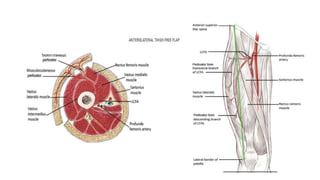
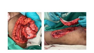
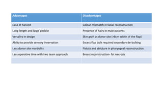
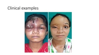
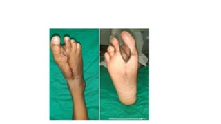
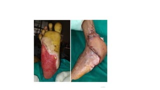
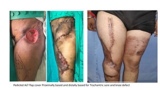
Anterolateral thigh (ALT) flap
Introduction
• The anterolateral thigh (ALT) free flap was first described by Song et al. in 1984
as a soft tissue flap that is perfused by septo-cutaneous branches of the lateral
circumflex femoral artery (LCFA).
• It is a Fasciocutaneous flap (type B – Septo-cutaneous perforator) or ( type C -
musculocutaneous perforator )
Work horse flap for reconstruction
• As a Pedicled flap
 Distally based (on distal minor pedicle) – for knee defect
 Proximally based –Trochanteric bed sore, Lower abdominal defects
Perineal reconstruction , Gluteal defect.
• As a Free Flap
 Buccal mucosa defect ,Buccal through & through defect , Pharyngo-
oesophageal reconstruction ,Lower lip ,Lateral & anterior skull base,
Scalp defects , breast reconstruction , Extremity reconstruction ,
Phalloplasty etc.
Flap supply
• ALT flap is supplied by either septocutaneaus vessels (87%) or
musculocutaneaus perforators (13%) from the descending branch of
LCFA.
o Length : 12 cm (range 8-16 cm )
o Diameter : 2.1 mm (range 2-2.5 mm)
Two venae comitantes accompanies the pedicle
Lateral femoral cutaneous nerve (L2-L3) provide the sensory innervation to the
area
Pre-op Evaluation
• Functional evaluation of knee extension
• Previous scar that may affect flap design
• Marking of perforator by Doppler
• Prior skin graft donor sites can be incorporated as part of the flap
Important landmark ASIS and
superior lateral border of patella
Perforators are located at this drawn line
• The ASIS to lateral patella (Septocutaneus)
• Posterior to this line (Musculocutaneaus)
Flap harvest
• Medial incision first and subfascial
approach.
• Rectus femoris is identified by its
bipinnate arrangement of fibers around
central raphe.
• Septum is identified between RF and VL.
• By doing medial retraction , Any Septo-
cutaneous Perforator should be visualized
by now.
• If No perforator is visualized the
deep fascia should be dissected off
the VL.
• And still no perforator is visualized
incision can be extended superiorly
or inferiorly to search for a
perforator.
• When the perforator is found ,then
only the dissection proceeds.
• The perforator is dissected to its
source pedicle .
• All Possible nerves to
vastus lateralis and the
muscle itself should be
preserved.
• Once the anterior
dissection is complete ,lateral
incision can be taken.
Flap modification
• Thin Flap
• Adipofascial flap
• Innervated flap
• Functional muscle flap
• Flow through flap
• Chimeric
Advantages Disadvantages
Ease of harvest Colour mismatch in facial reconstruction
Long length and large pedicle Presence of hairs in male patients
Versality in design Skin graft at donor site (>8cm width of the flap)
Ablity to provide sensory innervation Excess flap bulk required secondary de-bulking
Less donor site morbidity Fistula and stricture in pharyngeal reconstruction
Less operative time with two team approach Breast reconstruction- fat necrosis
Clinical examples
Pedicled ALT flap cover Proximally based and distally based for Trochantric sore and knee defect
Thank you

ALT flap presentation.pptx

  • 1.
  • 2.
    Introduction • The anterolateralthigh (ALT) free flap was first described by Song et al. in 1984 as a soft tissue flap that is perfused by septo-cutaneous branches of the lateral circumflex femoral artery (LCFA). • It is a Fasciocutaneous flap (type B – Septo-cutaneous perforator) or ( type C - musculocutaneous perforator )
  • 3.
    Work horse flapfor reconstruction • As a Pedicled flap  Distally based (on distal minor pedicle) – for knee defect  Proximally based –Trochanteric bed sore, Lower abdominal defects Perineal reconstruction , Gluteal defect. • As a Free Flap  Buccal mucosa defect ,Buccal through & through defect , Pharyngo- oesophageal reconstruction ,Lower lip ,Lateral & anterior skull base, Scalp defects , breast reconstruction , Extremity reconstruction , Phalloplasty etc.
  • 4.
    Flap supply • ALTflap is supplied by either septocutaneaus vessels (87%) or musculocutaneaus perforators (13%) from the descending branch of LCFA. o Length : 12 cm (range 8-16 cm ) o Diameter : 2.1 mm (range 2-2.5 mm) Two venae comitantes accompanies the pedicle Lateral femoral cutaneous nerve (L2-L3) provide the sensory innervation to the area
  • 6.
    Pre-op Evaluation • Functionalevaluation of knee extension • Previous scar that may affect flap design • Marking of perforator by Doppler • Prior skin graft donor sites can be incorporated as part of the flap
  • 7.
    Important landmark ASISand superior lateral border of patella Perforators are located at this drawn line • The ASIS to lateral patella (Septocutaneus) • Posterior to this line (Musculocutaneaus)
  • 8.
    Flap harvest • Medialincision first and subfascial approach. • Rectus femoris is identified by its bipinnate arrangement of fibers around central raphe. • Septum is identified between RF and VL. • By doing medial retraction , Any Septo- cutaneous Perforator should be visualized by now.
  • 9.
    • If Noperforator is visualized the deep fascia should be dissected off the VL. • And still no perforator is visualized incision can be extended superiorly or inferiorly to search for a perforator. • When the perforator is found ,then only the dissection proceeds. • The perforator is dissected to its source pedicle .
  • 10.
    • All Possiblenerves to vastus lateralis and the muscle itself should be preserved. • Once the anterior dissection is complete ,lateral incision can be taken.
  • 12.
    Flap modification • ThinFlap • Adipofascial flap • Innervated flap • Functional muscle flap • Flow through flap • Chimeric
  • 13.
    Advantages Disadvantages Ease ofharvest Colour mismatch in facial reconstruction Long length and large pedicle Presence of hairs in male patients Versality in design Skin graft at donor site (>8cm width of the flap) Ablity to provide sensory innervation Excess flap bulk required secondary de-bulking Less donor site morbidity Fistula and stricture in pharyngeal reconstruction Less operative time with two team approach Breast reconstruction- fat necrosis
  • 14.
  • 17.
    Pedicled ALT flapcover Proximally based and distally based for Trochantric sore and knee defect
  • 18.