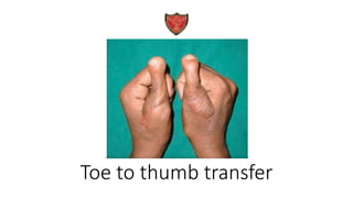
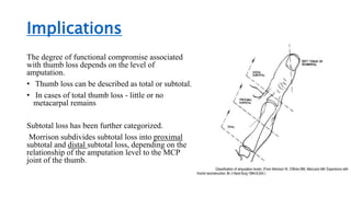
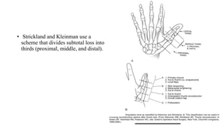
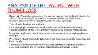
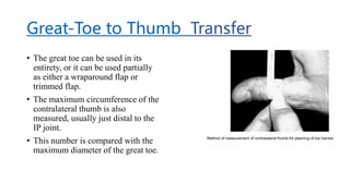
The document discusses toe to thumb transfer as an option for thumb reconstruction. It provides details on:
- The anatomy of the toe making it suitable for transfer, including its skeletal structure, tendons, skin, and nerves
- The history of toe to thumb transfer, including early experiments and the first microsurgical procedure
- Causes of thumb loss that may require reconstruction
- Preoperative assessment and considerations for candidates, including smoking cessation
- Surgical technique for both hand and toe dissection to prepare the structures for microvascular anastomosis
FDMA FLAP - The first dorsal metacarpal artery (FDMA) flap is used mainly for...Shubhanshu Gaurav
Also see the Dr Vinita puri & Dr Nilesh SHENDE Paper
First Dorsal Metacarpal Artery Flap a Workhorse for Reconstruction of Selected Small Defects of the Hand
Turkish Journal of Plastic Surgery 27(3):p 98-103, Jul–Sep 2019. | DOI: 10.4103/tjps.tjps_71_18
Best Ayurvedic medicine for Gas and IndigestionSwastikAyurveda
Here is the updated list of Top Best Ayurvedic medicine for Gas and Indigestion and those are Gas-O-Go Syp for Dyspepsia | Lavizyme Syrup for Acidity | Yumzyme Hepatoprotective Capsules etc
These lecture slides, by Dr Sidra Arshad, offer a quick overview of the physiological basis of a normal electrocardiogram.
Learning objectives:
1. Define an electrocardiogram (ECG) and electrocardiography
2. Describe how dipoles generated by the heart produce the waveforms of the ECG
3. Describe the components of a normal electrocardiogram of a typical bipolar lead (limb II)
4. Differentiate between intervals and segments
5. Enlist some common indications for obtaining an ECG
6. Describe the flow of current around the heart during the cardiac cycle
7. Discuss the placement and polarity of the leads of electrocardiograph
8. Describe the normal electrocardiograms recorded from the limb leads and explain the physiological basis of the different records that are obtained
9. Define mean electrical vector (axis) of the heart and give the normal range
10. Define the mean QRS vector
11. Describe the axes of leads (hexagonal reference system)
12. Comprehend the vectorial analysis of the normal ECG
13. Determine the mean electrical axis of the ventricular QRS and appreciate the mean axis deviation
14. Explain the concepts of current of injury, J point, and their significance
Study Resources:
1. Chapter 11, Guyton and Hall Textbook of Medical Physiology, 14th edition
2. Chapter 9, Human Physiology - From Cells to Systems, Lauralee Sherwood, 9th edition
3. Chapter 29, Ganong’s Review of Medical Physiology, 26th edition
4. Electrocardiogram, StatPearls - https://www.ncbi.nlm.nih.gov/books/NBK549803/
5. ECG in Medical Practice by ABM Abdullah, 4th edition
6. Chapter 3, Cardiology Explained, https://www.ncbi.nlm.nih.gov/books/NBK2214/
7. ECG Basics, http://www.nataliescasebook.com/tag/e-c-g-basics
New Drug Discovery and Development .....NEHA GUPTA
The "New Drug Discovery and Development" process involves the identification, design, testing, and manufacturing of novel pharmaceutical compounds with the aim of introducing new and improved treatments for various medical conditions. This comprehensive endeavor encompasses various stages, including target identification, preclinical studies, clinical trials, regulatory approval, and post-market surveillance. It involves multidisciplinary collaboration among scientists, researchers, clinicians, regulatory experts, and pharmaceutical companies to bring innovative therapies to market and address unmet medical needs.
CDSCO and Phamacovigilance {Regulatory body in India}NEHA GUPTA
The Central Drugs Standard Control Organization (CDSCO) is India's national regulatory body for pharmaceuticals and medical devices. Operating under the Directorate General of Health Services, Ministry of Health & Family Welfare, Government of India, the CDSCO is responsible for approving new drugs, conducting clinical trials, setting standards for drugs, controlling the quality of imported drugs, and coordinating the activities of State Drug Control Organizations by providing expert advice.
Pharmacovigilance, on the other hand, is the science and activities related to the detection, assessment, understanding, and prevention of adverse effects or any other drug-related problems. The primary aim of pharmacovigilance is to ensure the safety and efficacy of medicines, thereby protecting public health.
In India, pharmacovigilance activities are monitored by the Pharmacovigilance Programme of India (PvPI), which works closely with CDSCO to collect, analyze, and act upon data regarding adverse drug reactions (ADRs). Together, they play a critical role in ensuring that the benefits of drugs outweigh their risks, maintaining high standards of patient safety, and promoting the rational use of medicines.
Local Advanced Lung Cancer: Artificial Intelligence, Synergetics, Complex Sys...Oleg Kshivets
Overall life span (LS) was 1671.7±1721.6 days and cumulative 5YS reached 62.4%, 10 years – 50.4%, 20 years – 44.6%. 94 LCP lived more than 5 years without cancer (LS=2958.6±1723.6 days), 22 – more than 10 years (LS=5571±1841.8 days). 67 LCP died because of LC (LS=471.9±344 days). AT significantly improved 5YS (68% vs. 53.7%) (P=0.028 by log-rank test). Cox modeling displayed that 5YS of LCP significantly depended on: N0-N12, T3-4, blood cell circuit, cell ratio factors (ratio between cancer cells-CC and blood cells subpopulations), LC cell dynamics, recalcification time, heparin tolerance, prothrombin index, protein, AT, procedure type (P=0.000-0.031). Neural networks, genetic algorithm selection and bootstrap simulation revealed relationships between 5YS and N0-12 (rank=1), thrombocytes/CC (rank=2), segmented neutrophils/CC (3), eosinophils/CC (4), erythrocytes/CC (5), healthy cells/CC (6), lymphocytes/CC (7), stick neutrophils/CC (8), leucocytes/CC (9), monocytes/CC (10). Correct prediction of 5YS was 100% by neural networks computing (error=0.000; area under ROC curve=1.0).
- Video recording of this lecture in English language: https://youtu.be/kqbnxVAZs-0
- Video recording of this lecture in Arabic language: https://youtu.be/SINlygW1Mpc
- Link to download the book free: https://nephrotube.blogspot.com/p/nephrotube-nephrology-books.html
- Link to NephroTube website: www.NephroTube.com
- Link to NephroTube social media accounts: https://nephrotube.blogspot.com/p/join-nephrotube-on-social-media.html
2. Introduction
• Thumb plays a crucial role in proper functioning of the hand.
• At 9 months of age, the thumb begins to function as it will in adulthood.
• Thumb opposition- unique to primates, allows the human hand to perform power
grip and precision handling.
• The thumb itself is responsible for 40% to 50% of the overall hand functions .
3. Microsurgical toe transfer
• Excellent option for thumb reconstruction.
• The toe, after all, is a digit itself and therefore is similar anatomically to the
thumb.
• It’s skeletal structure consists of two phalanges motored by extensor and flexor
tendons ,covered with durable glabrous skin that is innervated by digital nerves. It
provides all the necessary components of good thumb function and excellent
appearance of the reconstructed thumb.
• With proper operative planning and meticulous technique, donor site morbidity
and functional compromise can be minimized.
5. History
• Buncke successfully performed toe to thumb transfer on a rhesus
monkey, proving the feasibility of this procedure in primates.
• Toe to thumb transplantation was first reported in 1891 by Nicolandi
using a pedicled technique.
• In 1969, Cobbett performed the first microsurgical thumb
reconstruction in humans with a transferred great toe.
6. Causes of Thumb Loss
• Congenital (partial or total) absence.
oFetal neurogenic injury
oMaternal viral infection, drugs (thalidomide )
oReduced oxygen tension
oDietary deficiency
• Loss due to malignant disease, such as melanoma.
• Loss due to trauma (M/C) including industrial, non-industrial,
and environmental insults.
7. CAMPBELL-REID CLASSIFICATION
• Group 1:- Amputation distal to the metacarpophalangeal joint, leaving an
adequate stump.
• Group 2:- Amputation of the thumb distal to or through the
metacarpophalangeal joint, leaving a stump of inadequate length.
• Group 3:- Amputation through the metacarpal, with preservation of some
functioning thenar muscles.
• Group 4:- Amputation at or near the carpometacarpal joint
8. Implications
The degree of functional compromise associated
with thumb loss depends on the level of
amputation.
• Thumb loss can be described as total or subtotal.
• In cases of total thumb loss - little or no
metacarpal remains
Subtotal loss has been further categorized.
Morrison subdivides subtotal loss into proximal
subtotal and distal subtotal loss, depending on the
relationship of the amputation level to the MCP
joint of the thumb.
9. • Strickland and Kleinman use a
scheme that divides subtotal loss into
thirds (proximal, middle, and distal).
10. ANALYSIS OF THE PATIENT WITH
THUMB LOSS
• History & Physical examination:- Toe transfer must pay particular attention to the
affected thumb or recipient site of the operation, assessment of the length,
mobility, gross sensibility, vascularity, and soft tissue coverage.
• Tests of hand function and anatomy.
• Plain radiographs demonstrate the skeletal extent of the thumb's deficiency.
Vascular Anatomy of the lower extremity must also be assessed preoperatively.
• In addition to physical examination, duplex ultrasonography or angiography can
be helpful.
• Pre-operative hand function to determine what functional needs the transferred toe
has to supply.
• Sensibility can be assessed by subjective measurement of light touch and two-
point discrimination and by Semmes-Weinstein monofilament testing.
11. Preoperative Consultation
Surgeon must carefully assess the patient in consultation to determine whether the
patient is an appropriate candidate for microvascular thumb reconstruction.
The procedure places significant physical demands on the patient and requires
intensive postoperative rehabilitation.
Stop smoking and tobacco free for at least 1 month before toe transfer.
For confirmation of abstinence from tobacco use, a urine nicotine level can be
checked within 24 hours of surgery. If the result is positive, surgery is postponed
until the patient is able to cease use of tobacco products.
12. TREATMENT GOALS
• Patient's occupation, hand dominance, and overall health are important.
• Microvascular thumb reconstruction places significant physiologic, psychological,
and socioeconomic demands on the patient.
• A patient undergoing this procedure must be able to modify daily activities to
allow the tissues time to heal.
• Patients who are unable or unwilling to comply with intensive hand therapy
should not be considered for toe to hand transfer because this procedure cannot
succeed without proper postoperative treatment.
13. Great-Toe to Thumb Transfer
• The great toe can be used in its
entirety, or it can be used partially
as either a wraparound flap or
trimmed flap.
• The maximum circumference of the
contralateral thumb is also
measured, usually just distal to the
IP joint.
• This number is compared with the
maximum diameter of the great toe.
14. • General anesthesia.
• Anaesthesia must diligently maintain the patient's body temperature to minimize
vasoconstriction.
• The patient's blood pressure should be maintained at an adequate level to maintain
perfusion to the toe flap.
• In general, a systolic pressure of at least 110 mm Hg is desirable. Blood pressure
should be managed with intravenous fluid when possible, and vasoconstrictive
agents should be avoided.
• Dissection of both the hand and foot is done under tourniquet control and loupe
magnification
15. • Ideally 2 separate surgical teams operate simultaneously to minimize operative
time.
• The goals of the hand dissection include careful incision planning identification
of the anatomy of the recipient hand and preparation of the skeletal, vascular, and
neural structures of the recipient hand for toe transfer.
• In general, an incision that designs proximally based radial and ulnar flaps is
preferred.
• The position of the radial artery should be identified by palpation or Doppler
examination and then marked on the skin. It provides proper orientation of
incision
16. A and B, Representative incision markings on recipient hands
17. • After Elevation of the skin flaps, the digital nerves are identified and dissected
free from surrounding soft tissues.
• Nerve ends are then tagged with 8-0 nylon suture for later identification.
• Tendons are next addressed if a dynamic toe transfer is planned (static toe
transfers, such as the wraparound procedure, do not require tendon repairs).
• Next, the donor arterial supply must be addressed. The artery is located by
extending the dorsal incision proximally toward the anatomic snuffbox, over the
dorsal web space of the thumb.
• Once it is identified, the artery is dissected free from surrounding soft tissues and
marked with a vessel loop.
• When an adequate vein is identified, it is dissected in a fashion similar to
dissection of the artery and marked with a vessel loop for later identification.
18. • If the toe is to be attached to the proximal phalanx of the thumb or to the thumb
metacarpal, the recipient structure will have to be prepared for osteosynthesis.
• Once it is identified, the distal end of the bone is dissected subperiosteally. An
osteotomy is then performed with an oscillating saw.
• If the toe is to be transferred to the proximal phalanx of the recipient thumb, the
osteotomy is made in a straight transverse fashion.
• At the completion of the hand dissection, the tourniquet is deflated to allow
reperfusion of the upper extremity, and hemostasis is achieved. The wounds are
then dressed with saline-moistened dressings until the donor toe is ready for
transfer.
19.
20. TOE DISSECTION
• Dissection is ideally carried out by a second operative team simultaneously with
the hand dissection.
• The course of the dorsalis pedis artery and dorsal superficial veins of the foot is
marked out on the dorsal skin of the foot preoperatively by a combination of
palpation and Doppler examination.
• Assessment of venous anatomy can be assisted by dependent positioning of the
extremity and the use of a "venous tourniquet" with a tourniquet pressure of
approximately 80 mmHg.
• The incision for the toe harvest is also designed. In general, an incision that
develops dorsal and plantar flaps, with a dorsal proximal extension for vessel
dissection, is used. This provides good access for dissection and develops flaps that
are complementary to the radial and ulnar flaps of the hand.
22. Exsanguination of the lower extremity and inflation of the tourniquet, incisions are
made.
Dissection is performed with the assistance of loupe magnification.
Skin flaps are elevated laterally and medially on the dorsum of the foot to expose
the underlying structures.
Where it is possible, small sensory nerves are preserved.
Next, the superficial saphenous venous system is examined, and one or more veins
of adequate size and length are identified as the donor vein.
23. • Next the dorsalis pedis artery is
identified proximally in the
dissection.
• This structure is dissected
proximally to provide adequate
length for anastomosis to the
recipient artery in the hand.
• During this dissection, the
extensor hallucis brevis muscle is
divided to allow adequate
exposure of the vessel.
• The final structure on the dorsum
of the foot that must be prepared
is the extensor hallucis longus
tendon and flexor pollicis longus
tendon in the hand.
24. • In great toe transfers, the branch to the second toe is divided just distal to the
branch point of the first dorsal metatarsal artery.
• Once the arterial and venous systems have been dissected, they can be tagged with
vessel loops for later identification.
• When re-establishment of dorsal sensory function of the transferred toe is desired,
the deep peroneal nerve, which runs adjacent to the dorsalis pedis artery, can be
included in the flap.
• With the neurovascular and tendinous dissections completed, attention is now
turned to the skeletal support of the toe.
25. • With the toe dissection completed, the lower extremity tourniquet is deflated,
allowing reperfusion of the toe.
• Because of arterial spasm resulting from dissection, the donor toe might remain
pale for up to 20 to 30 minutes after release of the tourniquet.
• Spasm can be minimized by topical application of compounds such as papaverine
or concentrated lidocaine (20%) to the vessels.
• Once reperfusion to the donor toe is established, it should be allowed to perfuse
for approximately 20 minutes before completion of the harvest and transfer to the
hand.
• With the recipient site prepared and the toe dissected, transfer of the toe to the
hand is next undertaken.
26. • The donor artery and then the vein are divided proximally and flushed with
lactated Ringer solution containing 10 units/mL of heparin.
• The transfer is begun with reconstruction of the skeletal framework.
• The joint should be pinned in a position of slight flexion.
• Many techniques exist for osteosynthesis, including the use of K-wires, rigid
fixation systems, and interosseous wiring.
• It is preferable to use rigid plate and screw fixation for osteosynthesis because of
its strength and allowance of early active range of motion in the reconstructed
thumb.
28. • With the skeletal reconstruction complete, repair of soft tissues is undertaken.
• It is preferable to repair tendinous structures first, then the nerves, followed by the
artery and finally the vein.
29. Donor and recipient skin flaps are brought into approximation and trimmed as
necessary.
It is preferable to use a single-layer, tension-free closure with interrupted
monofilament suture.
In children, absorbable suture, such as chromic gut, should be used to eliminate the
need for later suture removal.
In situations in which flaps cannot be closed without undue tension, a skin graft is
used for cover, provided that all vital structures, such as nerves, blood vessels, and
tendons, are covered by healthy vascularized soft tissue.
After placement of the usual sterile dressings, the extremity is placed into a well-
padded thumb spica splint with the tip of the toe flap visible for postoperative
monitoring.
30. 1.Wraparound Flap : Because the great toe is larger
than the normal thumb,
Various modifications of great-toe transfer for
thumb reconstruction have been developed to design
a better size match.
The wraparound technique was first described by
Morrison et al in 1980. In this procedure, only the
soft tissue and nail of the great toe are transferred to
the hand, without the metatarsal or proximal phalanx
of the great toe.
An iliac crest bone graft, if required, provides
skeletal support for the reconstructed thumb. Donor
site defect closed with cross toe skin flap.
31. 2.Trimmed great-toe flap -
The trimmed great-toe flap was first described in the
late 1980s.
This technique has the advantage of the wraparound
great- toe flap, namely, better size match of the
contralateral thumb, but avoids the disadvantage of lack
of mobility and growth potential.
The trimmed toe flap is harvested with both the
proximal and distal phalanges.
The circumferences of the soft tissues and the bone are
reduced to match the circumference of the contralateral
normal thumb.
With this technique, joints and epiphyses are preserved.
32. FLAP DISSECTION -
• For the trimmed great-toe technique are similar to those
for the wraparound technique.
• The medial proximally based skin flap is elevated off of
the underlying periosteum, with care being taken to
preserve the medial neurovascular structures of the toe.
• A proximally based flap consisting of periosteum and the
medial collateral ligament of the IP joint of the toe is then
elevated off of the underlying bone structures.
• The circumference of the phalanges is reduced by
making a longitudinal osteotomy in the sagittal plane.
• The toe is harvested and inset in a fashion similar to that
of the whole great-toe technique.
• Skin closure and management of the nail plate and
eponychial fold are similar to those of the wraparound
great-toe flap. The donor site closure is identical to that
for the wraparound flap.
33. Second-Toe to Thumb
Transfer of the second toe to the hand was first described in China in 1973.
The second toe's minimal donor site impact, excellent appearance, and superb
function after transfer into the thumb position make it the preferred digit for toe to
thumb transfer.
The use of the second toe minimizes donor site problems associated with toe
transfer, which can be both functionally and socially significant.
After transfer of the second toe, the overall contour of the foot is nearly normal.
absence of this toe is hardly noticeable on casual observation.
34. 1.Disadvantages of the donor - for
a thumb it is narrower and
weaker, and therefore potentially
less functional, than one
reconstructed with a great toe.
lack of soft tissue available for
coverage of interosseous muscles
and bone when a long length of
metatarsal is used.
35. POST OP
• Monitoring of the flap's vascular status postoperatively, includes implantable Doppler
devices, laser flowmetry, and pulse oximetry etc
• The most reliable method of evaluation is hourly visual inspection of the flap for color
and quality of capillary refill during the first 2 postoperative days.
• if flap perfusion is in question, urgent return to the operating room with exploration of
the anastomosis is imperative for flap salvage.
• The first 3 to 5 postoperative days are spent in a warm room to minimize
vasoconstriction, and adequate hydration of the patient is ensured.
• Anticoagulation of low-molecular weight dextran and aspirin are given
36. Intravenous antibiotics which are administered
intraoperatively, are also given during the first 5 to 7
postoperative days.
• At around postoperative day 5, the dressings on the
extremity are changed, and a well-padded splint is
reapplied.
• Sutures are removed from the hand and foot on or around
postoperative day 14.
• If pins were used for osteosynthesis or to support another
method of osteosynthesis such as interfragmentary
they are removed when there is clinical and radiographic
evidence of stability and healing of the bone.
37. EARLY COMPLICATIONS • Infection • Bleeding • Wound healing
complications • Thrombosis of microvascular anastomosis • Flap necrosis •
Rupture of nerve repairs • Rupture of tendon repairs
LATE COMPLICATIONS • Tendon adhesion • Joint stiffness • Flexion
contracture • Poor recovery of sensibility • Delayed union or non-union of
osteosynthesis
INCISION DESIGN DEPENDING ON CASE TO CASE DEPENDING ON THE NATURE OF INJURY
For dynamic
The advantages of this flap include – better size match with the opposite normal thumb; – provision of sensibility, – length, and stability to the reconstructed thumb; and – preservation of a portion of the great Toe. The disadvantages to this flap. – Because this is an essentially static transfer when the use of an iliac crest bone graft is required, it is useful only for thumb loss distal to the MCP joint. – If it were used more proximally, the limitation of motion in the thumb would be unacceptable. – Another disadvantage of this flap is its lack of potential for growth because of the absence of an epiphysis. – Resorption of the bone graft is another potential problem