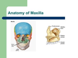
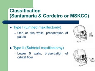
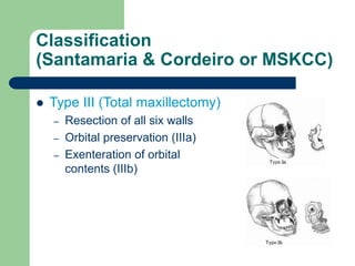
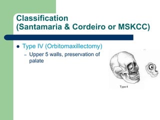
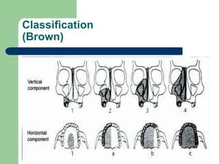
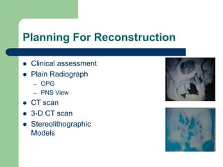
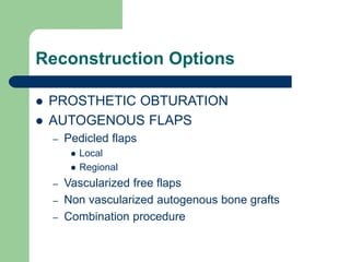
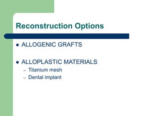
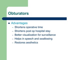
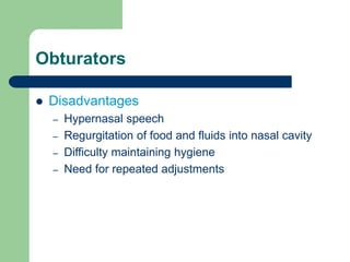
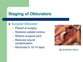
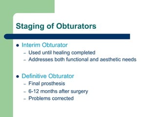
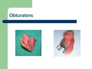
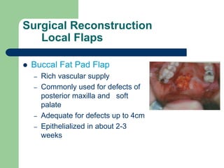
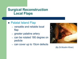
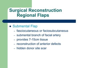
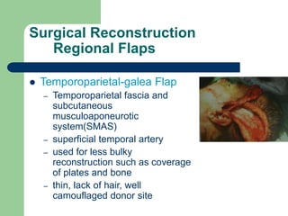
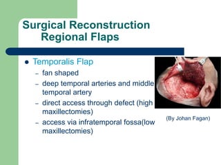
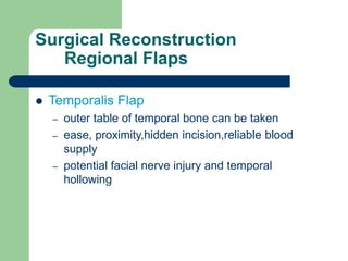
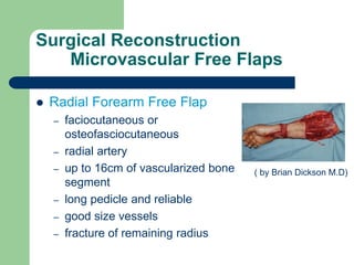
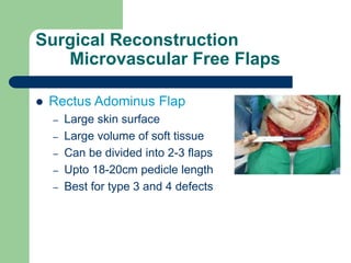
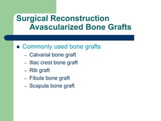
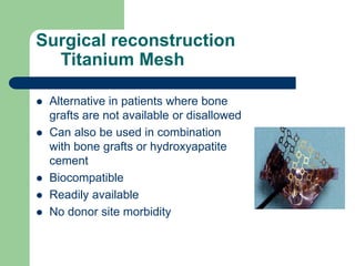
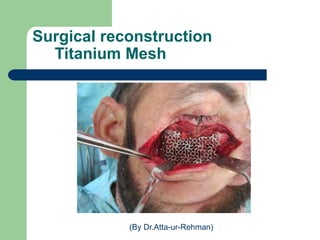
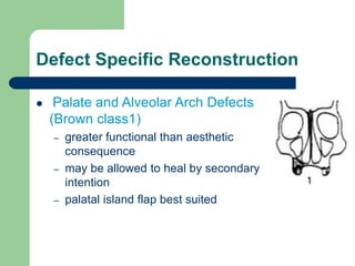
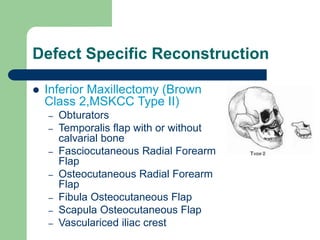
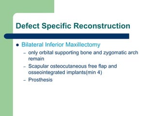
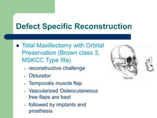
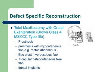
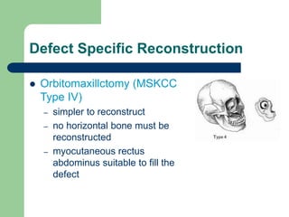
Reconstruction of Maxilla
by
Dr.Anjum Iqbal
Trainee Medical Officer
Oral & Maxillofacial Surgery
Khyber College of Dentistry.
Layout
 Anatomy of Maxilla
 Goals of Maxillary Reconstruction
 Classification of Maxillectomy Defects
 Planning and evaluation for reconstruction
 Reconstruction options
 Defect Specific Reconstruction
Anatomy of Maxilla
Goals of Maxillary Reconstruction
1. Obtain a healed wound.
2. Restore palatal competence and function.
3. Restore normal mastication and deglutition.
4. Support the eye.
5. Maintain a patent nasal airway.
6. Support and suspend facial soft tissues.
7. Restore the midfacial contour.
Classification Of Maxillectomy
Defects
Classification
(Santamaria & Cordeiro or MSKCC)
 Type I (Limited maxillectomy)
– One or two walls, preservation of
palate
 Type II (Subtotal maxillectomy)
– Lower 5 walls, preservation of
orbital floor
Classification
(Santamaria & Cordeiro or MSKCC)
 Type III (Total maxillectomy)
– Resection of all six walls
– Orbital preservation (IIIa)
– Exenteration of orbital
contents (IIIb)
Classification
(Santamaria & Cordeiro or MSKCC)
 Type IV (Orbitomaxillectomy)
– Upper 5 walls, preservation of
palate
Classification
(Brown)
Planning For Reconstruction
 Clinical assessment
 Plain Radiograph
– OPG
– PNS View
 CT scan
 3-D CT scan
 Stereolithographic
Models
Reconstruction Options
 PROSTHETIC OBTURATION
 AUTOGENOUS FLAPS
– Pedicled flaps
 Local
 Regional
– Vascularized free flaps
– Non vascularized autogenous bone grafts
– Combination procedure
Reconstruction Options
 ALLOGENIC GRAFTS
 ALLOPLASTIC MATERIALS
– Titanium mesh
– Dental implant
Prosthetic Obturation
Obturators
 Advantages
– Shortens operative time
– Shortens post op hospital stay
– Better visualization for surveillance
– Helps in speech and swallowing
– Restores aesthetics
Obturators
 Disadvantages
– Hypernasal speech
– Regurgitation of food and fluids into nasal cavity
– Difficulty maintaining hygiene
– Need for repeated adjustments
Staging of Obturators
 Surgical Obturator
– Placed at surgery
– Restores palatal contour
– Retains surgical pack
– Reduces wound
contamination
– Removed in 10-14 days
(By Dr.Muslim Khan)
Staging of Obturators
 Interim Obturator
– Used until healing completed
– Addresses both functional and aesthetic needs
 Definitive Obturator
– Final prosthesis
– 6-12 months after surgery
– Problems corrected
Obturators
Surgical Reconstruction
Local Flaps
Surgical Reconstruction
Local Flaps
 Buccal Fat Pad Flap
 Palatal Island Flap
 Nasolabial Flap
 Tongue Flap
 Uvula Flap
Surgical Reconstruction
Local Flaps
 Buccal Fat Pad Flap
– Rich vascular supply
– Commonly used for defects of
posterior maxilla and soft
palate
– Adequate for defects up to 4cm
– Epithelialized in about 2-3
weeks
Surgical Reconstruction
Local Flaps
 Palatal Island Flap
– versatile and reliable local
flap
– greater palatine artery
– can be rotated 180 degree on
pedicle
– can cover up to 15cm defects
(By Dr.Muslim Khan)
Surgical Reconstruction
Local Flaps
 Nasolabial Flap
– closure of oroantral fistulae and
defects of anterior floor of mouth
– facial and angular arteries
– up to 5cm width flap
– limited donor tissue, facial scarring
and second surgery (By Dr.Muslim Khan)
Surgical Reconstruction
Local Flaps
 Tongue Flap
– closure of residual cleft and fistulae
of hard palate
– lingual artery
– donor site morbidity, limited arc of
rotation, and small size
(By Dr.Muslim Khan)
Surgical Reconstruction
Regional Flaps
Surgical Reconstruction
Regional Flaps
 Submental Flap
 Temoproparietal-galea Flap
 Temporalis Flap
 Platysma Flap
 Masseter Flap
 Sternocleidomastoid Mastoid
 Trapezius Flap
Surgical Reconstruction
Regional Flaps
 Submental Flap
– fasciocutaneous or faciosubcutaneous
– submental branch of facial artery
– provides 7-15cm tissue
– reconstruction of anterior defects
– hidden donor site scar
Surgical Reconstruction
Regional Flaps
 Temporoparietal-galea Flap
– Temporoparietal fascia and
subcutaneous
musculoaponeurotic
system(SMAS)
– superficial temporal artery
– used for less bulky
reconstruction such as coverage
of plates and bone
– thin, lack of hair, well
camouflaged donor site
Surgical Reconstruction
Regional Flaps
 Temporalis Flap
– fan shaped
– deep temporal arteries and middle
temporal artery
– direct access through defect (high
maxillectomies)
– access via infratemporal fossa(low
maxillectomies)
(By Johan Fagan)
Surgical Reconstruction
Regional Flaps
 Temporalis Flap
– outer table of temporal bone can be taken
– ease, proximity,hidden incision,reliable blood
supply
– potential facial nerve injury and temporal
hollowing
Surgical Reconstruction
Regional Flaps
 Platysma Flap
– Myocutaneous
– submental and facial
arteries
– thin, pliable and easily
harvested
– less reliability (By Dr.Muslim Khan)
Surgical Reconstruction
Regional Flaps
 Masseter Flap
– masseteric artery
– useful for reconstruction of palatal defects
– limited volume, trismus
Surgical Reconstruction
Regional Flaps
 Sternocleidomastoid Flap
– myocutaneous or myo-osseus
– occipital, superior thyroid and supra scapular
arteries
– proximity to defect site, lack of requirement for
another incision
Surgical Reconstruction
Regional Flaps
 Trapezius Flap
– Myocutaneous
– may be used as composite flap with a portion of
clavicle or scapula
– transverse cervical artery, occipital, posterior
intercostal and dorsal scapular arteries
– adequate volume of well vascularized tissue
Surgical Reconstruction
Microvascular Free
Flaps
Surgical Reconstruction
Microvascular Free Flaps
 Radial Forearm Free Flap
 Radial Forearm Osteo-fascio-cutaneous Flap
 Rectus Abdominus Flap
 Fibula Osteo-cutaneous Flap
 Scapular Osteo-myocutaneous Flap
 Vascularized Iliac Crest
Surgical Reconstruction
Microvascular Free Flaps
 Radial Forearm Free Flap
– faciocutaneous or
osteofasciocutaneous
– radial artery
– up to 16cm of vascularized bone
segment
– long pedicle and reliable
– good size vessels
– fracture of remaining radius
( by Brian Dickson M.D)
Surgical Reconstruction
Microvascular Free Flaps
 Rectus Adominus Flap
– Large skin surface
– Large volume of soft tissue
– Can be divided into 2-3 flaps
– Upto 18-20cm pedicle length
– Best for type 3 and 4 defects
Surgical Reconstruction
Microvascular Free Flaps
 Fibula Osteo-cutaneous Flap
– peroneal artery and vein
– provides greatest length of
available bone
– usual pedicle length about 6-7cm
– provides sufficient bone for implant
placement
Surgical Reconstruction
Microvascular Free Flaps
 Scapular Osteo-myocutaneous Flap
– circumflex scapular artery
– pedicle length up to 20cm
– average thickness of bone about 3cm
– sufficient for implant placement
– inferior quality bone
– can be oriented vertically as well as horizontally
Surgical Reconstruction
Microvascular Free Flaps
 Vascularized Iliac Crest
– most successful
– deep circumflex iliac artery(DCIA)
– accompanying internal oblique
muscle provides excellent soft
tissue
– less donor site morbidity
Surgical Reconstruction
Avascularized Bone
Grafts
Surgical Reconstruction
Avascularized Bone Grafts
 Requirements Of Ideal Bone Grafts
– Stability
– Potential for graft integration
– Available in large quantities
– Moldable
No such ideal graft is available
Surgical Reconstruction
Avascularized Bone Grafts
 Commonly used bone grafts
– Calvarial bone graft
– Iliac crest bone graft
– Rib graft
– Fibula bone graft
– Scapula bone graft
Surgical Reconstruction
Titanium Mesh
Surgical reconstruction
Titanium Mesh
 Alternative in patients where bone
grafts are not available or disallowed
 Can also be used in combination
with bone grafts or hydroxyapatite
cement
 Biocompatible
 Readily available
 No donor site morbidity
Surgical reconstruction
Titanium Mesh
(By Dr.Atta-ur-Rehman)
Defect Specific
Reconstruction
Defect Specific Reconstruction
 Palate and Alveolar Arch Defects
(Brown class1)
– greater functional than aesthetic
consequence
– may be allowed to heal by secondary
intention
– palatal island flap best suited
Defect Specific Reconstruction
 Inferior Maxillectomy (Brown
Class 2,MSKCC Type II)
– Obturators
– Temporalis flap with or without
calvarial bone
– Fasciocutaneous Radial Forearm
Flap
– Osteocutaneous Radial Forearm
Flap
– Fibula Osteocutaneous Flap
– Scapula Osteocutaneous Flap
– Vasculariced iliac crest
Defect Specific Reconstruction
 Bilateral Inferior Maxillectomy
– only orbital supporting bone and zygomatic arch
remain
– Scapular osteocutaneous free flap and
osseointegrated implants(min 4)
– Prosthesis
Defect Specific Reconstruction
 Total Maxillectomy with Orbital
Preservation (Brown class 3,
MSKCC Type IIIa)
– reconstructive challenge
– Obturator
– Temporalis muscle flap
– Vascularized Osteocutaneous
free flaps are best
– followed by implants and
prosthesis
Defect Specific Reconstruction
 Total Maxillectomy with Orbital
Exenteration (Brown Class 4,
MSKCC Type IIIb)
– Prosthesis
– prosthesis with myocutaneous
flap e.g. rectus abdominus
– iliac crest myo-osseous flap
– Scapular osteocutaneous free
flap
– dental implants
Defect Specific Reconstruction
 Orbitomaxillctomy (MSKCC
Type IV)
– simpler to reconstruct
– no horizontal bone must be
reconstructed
– myocutaneous rectus
abdominus suitable to fill the
defect
THANK YOU

Reconstruction of maxilla