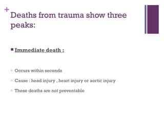
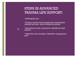
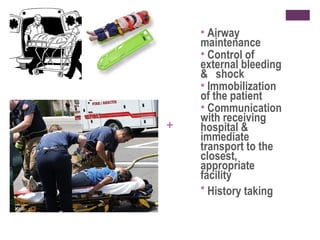
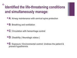
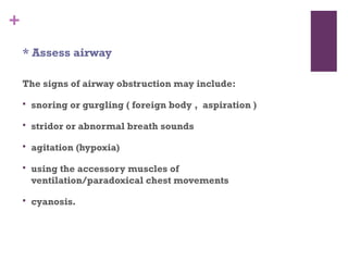
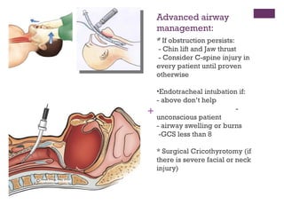
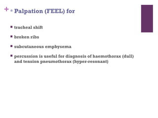
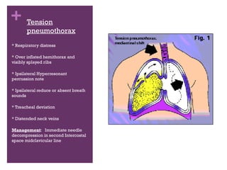
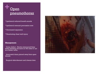
+

TRAUMA
PRIMARY AND SECONDARY SURVEY
+

CASE:
A 25 year old is brought
to casualty with history of
fall from a height of 20
feet. He landed on a pile
of bricks. Fortunately
there was no head injury.
He is complaining of
severe abdominal pain.
On examination he is
conscious; his pulse is
110/minute, BP 90/60
mmHg. There is no
external wound. However
has abrasions in left
upper quadrant and left
lower chest.
+

MECHANISMS OF
TRAUMA
Trauma can be classified
in type by causation and
by effect
Blunt , e.g. car bonnet
Penetrating , e.g. Knife
Blast , e.g. Bomb
Crush , e.g. building
collapse
Thermal
+

Deaths from trauma show three
peaks:
 Immediate

death :

o

Occurs within seconds

o

Cause : head injury , heart injury or aortic injury

o

These deaths are not preventable
+


Early death:

o

Begins an hour or two after injury

o

Cause: subdural and epidural hematomas , hemo or
pneumothorax , organ rupture or blood loss

o

Often preventable

o

This period is called the GOLDEN HOUR during which
prompt intervention can save a life
+
 Late

death:

o

Occurs many days after injury

o

Cause: sepsis or multi-organ failure

o

Prompt treatment of shock and hypoxemia during GOLDEN
HOUR can reduce these deaths
STEPS IN ADVANCED
TRAUMA LIFE SUPPORT:


Prehospital care

Primary survey with simultaneous resuscitation:
identify and treat what is killing the patient


+

Secondary survey : proceed to identify all other
injuries


Definitive care: develop a definitive management
plan

+

PREHOSPITAL CARE
Airway
maintenance
 Control of
external bleeding
& shock
 Immobilization
of the patient
 Communication
with receiving
hospital &
immediate
transport to the
closest,
appropriate
facility
 History taking


+
+

Prior to arrival:


Ensure senior emergency medical and nursing staff are aware of  all
available details of the case.



Call Trauma Team (Trauma surgeon , Anaesthetist , Nurses ,
Emergency physician , Respiratory therapist , Radiologist ,
Surgical subspecialists)



Delegate specific tasks to appropriate individuals.



Check the resuscitation equipment and prepare intravenous lines and
fluids.



If possible, estimate the patient's weight using the formula (Age + 4) x 2
(or 3 x Age for those over 9 years) and calculate:
1) The amount of fluid bolus at 20 ml/kg
2)The endotracheal tube size (age/ 4) + 4
3)Any other drugs likely to be needed 
+

On arrival:


Immediately perform a primary survey by assessing and
managing the patient's airway, with cervical spine
stabilisation, breathing and circulation.



Obtain a history, if possible, from the attendents or
ambulance officers e.g. type of trauma, speed of the vehicle,
height of the fall, restraints or safety equipment used,
whether other people were injured.



Obtain information regarding any treatment or interventions
to date.
+ PRIMARY

SURVEY
+

Identified the life-threatening conditions
and simultaneously manage:
 A: Airway maintenance with cervical spine protection
 B: Breathing and ventilation

 C: Circulation with hemorrhage control
 D: Disability ( Neurologic status )
 E: Exposure / Environmental control: Undress the patient &
prevent hypothermia
+

A : Airway and C- spine
* Talk to the patient


A patient who can speak clearly must have a clear airway



Unconscious patient may require airway and ventilatory
assistance.



The cervical spine must be protected during endotracheal
intubation if a head, neck or chest injury is suspected.



Airway obstruction is most commonly due to obstruction by
the tongue in the unconscious patient.



Hoarsness or pain with speaking indicate
laryngeal injury.
+
* Assess airway
The signs of airway obstruction may include:


snoring or gurgling ( foreign body , aspiration )



stridor or abnormal breath sounds



agitation (hypoxia)



using the accessory muscles of
ventilation/paradoxical chest movements



cyanosis.
+

* Consider need for advanced airway
management



Indications for advanced airway management techniques for
securing the airway include:

o

persisting airway obstruction

o

penetrating neck trauma with haematoma (expanding)

o

Apnoea

o

Hypoxia

o

severe head injury

o

chest trauma

o

maxillofacial injury
Advanced airway
management:
*
* If obstruction persists:
- Chin lift and Jaw thrust
- Consider C-spine injury in
every patient until proven
otherwise

+

•Endotracheal intubation if:
- above don’t help
unconscious patient
- airway swelling or burns
-GCS less than 8
* Surgical Cricothyrotomy (if
there is severe facial or neck
injury)
+

B: Breathing and ventilation
* Inspection (LOOK) of respiratory rate is
essential. Are any of the following present


Cyanosis



penetrating injury



presence of flail chest



sucking chest wounds



use of accessory muscles
+ * Palpation (FEEL) for


tracheal shift



broken ribs



subcutaneous emphysema



percussion is useful for diagnosis of haemothorax (dull)
and tension pneumothorax (hyper-resonant)
+ * Auscultation (LISTEN) for



pneumothorax (decreased breath sounds on site of
injury)



Detection of abnormal sounds in the chest.



Give 100% oxygen (if available, via self-inflating bag or
mask)
 injury





1.
2.
3.
4.

that may acutely impair ventilation

Tension pneumothorax
Flail chest with pulmonary contusion
Massive haemothorax
Open pneumothorax
+ Tension
pneumothorax
* Respiratory distress
* Over inflated hemithorax and
visibly splayed ribs
* Ipsilateral Hyperresonant
percussion note
* Ipsilateral reduce or absent breath
sounds
* Treacheal deviation
* Distended neck veins
Management: Immediate needle
decompression in second Intercostal
space midclavicular line
+ Open

pneumothorax

* Ipsilateral reduced breath sounds
* Ipsilateral resonant percussion note
* Decreased expansion
* Penetrating chest wall injury

Management:
Cover defect - Sterile waterproof three
sided dressing secured on two sides to act
as a flutter valve.
Intercostal drain placed away from open
wound.
Surgical debridement and closure later.
+ Massive

Hemothorax

* Hypotension due to blood loss
* Ipsilateral dullness to percussion
note
* Ipsilateral absent or reduced
breath sounds
* Ipsilateral decreased chest
movements
Management:
Infusion of fluids through large bore
IV cannula before draining
Large bore intercostal drain for
adults
+ Flial Chest
* Segment of chest looses bony
continuity with thoracic cage
* Moves paradoxically with
respiration and reduces tidal volume
Management: Analgesia for pain
Fluid management
Ventilatory support
C: Circulation and hemorrhagic
+
control
* Hemorrhagic control


Direct pressure for external hemorrhage



No tourniquet unless other methods are not effective in
controlling bleeding



Long bones splinted with external fixation



Pelvic binding or pneumatic anti-shock garment



Watch out for hypothermia, acidosis and coagulopathy
+
* Assessment for hypovolaemia


Check skin: color , clamminess and capillary refill time



Heart rate



Blood pressure



Pulse pressure



Conscious level



Connect an automatic BP recorder and ECG

Hypovolaemia is the commonest cause of shock in trauma
patients
+
*Vascular cannulization


Two Large bore IV cannulas: peripheral i.e. Femoral Vein
Central – Subclavian or Internal Jugular
Intraosseous in children
Draw

20ml blood for grouping and cross matching , analysis of
electrolytes and full blood count
+ * Fluid resuscitation


Bolus of warm crystalloids



Surgical control of hemorrhage is better than aggressive
fluid resuscitation



Fluid resuscitation inhibits platelet aggregation , dilutes
clotting factors and raises BP



Altered cardiovascular response to hemorrhage in trauma
pts



Enough warm crystalloids to maintain a radial pulse



Blood may also be required
+

D : Disability


Glasgow Coma Scale



Pupilary reflexes

Monitor frequently to detect deterioration

Common causes for
deterioration


Hypoxia



Hypovolaemia



Hypoglycemia



Raised intracranial pressure
+

E : Exposure



Clothes should be cut to remove



Pt kept warm and covered with blankets



Log roll



Assess spine from base of skull to coccyx



Examine back for any signs of injury



Digital Rectal Examination:
Boney fragments
Rectal wall
Bleeding
Prostate
+

SECONDARY SURVEY
+

HISTORY


A. Allergies



M. Medications currently used + tetanus status



P. Past illness / pregnancy



L. Last meal / LMP



E. Events / Environment related to injury
+

HISTORY : MECHANISM OF INJURY
 Blunt
 Automobile collisions
 Seat belt usage
 Steering wheel deformation
 Direction of impact
 Ejection of passenger form the vehicle
 Burns and Cold injury
 Inhalation injury and
 Hazardous

CO. intoxication in fire field

environment
+

 Penetrating
 Anatomy factors
 Energy transfer factor
 Velocity and caliber of bullet
 Trajectory
 Distance
PHYSICAL
EXAMINATION
+
+

HEAD


Scalp: lacerations, bruising, depressions or irregularities in
the skull, Battles sign (bruising behind the ear indicative of a
base of skull fracture).



Mouth: lacerations to the lips, gums, tongue or palate.



Teeth: subluxed, loose, missing or fractured.



Nose: deformities, bleeding, nasal septal haematoma, CSF
leak



Ears: bleeding, blood behind tympanic membrane.



Eyes: foreign body, subconjunctival haemmorhage,
hyphaema, irregular iris, penetrating injury, contact lenses.



Jaw: pain, trismus, malocclusion.
+

NECK


Cervical spine: pain, tenderness, deformity, inability to
move neck;



Soft tissues: bruising, pain and tenderness;



Trachea: deviation, crepitus;



Neck veins: distention.
+

CHEST


Chest wall: bruising, lacerations, penetrating injury,
tenderness, flail segment.



Lung fields: percussion note, lack of breath sounds,
wheezing, crepitations.



Heart: Apex beat, presence and quality of heart sounds.
+

ABDOMEN


Abdo wall: bruising, lacerations, penetrating injury,
tenderness.



Viscera: splenic, hepatic or renal tenderness, bladder
tenderness or enlargement.



Bowel: abdominal tenderness or rebound, absent bowel
sounds.



Pelvis: pain on springing.
+

LIMBS


Soft tissues: bruising, lacerations, muscle, nerve or tendon
damage.



Bones: tenderness, deformities, open fractures.



Joints: penetrating injuries, ligament injuries.
+

BACK
 Soft

tissues: bruising, lacerations

 Bones: tenderness, space

between vertebrae.
+

BUTTOCKS AND PERINIEUM
Soft

tissues: bruising,
lacerations. 
+

GENITALIA
 Soft

tissues: bruising, lacerations.

 Urethra: bleeding.
 Introitus: bleeding.
+

NEUROLOGIC
 Determine

GCS score
 Re-evaluate pupils
 Sensory / motor evaluation
 Maintain immobilization
 Prevent secondary CNS injury ( keep stable vital
signs, avoid increased ICP and treat IICP )
 Early neurosurgical consultation
+



INVESTIGATIONS

CBC
Urine output



Urinanalysis



Xray



CT



MRI
+

Trauma

  • 1.
  • 2.
    + CASE: A 25 yearold is brought to casualty with history of fall from a height of 20 feet. He landed on a pile of bricks. Fortunately there was no head injury. He is complaining of severe abdominal pain. On examination he is conscious; his pulse is 110/minute, BP 90/60 mmHg. There is no external wound. However has abrasions in left upper quadrant and left lower chest.
  • 3.
    + MECHANISMS OF TRAUMA Trauma canbe classified in type by causation and by effect Blunt , e.g. car bonnet Penetrating , e.g. Knife Blast , e.g. Bomb Crush , e.g. building collapse Thermal
  • 4.
    + Deaths from traumashow three peaks:  Immediate death : o Occurs within seconds o Cause : head injury , heart injury or aortic injury o These deaths are not preventable
  • 5.
    +  Early death: o Begins anhour or two after injury o Cause: subdural and epidural hematomas , hemo or pneumothorax , organ rupture or blood loss o Often preventable o This period is called the GOLDEN HOUR during which prompt intervention can save a life
  • 6.
    +  Late death: o Occurs manydays after injury o Cause: sepsis or multi-organ failure o Prompt treatment of shock and hypoxemia during GOLDEN HOUR can reduce these deaths
  • 7.
    STEPS IN ADVANCED TRAUMALIFE SUPPORT:  Prehospital care Primary survey with simultaneous resuscitation: identify and treat what is killing the patient  + Secondary survey : proceed to identify all other injuries  Definitive care: develop a definitive management plan 
  • 8.
  • 9.
    Airway maintenance  Control of externalbleeding & shock  Immobilization of the patient  Communication with receiving hospital & immediate transport to the closest, appropriate facility  History taking  +
  • 10.
    + Prior to arrival:  Ensuresenior emergency medical and nursing staff are aware of  all available details of the case.  Call Trauma Team (Trauma surgeon , Anaesthetist , Nurses , Emergency physician , Respiratory therapist , Radiologist , Surgical subspecialists)  Delegate specific tasks to appropriate individuals.  Check the resuscitation equipment and prepare intravenous lines and fluids.  If possible, estimate the patient's weight using the formula (Age + 4) x 2 (or 3 x Age for those over 9 years) and calculate: 1) The amount of fluid bolus at 20 ml/kg 2)The endotracheal tube size (age/ 4) + 4 3)Any other drugs likely to be needed 
  • 11.
    + On arrival:  Immediately performa primary survey by assessing and managing the patient's airway, with cervical spine stabilisation, breathing and circulation.  Obtain a history, if possible, from the attendents or ambulance officers e.g. type of trauma, speed of the vehicle, height of the fall, restraints or safety equipment used, whether other people were injured.  Obtain information regarding any treatment or interventions to date.
  • 12.
  • 13.
    + Identified the life-threateningconditions and simultaneously manage:  A: Airway maintenance with cervical spine protection  B: Breathing and ventilation  C: Circulation with hemorrhage control  D: Disability ( Neurologic status )  E: Exposure / Environmental control: Undress the patient & prevent hypothermia
  • 14.
    + A : Airwayand C- spine * Talk to the patient  A patient who can speak clearly must have a clear airway  Unconscious patient may require airway and ventilatory assistance.  The cervical spine must be protected during endotracheal intubation if a head, neck or chest injury is suspected.  Airway obstruction is most commonly due to obstruction by the tongue in the unconscious patient.  Hoarsness or pain with speaking indicate laryngeal injury.
  • 15.
    + * Assess airway Thesigns of airway obstruction may include:  snoring or gurgling ( foreign body , aspiration )  stridor or abnormal breath sounds  agitation (hypoxia)  using the accessory muscles of ventilation/paradoxical chest movements  cyanosis.
  • 16.
    + * Consider needfor advanced airway management  Indications for advanced airway management techniques for securing the airway include: o persisting airway obstruction o penetrating neck trauma with haematoma (expanding) o Apnoea o Hypoxia o severe head injury o chest trauma o maxillofacial injury
  • 17.
    Advanced airway management: * * Ifobstruction persists: - Chin lift and Jaw thrust - Consider C-spine injury in every patient until proven otherwise + •Endotracheal intubation if: - above don’t help unconscious patient - airway swelling or burns -GCS less than 8 * Surgical Cricothyrotomy (if there is severe facial or neck injury)
  • 18.
    + B: Breathing andventilation * Inspection (LOOK) of respiratory rate is essential. Are any of the following present  Cyanosis  penetrating injury  presence of flail chest  sucking chest wounds  use of accessory muscles
  • 19.
    + * Palpation(FEEL) for  tracheal shift  broken ribs  subcutaneous emphysema  percussion is useful for diagnosis of haemothorax (dull) and tension pneumothorax (hyper-resonant)
  • 20.
    + * Auscultation(LISTEN) for  pneumothorax (decreased breath sounds on site of injury)  Detection of abnormal sounds in the chest.  Give 100% oxygen (if available, via self-inflating bag or mask)  injury     1. 2. 3. 4. that may acutely impair ventilation Tension pneumothorax Flail chest with pulmonary contusion Massive haemothorax Open pneumothorax
  • 21.
    + Tension pneumothorax * Respiratorydistress * Over inflated hemithorax and visibly splayed ribs * Ipsilateral Hyperresonant percussion note * Ipsilateral reduce or absent breath sounds * Treacheal deviation * Distended neck veins Management: Immediate needle decompression in second Intercostal space midclavicular line
  • 22.
    + Open pneumothorax * Ipsilateralreduced breath sounds * Ipsilateral resonant percussion note * Decreased expansion * Penetrating chest wall injury Management: Cover defect - Sterile waterproof three sided dressing secured on two sides to act as a flutter valve. Intercostal drain placed away from open wound. Surgical debridement and closure later.
  • 23.
    + Massive Hemothorax * Hypotensiondue to blood loss * Ipsilateral dullness to percussion note * Ipsilateral absent or reduced breath sounds * Ipsilateral decreased chest movements Management: Infusion of fluids through large bore IV cannula before draining Large bore intercostal drain for adults
  • 24.
    + Flial Chest *Segment of chest looses bony continuity with thoracic cage * Moves paradoxically with respiration and reduces tidal volume Management: Analgesia for pain Fluid management Ventilatory support
  • 25.
    C: Circulation andhemorrhagic + control * Hemorrhagic control  Direct pressure for external hemorrhage  No tourniquet unless other methods are not effective in controlling bleeding  Long bones splinted with external fixation  Pelvic binding or pneumatic anti-shock garment  Watch out for hypothermia, acidosis and coagulopathy
  • 26.
    + * Assessment forhypovolaemia  Check skin: color , clamminess and capillary refill time  Heart rate  Blood pressure  Pulse pressure  Conscious level  Connect an automatic BP recorder and ECG Hypovolaemia is the commonest cause of shock in trauma patients
  • 27.
    + *Vascular cannulization  Two Largebore IV cannulas: peripheral i.e. Femoral Vein Central – Subclavian or Internal Jugular Intraosseous in children Draw 20ml blood for grouping and cross matching , analysis of electrolytes and full blood count
  • 28.
    + * Fluidresuscitation  Bolus of warm crystalloids  Surgical control of hemorrhage is better than aggressive fluid resuscitation  Fluid resuscitation inhibits platelet aggregation , dilutes clotting factors and raises BP  Altered cardiovascular response to hemorrhage in trauma pts  Enough warm crystalloids to maintain a radial pulse  Blood may also be required
  • 29.
    + D : Disability  GlasgowComa Scale  Pupilary reflexes Monitor frequently to detect deterioration Common causes for deterioration  Hypoxia  Hypovolaemia  Hypoglycemia  Raised intracranial pressure
  • 30.
    + E : Exposure  Clothesshould be cut to remove  Pt kept warm and covered with blankets  Log roll  Assess spine from base of skull to coccyx  Examine back for any signs of injury  Digital Rectal Examination: Boney fragments Rectal wall Bleeding Prostate
  • 31.
  • 32.
    + HISTORY  A. Allergies  M. Medicationscurrently used + tetanus status  P. Past illness / pregnancy  L. Last meal / LMP  E. Events / Environment related to injury
  • 33.
    + HISTORY : MECHANISMOF INJURY  Blunt  Automobile collisions  Seat belt usage  Steering wheel deformation  Direction of impact  Ejection of passenger form the vehicle  Burns and Cold injury  Inhalation injury and  Hazardous CO. intoxication in fire field environment
  • 34.
    +  Penetrating  Anatomyfactors  Energy transfer factor  Velocity and caliber of bullet  Trajectory  Distance
  • 35.
  • 36.
    + HEAD  Scalp: lacerations, bruising,depressions or irregularities in the skull, Battles sign (bruising behind the ear indicative of a base of skull fracture).  Mouth: lacerations to the lips, gums, tongue or palate.  Teeth: subluxed, loose, missing or fractured.  Nose: deformities, bleeding, nasal septal haematoma, CSF leak  Ears: bleeding, blood behind tympanic membrane.  Eyes: foreign body, subconjunctival haemmorhage, hyphaema, irregular iris, penetrating injury, contact lenses.  Jaw: pain, trismus, malocclusion.
  • 37.
    + NECK  Cervical spine: pain,tenderness, deformity, inability to move neck;  Soft tissues: bruising, pain and tenderness;  Trachea: deviation, crepitus;  Neck veins: distention.
  • 38.
    + CHEST  Chest wall: bruising,lacerations, penetrating injury, tenderness, flail segment.  Lung fields: percussion note, lack of breath sounds, wheezing, crepitations.  Heart: Apex beat, presence and quality of heart sounds.
  • 39.
    + ABDOMEN  Abdo wall: bruising,lacerations, penetrating injury, tenderness.  Viscera: splenic, hepatic or renal tenderness, bladder tenderness or enlargement.  Bowel: abdominal tenderness or rebound, absent bowel sounds.  Pelvis: pain on springing.
  • 40.
    + LIMBS  Soft tissues: bruising,lacerations, muscle, nerve or tendon damage.  Bones: tenderness, deformities, open fractures.  Joints: penetrating injuries, ligament injuries.
  • 41.
    + BACK  Soft tissues: bruising,lacerations  Bones: tenderness, space between vertebrae.
  • 42.
  • 43.
    + GENITALIA  Soft tissues: bruising,lacerations.  Urethra: bleeding.  Introitus: bleeding.
  • 44.
    + NEUROLOGIC  Determine GCS score Re-evaluate pupils  Sensory / motor evaluation  Maintain immobilization  Prevent secondary CNS injury ( keep stable vital signs, avoid increased ICP and treat IICP )  Early neurosurgical consultation
  • 45.
  • 46.