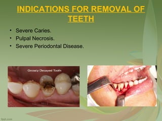
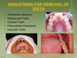
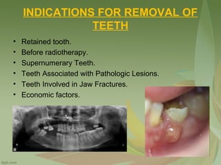
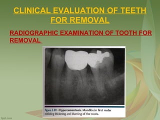
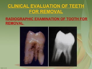
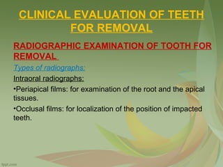
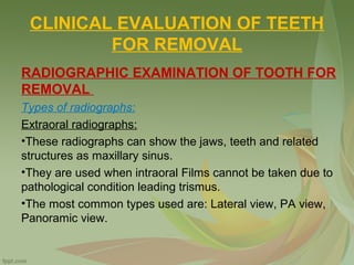
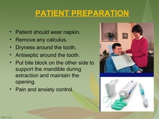
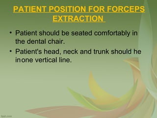
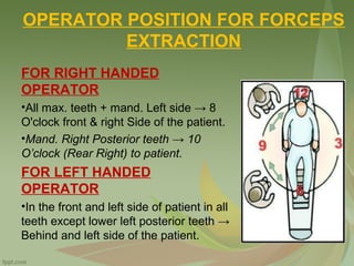
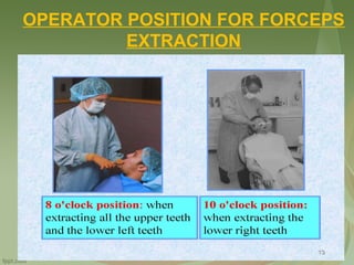
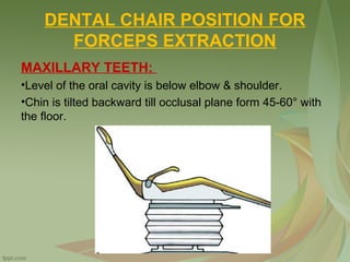
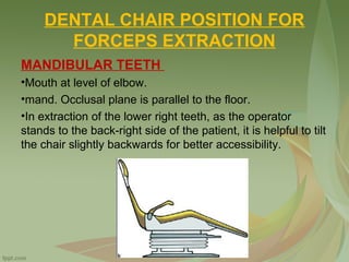
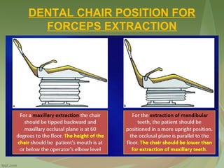
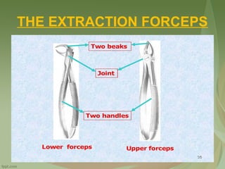
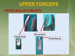
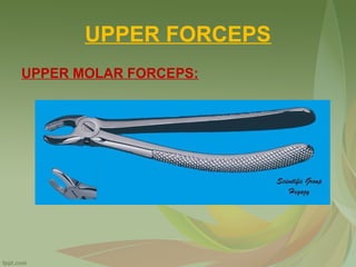
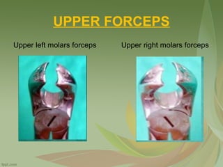
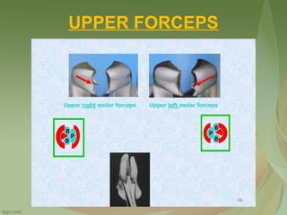
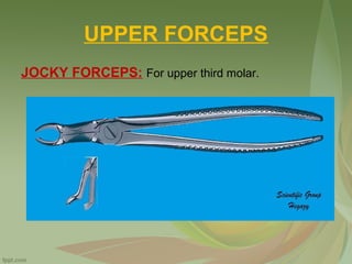
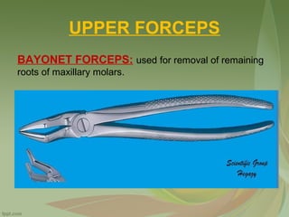
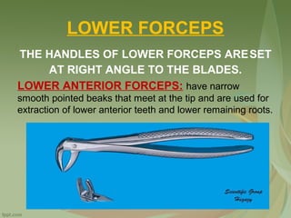
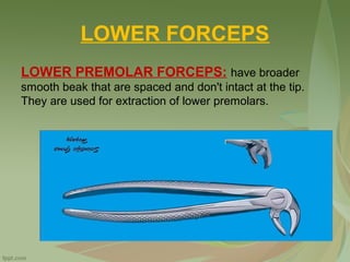
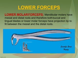
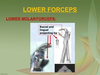
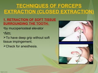
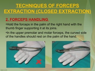
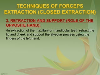
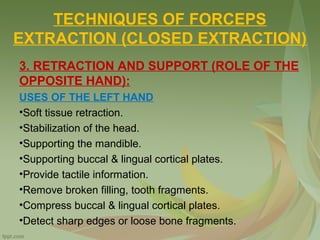
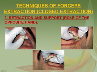
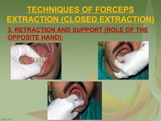
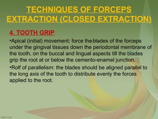
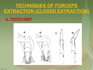
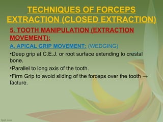
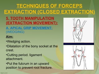
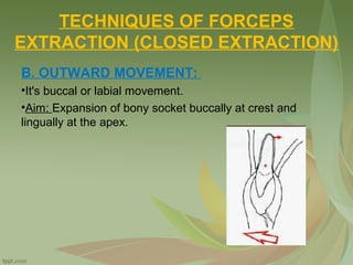
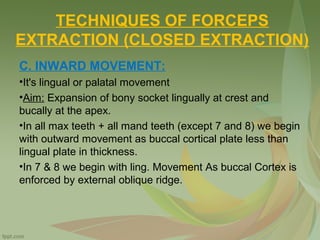
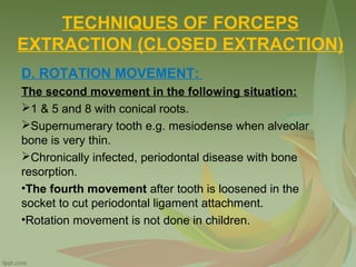
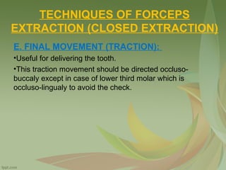
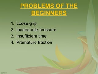
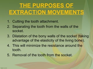
EXODONTIA
Dr. Saleh Bakry
Assistant Professor of Oral and Maxillofacial Surgery
INDICATIONS FOR REMOVAL OF
TEETH
• Severe Caries.
• Pulpal Necrosis.
• Severe Periodontal Disease.
INDICATIONS FOR REMOVAL OF
TEETH
• Orthodontic Reasons.
• Malopposed Teeth.
• Cracked Teeth.
• Preprosthetic Extractions.
• Impacted Teeth.
INDICATIONS FOR REMOVAL OF
TEETH
• Retained tooth.
• Before radiotherapy.
• Supernumerary Teeth.
• Teeth Associated with Pathologic Lesions.
• Teeth Involved in Jaw Fractures.
• Economic factors.
CONTRAINDICATIONS FOR THE
REMOVAL OF TEETH
1. Systemic Contraindications
•Severe uncontrolled metabolic diseases and cardiac
diseases.
•Severe uncontrolled hypertension.
•Severe bleeding disorder.
•Drugs as corticosteroids, immunosuppressive, and cancer
chemotherapeutic agents.
•Pregnancy during the first and third trimester.
•Patient taking Bisphosphonate drug because the drug affect
osteoblast function and bone metabolism so these patients
consider as the absolute contraindication for extraction.
CONTRAINDICATIONS FOR THE
REMOVAL OF TEETH
2. Local Contraindications
•Extractions performed in an area of radiation may result in
osteoradionecrosis.
•Teeth that is located within an area of tumor, especially a
malignant tumor.
•Severe pericoronitis around an impacted mandibular third
molar.
•Teeth with acute dentoalveolar abscess.
CONTRAINDICATIONS FOR THE
REMOVAL OF TEETH
2. Local Contraindications
CLINICAL EVALUATION OF TEETH
FOR REMOVAL
Examined the following:
•The mouth opening (Incisal opening).
•The upper and the lower teeth on the side of complain.
•Soft tissues inflammation and swelling.
•Access to Tooth.
•Mobility of Tooth.
CLINICAL EVALUATION OF TEETH
FOR REMOVAL
RADIOGRAPHIC EXAMINATION OF TOOTH FOR
REMOVAL
Determine the followings:
•Roots: number, curvature and root canal treatment.
•Bone: height, density, apical pathology.
•Vital structures: inferior dental canal and maxillary sinus.
CLINICAL EVALUATION OF TEETH
FOR REMOVAL
RADIOGRAPHIC EXAMINATION OF TOOTH FOR
REMOVAL
CLINICAL EVALUATION OF TEETH
FOR REMOVAL
RADIOGRAPHIC EXAMINATION OF TOOTH FOR
REMOVAL
CLINICAL EVALUATION OF TEETH
FOR REMOVAL
RADIOGRAPHIC EXAMINATION OF TOOTH FOR
REMOVAL
Types of radiographs:
Intraoral radiographs:
•Periapical films: for examination of the root and the apical
tissues.
•Occlusal films: for localization of the position of impacted
teeth.
CLINICAL EVALUATION OF TEETH
FOR REMOVAL
RADIOGRAPHIC EXAMINATION OF TOOTH FOR
REMOVAL
Types of radiographs:
Extraoral radiographs:
•These radiographs can show the jaws, teeth and related
structures as maxillary sinus.
•They are used when intraoral Films cannot be taken due to
pathological condition leading trismus.
•The most common types used are: Lateral view, PA view,
Panoramic view.
ANATOMICAL FACTORS
INFLUENCING FORCEPS MOVEMENT
ROOT PATTERN OF UPPER TOOTH
1 → Straight conical root & circular cross section.
2 → Straight root which tapers to a curved apex with oval
cross section
3 → Long root, triangular in cross section and may have
apical curvature
4 → It has 2 tapered BP roots (easily fracture) Õ Two thin
and divergent root and conical neck.
5 → Has one root
ANATOMICAL FACTORS
INFLUENCING FORCEPS MOVEMENT
ROOT PATTERN OF UPPER TOOTH
6 → Have 3 divergent roots, 1 palatal and 2 thin buccal
roots. It is firmly embedded in the alveolar bone Õ difficult
in extraction due to the presence of the base of the malar
bone on its roots.
7 → Have 3 divergent roots, 1 palatal and 2 thin buccal
roots. It is firmly embedded in the alveolar bone
8 → The roots varied in shape and number, but frequently
have conical roots.
ANATOMICAL FACTORS
INFLUENCING FORCEPS MOVEMENT
ROOT PATTERN OF LOWER TOOTH
1, 2, 3 → Their roots are long and very thin.
4 → Straight single root
5 → It has conical root, rounded cross section.
6 → Mesial and distal roots.
7 → Mesial and distal roots.
8 → Root pattern is variable
COMPARATIVE THICKNESS OF
ALVEOLAR BONE ON THE BUCCAL
AND LINGUAL (PALATAL) SIDES:
MAXILLARY ALVEOLAR BONE:
•Buccal side is thin with little trabeculation, while palatal
side is thicker with more trabeculation.
•When using B-L movement, stress should be placed on
buccal movement since buccal bone will be easier to
expand.
•Maxillary teeth are delivered to the buccal side.
COMPARATIVE THICKNESS OF
ALVEOLAR BONE ON THE BUCCAL
AND LINGUAL (PALATAL) SIDES:
MANDIBULAR ALVEOLAR BONE:
•Anterior buccal bone is usually thinner thanlingual bone,
stress should he placed on buccal movement.
•In mandibular second and third molars, the buccal bone is
thicker (due to presence of external oblique ridge) than
lingual bone, stress is should be placed on lingual
movement.
PROCEDURE FOR CLOSED
EXTRACTION (GENERAL PRINCIPLES)
1. Adequate access and visualization.
2. Select the correct forceps.
3. Correct grasp of the forceps.
4. Firm apical grip, parallel to the long axis.
5. Steady controlled force for sufficient time.
6. Avoid injury to adjacent structures.
“5” GENERAL STEPS OF FORCEPS
EXTRACTION
1- Loosening of soft tissue attachment
•Assure profound anesthesia.
•More apical grip.
2- Luxation with an Elevator
•Straight elevator.
3- Adaptation of the forceps to the tooth (grip)
4- Luxation of the Tooth
•Start with the side of least resistance.
•Slow, steady, gradual force in B-L direction.
•Allow bone to expand.
5- Removal of the Tooth from the Socket
•Slight traction force (Buccally)
TECHNIQUE OF EXTRACTION
OPERATOR PREPARATION
• Consider that all the patients having
blood-borne diseases.
• Wear surgical gloves, surgical mask,
and eyewear with side shields.
• Wear long-sleeved gowns.
• If the surgeon has long hair, it is
essential that the hair be held in
position with barrettes or other holding
devices or be covered with a surgical
cap.
OPERATOR PREPARATION
PATIENT PREPARATION
• Patient should wear napkin.
• Remove any calculus.
• Dryness around the tooth.
• Antiseptic around the tooth.
• Put bite block on the other side to
support the mandible during
extraction and maintain the
opening.
• Pain and anxiety control.
PATIENT POSITION FOR FORCEPS
EXTRACTION
• Patient should be seated comfortably in
the dental chair.
• Patient's head, neck and trunk should he
inone vertical line.
OPERATOR POSITION FOR FORCEPS
EXTRACTION
FOR RIGHT HANDED
OPERATOR
•All max. teeth + mand. Left side → 8
O'clock front & right Side of the patient.
•Mand. Right Posterior teeth → 10
O’clock (Rear Right) to patient.
FOR LEFT HANDED
OPERATOR
•In the front and left side of patient in all
teeth except lower left posterior teeth →
Behind and left side of the patient.
OPERATOR POSITION FOR FORCEPS
EXTRACTION
DENTAL CHAIR POSITION FOR
FORCEPS EXTRACTION
MAXILLARY TEETH:
•Level of the oral cavity is below elbow & shoulder.
•Chin is tilted backward till occlusal plane form 45-60° with
the floor.
DENTAL CHAIR POSITION FOR
FORCEPS EXTRACTION
MANDIBULAR TEETH
•Mouth at level of elbow.
•mand. Occlusal plane is parallel to the floor.
•In extraction of the lower right teeth, as the operator
stands to the back-right side of the patient, it is helpful to tilt
the chair slightly backwards for better accessibility.
DENTAL CHAIR POSITION FOR
FORCEPS EXTRACTION
THE EXTRACTION FORCEPS
Consists of:
•Two handles.
•One joint.
•Two blades.
THE EXTRACTION FORCEPS
REQUIREMENTS OF THE DENTAL FORCEPS:
•They must be made of light and strong metal to be easily
used and resist breakage.
•Must be made of non-corrosive metal to be sterilized
without rusting.
•Constructed of stainless steel or cobalt chromium alloy.
THE EXTRACTION FORCEPS
REQUIREMENTS OF THE DENTAL FORCEPS:
THE HANDLES:
•Must be serrated to avoid slippage.
•Must be of suitable size to rest comfortably in the operator
hand.
THE EXTRACTION FORCEPS
REQUIREMENTS OF THE DENTAL FORCEPS:
THE BLADES:
•Must be sharp enough (not knife sharp) to he introduced
under the free gingival margin without causing contusion of
the soft tissues.
•Must be of proper angulations with a space to accommodate,
the bulbous crown.
THE EXTRACTION FORCEPS
REQUIREMENTS OF THE DENTAL FORCEPS:
THE JOINTS:
•Must be heavy and strong.
•Have free movement for easy manipulation.
UPPER FORCEPS
THEIR HANDLES AND BLADES ARE PRESENT
IN THE SAME LINE.
UPPER ANTERIOR FORCEPS: For anterior
maxillary teeth.
UPPER FORCEPS
UPPER PREMOLAR FORCEPS: their handles are
angled in an opposite direction relative to the blades.
UPPER FORCEPS
UPPER MOLAR FORCEPS:
•Maxillary first and second molars have two buccal roots
and a single palatal root.
•The buccal blade is so designed that its projecting tip fits
between the buccal roots, while the palatal blade is smooth
with no projecting tip.
•The handles are also curved as upper premolar forceps, to
offer correct alignment of the blades on the tooth. Have
right and left.
UPPER FORCEPS
UPPER MOLAR FORCEPS:
UPPER FORCEPS
UPPER MOLAR FORCEPS:
UPPER FORCEPS
Upper left molars forceps Upper right molars forceps
UPPER FORCEPS
UPPER FORCEPS
JOCKY FORCEPS: For upper third molar.
UPPER FORCEPS
BAYONET FORCEPS: used for removal of remaining
roots of maxillary molars.
LOWER FORCEPS
THE HANDLES OF LOWER FORCEPS ARESET
AT RIGHT ANGLE TO THE BLADES.
LOWER ANTERIOR FORCEPS: have narrow
smooth pointed beaks that meet at the tip and are used for
extraction of lower anterior teeth and lower remaining roots.
LOWER FORCEPS
LOWER PREMOLAR FORCEPS: have broader
smooth beak that are spaced and don't intact at the tip.
They are used for extraction of lower premolars.
LOWER FORCEPS
LOWER MOLARFORCEPS: Mandibular molars have
mesial and distal roots and therefore bothbuccal and
lingual blades or lower molar forceps have projection tip to
fit between the mesial and the distal roots.
LOWER FORCEPS
LOWER MOLARFORCEPS:
TECHNIQUES OF FORCEPS
EXTRACTION (CLOSED EXTRACTION)
1. RETRACTION OF SOFT TISSUE
SURROUNDING THE TOOTH:
•by mucoperiosteal elevator
•Aim:
To have deep grip without soft
tissue impingement.
Check for anesthesia.
TECHNIQUES OF FORCEPS
EXTRACTION (CLOSED EXTRACTION)
2. FORCEPS HANDLING
•Hold the forceps in the palm of the right hand with the
thumb finger supporting it at its joins.
•In the upper premolar and molar forceps, the curved side
of the handles should rest on the palm of the hand.
2. FORCEPS HANDLING
2. FORCEPS HANDLING
TECHNIQUES OF FORCEPS
EXTRACTION (CLOSED EXTRACTION)
3. RETRACTION AND SUPPORT (ROLE OF THE
OPPOSITE HAND):
•In extraction of the maxillary or mandibular teeth retract the
lip and cheek and support the alveolar process using the
fingers of the left hand.
TECHNIQUES OF FORCEPS
EXTRACTION (CLOSED EXTRACTION)
3. RETRACTION AND SUPPORT (ROLE OF THE
OPPOSITE HAND):
USES OF THE LEFT HAND
•Soft tissue retraction.
•Stabilization of the head.
•Supporting the mandible.
•Supporting buccal & lingual cortical plates.
•Provide tactile information.
•Remove broken filling, tooth fragments.
•Compress buccal & lingual cortical plates.
•Detect sharp edges or loose bone fragments.
TECHNIQUES OF FORCEPS
EXTRACTION (CLOSED EXTRACTION)
3. RETRACTION AND SUPPORT (ROLE OF THE
OPPOSITE HAND):
TECHNIQUES OF FORCEPS
EXTRACTION (CLOSED EXTRACTION)
3. RETRACTION AND SUPPORT (ROLE OF THE
OPPOSITE HAND):
TECHNIQUES OF FORCEPS
EXTRACTION (CLOSED EXTRACTION)
4. TOOTH GRIP
•Apical (initial) movement: force theblades of the forceps
under the gingival tissues down the periodontal membrane of
the tooth, on the buccal and lingual aspects till the blades
grip the root at or below the cemento-enamel junction.
•Rolf of parallelism: the blades should be aligned parallel to
the long axis of the tooth to distribute evenly the forces
applied to the root.
TECHNIQUES OF FORCEPS
EXTRACTION (CLOSED EXTRACTION)
4. TOOTH GRIP
TECHNIQUES OF FORCEPS
EXTRACTION (CLOSED EXTRACTION)
5. TOOTH MANIPULATION (EXTRACTION
MOVEMENT):
A. APICAL GRIP MOVEMENT: (WEDGING)
•Deep grip at C.E.J. or root surface extending to crestal
bone.
•Parallel to long axis of the tooth.
•Firm Grip to avoid sliding of the forceps over the tooth →
facture.
TECHNIQUES OF FORCEPS
EXTRACTION (CLOSED EXTRACTION)
5. TOOTH MANIPULATION
(EXTRACTION MOVEMENT):
A. APICAL GRIP MOVEMENT:
(WEDGING)
Aim:
•Wedging action.
•Dilatation of the bony socket at the
crest.
•Cutting period. ligament
attachment.
•Put the fulcrum in an upward
position to prevent root fracture.
TECHNIQUES OF FORCEPS
EXTRACTION (CLOSED EXTRACTION)
B. OUTWARD MOVEMENT:
•It's buccal or labial movement.
•Aim: Expansion of bony socket buccally at crest and
lingually at the apex.
TECHNIQUES OF FORCEPS
EXTRACTION (CLOSED EXTRACTION)
C. INWARD MOVEMENT:
•It's lingual or palatal movement
•Aim: Expansion of bony socket lingually at crest and
bucally at the apex.
•In all max teeth + all mand teeth (except 7 and 8) we begin
with outward movement as buccal cortical plate less than
lingual plate in thickness.
•In 7 & 8 we begin with ling. Movement As buccal Cortex is
enforced by external oblique ridge.
TECHNIQUES OF FORCEPS
EXTRACTION (CLOSED EXTRACTION)
D. ROTATION MOVEMENT:
The second movement in the following situation:
1 & 5 and 8 with conical roots.
Supernumerary tooth e.g. mesiodense when alveolar
bone is very thin.
Chronically infected, periodontal disease with bone
resorption.
•The fourth movement after tooth is loosened in the
socket to cut periodontal ligament attachment.
•Rotation movement is not done in children.
TECHNIQUES OF FORCEPS
EXTRACTION (CLOSED EXTRACTION)
E. FINAL MOVEMENT (TRACTION):
•Useful for delivering the tooth.
•This traction movement should be directed occluso-
buccaly except in case of lower third molar which is
occluso-lingualy to avoid the check.
PROBLEMS OF THE
BEGINNERS
1. Loose grip
2. Inadequate pressure
3. Insufficient time
4. Premature traction
ROLE OF THE ASSISTANT
1. Soft tissue reflection.
2. Suction.
3. Protection.
4. Support the mandible.
5. Psychological support for the patient.
THE PURPOSES OF
EXTRACTION MOVEMENTS
1. Cutting the tooth attachment.
2. Separating the tooth from the walls of the
socket.
3. Dilatation of the bony walls of the socket (taking
advantage of the elasticity of the living bone).
4. This will minimize the resistance around the
tooth.
5. Removal of the tooth from the socket.
FACTORS COMPLICATING
TEETH EXTRACTION
1. Restricted area (by lips and cheek).
2. Tongue movements.
3. mandibular movements (TMJ).
4. Tooth may be aspirated through the pharynx or larynx →
bronchi → lung leading to suffocation and lung abscess.
5. Proximity to vital structures e.g. Max. sinus, inferior
alveolar nerve.
6. Oral cavity is filled with saliva that contains M.0 that may
cause infection.
EXTRACTION OF DECIDUOUS
TEETH
FACTORS COMPLICATING EXTRACTION:
1.Limited access.
2.Root resorption → easy to fracture.
3.Resorbed root → irregular surface.
4.Fractured root → sharp shiny margin.
5.Tooth buds of premolars may be engaged by the roots of
deciduous molars.
EXTRACTION OF DECIDUOUS
TEETH
• We use the same technique but by pediatric forceps.
• Extraction technique: Use pediatric forceps.
• Avoid rotation movement → Fracture root & Injury to
successors.
• Avoid curettage injury to successors.
SEQUENCE OF EXTRACTION
• Chief complaint.
• Posterior before anterior teeth for better vision.
• Maxillary teeth before mandibular teeth:
 To Avoid fragment coming from maxillary teeth to
mandibular teeth.
 Because infiltration anesthesia has shorter duration than
nerve block.
 Maxillary teeth are easier in extraction.
• 6 and 3 are the last teeth to be extracted to have the
advantages of proper plate expansion of the adjacent
teeth.
POST EXTRACTION CARE
IMMEDIATE POST-EXTRACTION CARE:
•Examination of the socket for any tooth fragment, broken
filling or pieces of calculi.
•Squeeze the socket to decrease the socket orifice to:
•Allow proper organization of the blood clot in the socket to
prevent postoperative pain.
•Prevent saliva to enter the socket.
•Promotes rapid healing.
•To prevent bony undercuts.
•Clean the patient's lips and face from any blood spots.
•Dismiss the patients after examining the socket for
abnormal.
POST EXTRACTION CARE
POSTOPERATIVE INSTRUCTIONS: ON A
WRITTEN SLIP.
•Advise the patient to keep biting, pressure on the gauze for
about one hour.
•No mouth washes for at least 12 hours after extraction.
•Avoid any hot food or drinks for the rest of the day to prevent
bleeding.
•Avoid smoking.
•Do chewing away from the extraction site.
POST EXTRACTION CARE
POSTOPERATIVE INSTRUCTIONS: ON A
WRITTEN SLIP.
•Avoid brushing the teeth near the extraction site for one day.
•Use warm salt water as a mouth wash, it is started 24 hours
after the extraction.
•Avoid milk and milk product in the day of extraction as the
milk is consider a media for bacterial proliferation.
•Analgesics, for pain relief.
•Antibiotics describe only in case of preoperative infection.
THANK YOU

Exodontia

  • 1.
    EXODONTIA Dr. Saleh Bakry AssistantProfessor of Oral and Maxillofacial Surgery
  • 2.
    INDICATIONS FOR REMOVALOF TEETH • Severe Caries. • Pulpal Necrosis. • Severe Periodontal Disease.
  • 3.
    INDICATIONS FOR REMOVALOF TEETH • Orthodontic Reasons. • Malopposed Teeth. • Cracked Teeth. • Preprosthetic Extractions. • Impacted Teeth.
  • 4.
    INDICATIONS FOR REMOVALOF TEETH • Retained tooth. • Before radiotherapy. • Supernumerary Teeth. • Teeth Associated with Pathologic Lesions. • Teeth Involved in Jaw Fractures. • Economic factors.
  • 5.
    CONTRAINDICATIONS FOR THE REMOVALOF TEETH 1. Systemic Contraindications •Severe uncontrolled metabolic diseases and cardiac diseases. •Severe uncontrolled hypertension. •Severe bleeding disorder. •Drugs as corticosteroids, immunosuppressive, and cancer chemotherapeutic agents. •Pregnancy during the first and third trimester. •Patient taking Bisphosphonate drug because the drug affect osteoblast function and bone metabolism so these patients consider as the absolute contraindication for extraction.
  • 6.
    CONTRAINDICATIONS FOR THE REMOVALOF TEETH 2. Local Contraindications •Extractions performed in an area of radiation may result in osteoradionecrosis. •Teeth that is located within an area of tumor, especially a malignant tumor. •Severe pericoronitis around an impacted mandibular third molar. •Teeth with acute dentoalveolar abscess.
  • 7.
    CONTRAINDICATIONS FOR THE REMOVALOF TEETH 2. Local Contraindications
  • 8.
    CLINICAL EVALUATION OFTEETH FOR REMOVAL Examined the following: •The mouth opening (Incisal opening). •The upper and the lower teeth on the side of complain. •Soft tissues inflammation and swelling. •Access to Tooth. •Mobility of Tooth.
  • 9.
    CLINICAL EVALUATION OFTEETH FOR REMOVAL RADIOGRAPHIC EXAMINATION OF TOOTH FOR REMOVAL Determine the followings: •Roots: number, curvature and root canal treatment. •Bone: height, density, apical pathology. •Vital structures: inferior dental canal and maxillary sinus.
  • 10.
    CLINICAL EVALUATION OFTEETH FOR REMOVAL RADIOGRAPHIC EXAMINATION OF TOOTH FOR REMOVAL
  • 11.
    CLINICAL EVALUATION OFTEETH FOR REMOVAL RADIOGRAPHIC EXAMINATION OF TOOTH FOR REMOVAL
  • 12.
    CLINICAL EVALUATION OFTEETH FOR REMOVAL RADIOGRAPHIC EXAMINATION OF TOOTH FOR REMOVAL Types of radiographs: Intraoral radiographs: •Periapical films: for examination of the root and the apical tissues. •Occlusal films: for localization of the position of impacted teeth.
  • 13.
    CLINICAL EVALUATION OFTEETH FOR REMOVAL RADIOGRAPHIC EXAMINATION OF TOOTH FOR REMOVAL Types of radiographs: Extraoral radiographs: •These radiographs can show the jaws, teeth and related structures as maxillary sinus. •They are used when intraoral Films cannot be taken due to pathological condition leading trismus. •The most common types used are: Lateral view, PA view, Panoramic view.
  • 14.
    ANATOMICAL FACTORS INFLUENCING FORCEPSMOVEMENT ROOT PATTERN OF UPPER TOOTH 1 → Straight conical root & circular cross section. 2 → Straight root which tapers to a curved apex with oval cross section 3 → Long root, triangular in cross section and may have apical curvature 4 → It has 2 tapered BP roots (easily fracture) Õ Two thin and divergent root and conical neck. 5 → Has one root
  • 15.
    ANATOMICAL FACTORS INFLUENCING FORCEPSMOVEMENT ROOT PATTERN OF UPPER TOOTH 6 → Have 3 divergent roots, 1 palatal and 2 thin buccal roots. It is firmly embedded in the alveolar bone Õ difficult in extraction due to the presence of the base of the malar bone on its roots. 7 → Have 3 divergent roots, 1 palatal and 2 thin buccal roots. It is firmly embedded in the alveolar bone 8 → The roots varied in shape and number, but frequently have conical roots.
  • 16.
    ANATOMICAL FACTORS INFLUENCING FORCEPSMOVEMENT ROOT PATTERN OF LOWER TOOTH 1, 2, 3 → Their roots are long and very thin. 4 → Straight single root 5 → It has conical root, rounded cross section. 6 → Mesial and distal roots. 7 → Mesial and distal roots. 8 → Root pattern is variable
  • 17.
    COMPARATIVE THICKNESS OF ALVEOLARBONE ON THE BUCCAL AND LINGUAL (PALATAL) SIDES: MAXILLARY ALVEOLAR BONE: •Buccal side is thin with little trabeculation, while palatal side is thicker with more trabeculation. •When using B-L movement, stress should be placed on buccal movement since buccal bone will be easier to expand. •Maxillary teeth are delivered to the buccal side.
  • 18.
    COMPARATIVE THICKNESS OF ALVEOLARBONE ON THE BUCCAL AND LINGUAL (PALATAL) SIDES: MANDIBULAR ALVEOLAR BONE: •Anterior buccal bone is usually thinner thanlingual bone, stress should he placed on buccal movement. •In mandibular second and third molars, the buccal bone is thicker (due to presence of external oblique ridge) than lingual bone, stress is should be placed on lingual movement.
  • 19.
    PROCEDURE FOR CLOSED EXTRACTION(GENERAL PRINCIPLES) 1. Adequate access and visualization. 2. Select the correct forceps. 3. Correct grasp of the forceps. 4. Firm apical grip, parallel to the long axis. 5. Steady controlled force for sufficient time. 6. Avoid injury to adjacent structures.
  • 20.
    “5” GENERAL STEPSOF FORCEPS EXTRACTION 1- Loosening of soft tissue attachment •Assure profound anesthesia. •More apical grip. 2- Luxation with an Elevator •Straight elevator. 3- Adaptation of the forceps to the tooth (grip) 4- Luxation of the Tooth •Start with the side of least resistance. •Slow, steady, gradual force in B-L direction. •Allow bone to expand. 5- Removal of the Tooth from the Socket •Slight traction force (Buccally)
  • 21.
  • 22.
    OPERATOR PREPARATION • Considerthat all the patients having blood-borne diseases. • Wear surgical gloves, surgical mask, and eyewear with side shields. • Wear long-sleeved gowns. • If the surgeon has long hair, it is essential that the hair be held in position with barrettes or other holding devices or be covered with a surgical cap.
  • 23.
  • 24.
  • 25.
    PATIENT PREPARATION • Patientshould wear napkin. • Remove any calculus. • Dryness around the tooth. • Antiseptic around the tooth. • Put bite block on the other side to support the mandible during extraction and maintain the opening. • Pain and anxiety control.
  • 26.
    PATIENT POSITION FORFORCEPS EXTRACTION • Patient should be seated comfortably in the dental chair. • Patient's head, neck and trunk should he inone vertical line.
  • 27.
    OPERATOR POSITION FORFORCEPS EXTRACTION FOR RIGHT HANDED OPERATOR •All max. teeth + mand. Left side → 8 O'clock front & right Side of the patient. •Mand. Right Posterior teeth → 10 O’clock (Rear Right) to patient. FOR LEFT HANDED OPERATOR •In the front and left side of patient in all teeth except lower left posterior teeth → Behind and left side of the patient.
  • 28.
    OPERATOR POSITION FORFORCEPS EXTRACTION
  • 29.
    DENTAL CHAIR POSITIONFOR FORCEPS EXTRACTION MAXILLARY TEETH: •Level of the oral cavity is below elbow & shoulder. •Chin is tilted backward till occlusal plane form 45-60° with the floor.
  • 30.
    DENTAL CHAIR POSITIONFOR FORCEPS EXTRACTION MANDIBULAR TEETH •Mouth at level of elbow. •mand. Occlusal plane is parallel to the floor. •In extraction of the lower right teeth, as the operator stands to the back-right side of the patient, it is helpful to tilt the chair slightly backwards for better accessibility.
  • 31.
    DENTAL CHAIR POSITIONFOR FORCEPS EXTRACTION
  • 32.
  • 33.
    THE EXTRACTION FORCEPS Consistsof: •Two handles. •One joint. •Two blades.
  • 34.
    THE EXTRACTION FORCEPS REQUIREMENTSOF THE DENTAL FORCEPS: •They must be made of light and strong metal to be easily used and resist breakage. •Must be made of non-corrosive metal to be sterilized without rusting. •Constructed of stainless steel or cobalt chromium alloy.
  • 35.
    THE EXTRACTION FORCEPS REQUIREMENTSOF THE DENTAL FORCEPS: THE HANDLES: •Must be serrated to avoid slippage. •Must be of suitable size to rest comfortably in the operator hand.
  • 36.
    THE EXTRACTION FORCEPS REQUIREMENTSOF THE DENTAL FORCEPS: THE BLADES: •Must be sharp enough (not knife sharp) to he introduced under the free gingival margin without causing contusion of the soft tissues. •Must be of proper angulations with a space to accommodate, the bulbous crown.
  • 37.
    THE EXTRACTION FORCEPS REQUIREMENTSOF THE DENTAL FORCEPS: THE JOINTS: •Must be heavy and strong. •Have free movement for easy manipulation.
  • 38.
    UPPER FORCEPS THEIR HANDLESAND BLADES ARE PRESENT IN THE SAME LINE. UPPER ANTERIOR FORCEPS: For anterior maxillary teeth.
  • 39.
    UPPER FORCEPS UPPER PREMOLARFORCEPS: their handles are angled in an opposite direction relative to the blades.
  • 40.
    UPPER FORCEPS UPPER MOLARFORCEPS: •Maxillary first and second molars have two buccal roots and a single palatal root. •The buccal blade is so designed that its projecting tip fits between the buccal roots, while the palatal blade is smooth with no projecting tip. •The handles are also curved as upper premolar forceps, to offer correct alignment of the blades on the tooth. Have right and left.
  • 41.
  • 42.
  • 43.
    UPPER FORCEPS Upper leftmolars forceps Upper right molars forceps
  • 44.
  • 45.
    UPPER FORCEPS JOCKY FORCEPS:For upper third molar.
  • 46.
    UPPER FORCEPS BAYONET FORCEPS:used for removal of remaining roots of maxillary molars.
  • 47.
    LOWER FORCEPS THE HANDLESOF LOWER FORCEPS ARESET AT RIGHT ANGLE TO THE BLADES. LOWER ANTERIOR FORCEPS: have narrow smooth pointed beaks that meet at the tip and are used for extraction of lower anterior teeth and lower remaining roots.
  • 48.
    LOWER FORCEPS LOWER PREMOLARFORCEPS: have broader smooth beak that are spaced and don't intact at the tip. They are used for extraction of lower premolars.
  • 49.
    LOWER FORCEPS LOWER MOLARFORCEPS:Mandibular molars have mesial and distal roots and therefore bothbuccal and lingual blades or lower molar forceps have projection tip to fit between the mesial and the distal roots.
  • 50.
  • 51.
    TECHNIQUES OF FORCEPS EXTRACTION(CLOSED EXTRACTION) 1. RETRACTION OF SOFT TISSUE SURROUNDING THE TOOTH: •by mucoperiosteal elevator •Aim: To have deep grip without soft tissue impingement. Check for anesthesia.
  • 52.
    TECHNIQUES OF FORCEPS EXTRACTION(CLOSED EXTRACTION) 2. FORCEPS HANDLING •Hold the forceps in the palm of the right hand with the thumb finger supporting it at its joins. •In the upper premolar and molar forceps, the curved side of the handles should rest on the palm of the hand.
  • 53.
  • 54.
  • 55.
    TECHNIQUES OF FORCEPS EXTRACTION(CLOSED EXTRACTION) 3. RETRACTION AND SUPPORT (ROLE OF THE OPPOSITE HAND): •In extraction of the maxillary or mandibular teeth retract the lip and cheek and support the alveolar process using the fingers of the left hand.
  • 56.
    TECHNIQUES OF FORCEPS EXTRACTION(CLOSED EXTRACTION) 3. RETRACTION AND SUPPORT (ROLE OF THE OPPOSITE HAND): USES OF THE LEFT HAND •Soft tissue retraction. •Stabilization of the head. •Supporting the mandible. •Supporting buccal & lingual cortical plates. •Provide tactile information. •Remove broken filling, tooth fragments. •Compress buccal & lingual cortical plates. •Detect sharp edges or loose bone fragments.
  • 57.
    TECHNIQUES OF FORCEPS EXTRACTION(CLOSED EXTRACTION) 3. RETRACTION AND SUPPORT (ROLE OF THE OPPOSITE HAND):
  • 58.
    TECHNIQUES OF FORCEPS EXTRACTION(CLOSED EXTRACTION) 3. RETRACTION AND SUPPORT (ROLE OF THE OPPOSITE HAND):
  • 59.
    TECHNIQUES OF FORCEPS EXTRACTION(CLOSED EXTRACTION) 4. TOOTH GRIP •Apical (initial) movement: force theblades of the forceps under the gingival tissues down the periodontal membrane of the tooth, on the buccal and lingual aspects till the blades grip the root at or below the cemento-enamel junction. •Rolf of parallelism: the blades should be aligned parallel to the long axis of the tooth to distribute evenly the forces applied to the root.
  • 60.
    TECHNIQUES OF FORCEPS EXTRACTION(CLOSED EXTRACTION) 4. TOOTH GRIP
  • 61.
    TECHNIQUES OF FORCEPS EXTRACTION(CLOSED EXTRACTION) 5. TOOTH MANIPULATION (EXTRACTION MOVEMENT): A. APICAL GRIP MOVEMENT: (WEDGING) •Deep grip at C.E.J. or root surface extending to crestal bone. •Parallel to long axis of the tooth. •Firm Grip to avoid sliding of the forceps over the tooth → facture.
  • 62.
    TECHNIQUES OF FORCEPS EXTRACTION(CLOSED EXTRACTION) 5. TOOTH MANIPULATION (EXTRACTION MOVEMENT): A. APICAL GRIP MOVEMENT: (WEDGING) Aim: •Wedging action. •Dilatation of the bony socket at the crest. •Cutting period. ligament attachment. •Put the fulcrum in an upward position to prevent root fracture.
  • 63.
    TECHNIQUES OF FORCEPS EXTRACTION(CLOSED EXTRACTION) B. OUTWARD MOVEMENT: •It's buccal or labial movement. •Aim: Expansion of bony socket buccally at crest and lingually at the apex.
  • 64.
    TECHNIQUES OF FORCEPS EXTRACTION(CLOSED EXTRACTION) C. INWARD MOVEMENT: •It's lingual or palatal movement •Aim: Expansion of bony socket lingually at crest and bucally at the apex. •In all max teeth + all mand teeth (except 7 and 8) we begin with outward movement as buccal cortical plate less than lingual plate in thickness. •In 7 & 8 we begin with ling. Movement As buccal Cortex is enforced by external oblique ridge.
  • 65.
    TECHNIQUES OF FORCEPS EXTRACTION(CLOSED EXTRACTION) D. ROTATION MOVEMENT: The second movement in the following situation: 1 & 5 and 8 with conical roots. Supernumerary tooth e.g. mesiodense when alveolar bone is very thin. Chronically infected, periodontal disease with bone resorption. •The fourth movement after tooth is loosened in the socket to cut periodontal ligament attachment. •Rotation movement is not done in children.
  • 66.
    TECHNIQUES OF FORCEPS EXTRACTION(CLOSED EXTRACTION) E. FINAL MOVEMENT (TRACTION): •Useful for delivering the tooth. •This traction movement should be directed occluso- buccaly except in case of lower third molar which is occluso-lingualy to avoid the check.
  • 67.
    PROBLEMS OF THE BEGINNERS 1.Loose grip 2. Inadequate pressure 3. Insufficient time 4. Premature traction
  • 68.
    ROLE OF THEASSISTANT 1. Soft tissue reflection. 2. Suction. 3. Protection. 4. Support the mandible. 5. Psychological support for the patient.
  • 69.
    THE PURPOSES OF EXTRACTIONMOVEMENTS 1. Cutting the tooth attachment. 2. Separating the tooth from the walls of the socket. 3. Dilatation of the bony walls of the socket (taking advantage of the elasticity of the living bone). 4. This will minimize the resistance around the tooth. 5. Removal of the tooth from the socket.
  • 70.
    FACTORS COMPLICATING TEETH EXTRACTION 1.Restricted area (by lips and cheek). 2. Tongue movements. 3. mandibular movements (TMJ). 4. Tooth may be aspirated through the pharynx or larynx → bronchi → lung leading to suffocation and lung abscess. 5. Proximity to vital structures e.g. Max. sinus, inferior alveolar nerve. 6. Oral cavity is filled with saliva that contains M.0 that may cause infection.
  • 71.
    EXTRACTION OF DECIDUOUS TEETH FACTORSCOMPLICATING EXTRACTION: 1.Limited access. 2.Root resorption → easy to fracture. 3.Resorbed root → irregular surface. 4.Fractured root → sharp shiny margin. 5.Tooth buds of premolars may be engaged by the roots of deciduous molars.
  • 72.
    EXTRACTION OF DECIDUOUS TEETH •We use the same technique but by pediatric forceps. • Extraction technique: Use pediatric forceps. • Avoid rotation movement → Fracture root & Injury to successors. • Avoid curettage injury to successors.
  • 73.
    SEQUENCE OF EXTRACTION •Chief complaint. • Posterior before anterior teeth for better vision. • Maxillary teeth before mandibular teeth:  To Avoid fragment coming from maxillary teeth to mandibular teeth.  Because infiltration anesthesia has shorter duration than nerve block.  Maxillary teeth are easier in extraction. • 6 and 3 are the last teeth to be extracted to have the advantages of proper plate expansion of the adjacent teeth.
  • 74.
    POST EXTRACTION CARE IMMEDIATEPOST-EXTRACTION CARE: •Examination of the socket for any tooth fragment, broken filling or pieces of calculi. •Squeeze the socket to decrease the socket orifice to: •Allow proper organization of the blood clot in the socket to prevent postoperative pain. •Prevent saliva to enter the socket. •Promotes rapid healing. •To prevent bony undercuts. •Clean the patient's lips and face from any blood spots. •Dismiss the patients after examining the socket for abnormal.
  • 75.
    POST EXTRACTION CARE POSTOPERATIVEINSTRUCTIONS: ON A WRITTEN SLIP. •Advise the patient to keep biting, pressure on the gauze for about one hour. •No mouth washes for at least 12 hours after extraction. •Avoid any hot food or drinks for the rest of the day to prevent bleeding. •Avoid smoking. •Do chewing away from the extraction site.
  • 76.
    POST EXTRACTION CARE POSTOPERATIVEINSTRUCTIONS: ON A WRITTEN SLIP. •Avoid brushing the teeth near the extraction site for one day. •Use warm salt water as a mouth wash, it is started 24 hours after the extraction. •Avoid milk and milk product in the day of extraction as the milk is consider a media for bacterial proliferation. •Analgesics, for pain relief. •Antibiotics describe only in case of preoperative infection.
  • 77.