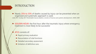
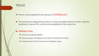
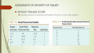
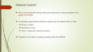
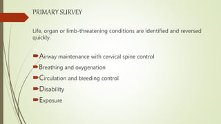
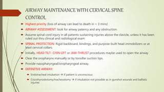
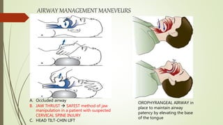
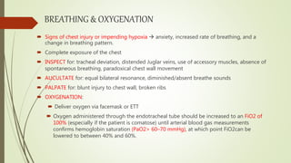
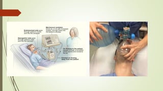
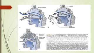
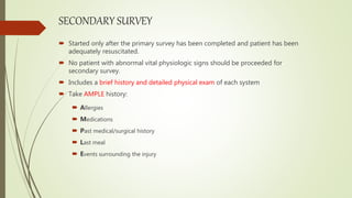
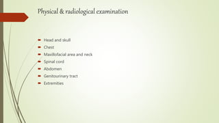
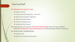
ADVANCE TRAUMA LIFE SUPPORT
(ATLS)
INITIAL MANAGEMENT OF A TRAUMA PATIENT
BY: DR.HASSAN HAFEEZ KHAN
FCPS RESIDENT OMFS
TABLE OF CONTENTS
 INTRODUCTION
 TRIAGE
 ASSESSMENT OF SEVERITY OF INJURY
 PRIMARY SURVEY
 SECONDARY SURVEY
 DEFINTIVE CARE
INTRODUCTION
 Nearly 25% to 33% of deaths caused by injury can be prevented when an
organized and systematic approach is used.
Cales RH, Trunkey DD. Preventable trauma deaths: a review of trauma care systems development. JAMA 1985
 GOLDEN HOUR: the first hour after the traumatic injury where emergency
treatment is most likely to be successful
 ATLS consists of:
 Rapid primary evaluation
 Resuscitation of vital functions
 Detailed secondary assessment
 Initiation of definitive care.
TRIAGE
 French word; applied to the process of SORTING OUT.
 The process of categorizing victims or mass casualties based on their need for
treatment, chances for survival and availability of the resources.
 PRIMARY GOAL:
 Prevent avoidable deaths
 Ensure proper emergency care within minimal time frame
 Avoid misuse of the resources on hopeless cases.
ASSESSMENT OF SEVERITY OF INJURY
 REVISED TRAUMA SCORE
 Quickly assess the severity and extent of injury to the vital systems.
PRIMARY SURVEY
 First, most important thing, when you encounter a trauma patient is to
speak to him/her.
 A complete appropriate sentence spoken by the patient tells us that:
 Airway is patent
 Breathing is intact
 There is adequate cerebral circulation
 If patient is not able to speak, proceed with the ABCDE
PRIMARY SURVEY
Life, organ or limb-threatening conditions are identified and reversed
quickly.
Airway maintenance with cervical spine control
Breathing and oxygenation
Circulation and bleeding control
Disability
Exposure
AIRWAY MAINTENANCE WITH CERVICAL SPINE
CONTROL
 Highest priority (loss of airway can lead to death in < 3 mins)
 AIRWAY ASSESSMENT: look for airway patency and any obstruction.
 Assume spinal cord injury in all patients sustaining injuries above the clavicle, unless it has been
ruled out thru clinical and radiological exam
 SPINAL PROTECTION: Rigid backboard, bindings, and purpose-built head immobilizers or at
least cervical collars.
 Initially, HEAD TILT- CHIN LIFT or JAW-THRUST procedures maybe used to open the airway
 Clear the oropharynx manually or by tonsillar suction tips.
 Provide nasopharyngeal/oropharyngeal airway.
 DEFINITIVE ARIWAY:
 Endotracheal intubation  if patient is unconscious
 Cricothyroidotomy/tracheostomy  if intubation not possible as in gunshot wounds and ballistic
injuries
AIRWAY MANAGEMENT MANEVEURS
A. Occluded airway
B. JAW THRUST  SAFEST method of jaw
manipulation in a patient with suspected
CERVICAL SPINE INJURY
C. HEAD TILT-CHIN LIFT
OROPHYRANGEAL AIRWAY in
place to maintain airway
patency by elevating the base
of the tongue
BREATHING & OXYGENATION
 Signs of chest injury or impending hypoxia  anxiety, increased rate of breathing, and a
change in breathing pattern.
 Complete exposure of the chest
 INSPECT for: tracheal deviation, distended Juglar veins, use of accessory muscles, absence of
spontaneous breathing, paradoxical chest wall movement
 AUCULTATE for: equal bilateral resonance, diminished/absent breathe sounds
 PALPATE for: blunt injury to chest wall, broken ribs
 OXYGENATION:
 Deliver oxygen via facemask or ETT
 Oxygen administered through the endotracheal tube should be increased to an FiO2 of
100% (especially if the patient is comatose) until arterial blood gas measurements
confirms hemoglobin saturation (PaO2> 60–70 mmHg), at which point FiO2can be
lowered to between 40% and 60%.
1. OPEN PNEUMOTHORAX
2. Tension pneumothorax
3.HEMOTHORAX
 Fluid collections > 200 to 300 mL can usually be seen on a good upright chest radiograph
as blunting of the costophrenic angle.
 Rx: transfusion of fluids or blood products, control of the airway and support of the
ventilation as required and drainage of the accumulated blood.
TUBE THORACOTOMY
A moderate-sized chest tube (32–40 Fr in adults or 26–30 Fr in children) is
generally placed in the fifth intercostal space midaxillary line.
4. FLAIL CHEST
 Occurs when three or more adjacent ribs are fractured in at least two locations,
resulting in a freely moving segment of the chest wall during respirations.
 The pulmonary contusion under-lying major chest wall injuries may be the primary
cause of hypoxia and morbidity in patients with flail chest.
 Three treatment considerations—pain management, supplemental oxygen delivery,
and ventilation control.
3. CIRCULATION AND BLEEDING CONTROL
 Most common cause of shock in the traumatized patient is
hypovolemia caused by hemorrhage.
 Clinical signs of shock:
 Altered level of consciousness
 Tachycardia (HR>100)  most common sign
 Arterial hypotension (SBP<120mmHg)
 Inadequate tissue perfusion
Pale, cool clammy skin
Delayed capillary refill (blanch test normal < 3 secs)
Decreased urine output (< 0.5 ml/kg/hr.)
HYPOVOLEMIC SHOCK
GENERAL MANAGEMENT PRINCIPLE
 A minimum of two large-bore (14–16 gauge) intravenous catheters should be
placed peripherally in the basilic or cephalic veins in the antecubital fossa of
both arms.
 Control bleeding by:
 Applying direct pressure
 Elevate the bleeding site
 Suturing the lacerations with 2.0 non-resorbable suture without esthetic
consideration.
 Immobilize the fracture
 Restore circulating volume
 Crystalloid resuscitation initially with lactated Ringer’s solution or 0.9% normal saline 2L
bolus over 10-15 mins (20ml/kg for children)
 Administer blood products
DISABILITY OR DEGREE OF CONCIOUSNESS
 GCS: Three variables included:
 Best motor response  reflects CNS function
 Best verbal response  shows CNS’s ability to integrate
information
 Eye opening  reflects brainstem activity
 AVPU scale:
 Alert
 Responds to Vocal stimuli
 Responds to Painful stimuli
 Unresponsive
EXPOSURE
 Completely disrobe the patient to avoid missing on any injury.
 Logroll the patient to examine patients back
 Avoid hypothermia
SECONDARY SURVEY
 Started only after the primary survey has been completed and patient has been
adequately resuscitated.
 No patient with abnormal vital physiologic signs should be proceeded for
secondary survey.
 Includes a brief history and detailed physical exam of each system
 Take AMPLE history:
 Allergies
 Medications
 Past medical/surgical history
 Last meal
 Events surrounding the injury
Physical & radiological examination
 Head and skull
 Chest
 Maxillofacial area and neck
 Spinal cord
 Abdomen
 Genitourinary tract
 Extremities
Head and skull
 Indications for brain CT scan:
 Seizure activity
 Unconsciousness lasting for > few mins
 Abnormal neurologic evaluation
 Abnormal mental status
 History of head trauma
 Suspected skull fracture
 Head injuries can result in intracranial hemorrhage that can cause cerebral
ischemia by elevating intracranial pressure and compressing vascular structures.
 NEUROLOGIC EXAMINATION:
 Evaluate LOC
 Motor and cranial nerve functions
MAXILLOFACIAL AREA
 PHYSICAL EXAMINATION:
 Soft tissue lacerations (examined for disruption of vital structures)
 Elevate eyelids (examine for neurologic or ocular damage)
 Facial symmetry and discoloration
 PALPATE the bony landmarks for any step-off or irregularity; beginning
with
Supra, infra and lateral orbital rims
Malar eminences
Zygomatic arches and nasal bones
 INSPECT oral cavity
Lost teeth
Lacerations
occlusion
DEFINITIVE CARE
 Secondary survey followed by radiographic evaluation
 CT scans or MRI
 Relevant laboratory investigations
 Consultation/referral
General surgery
Neurosurgery
Ophthalmic surgery
Plastic or vascular surgery
 Transport to definitive care
 Operating room
 ICU
 Higher level facility
THANK YOU
REFERENCE:
Peterson’s Principles of Oral & Maxillofacial Surgery vol:1 3rd Ed

Advance trauma life support (atls)

  • 1.
    ADVANCE TRAUMA LIFESUPPORT (ATLS) INITIAL MANAGEMENT OF A TRAUMA PATIENT BY: DR.HASSAN HAFEEZ KHAN FCPS RESIDENT OMFS
  • 2.
    TABLE OF CONTENTS INTRODUCTION  TRIAGE  ASSESSMENT OF SEVERITY OF INJURY  PRIMARY SURVEY  SECONDARY SURVEY  DEFINTIVE CARE
  • 3.
    INTRODUCTION  Nearly 25%to 33% of deaths caused by injury can be prevented when an organized and systematic approach is used. Cales RH, Trunkey DD. Preventable trauma deaths: a review of trauma care systems development. JAMA 1985  GOLDEN HOUR: the first hour after the traumatic injury where emergency treatment is most likely to be successful  ATLS consists of:  Rapid primary evaluation  Resuscitation of vital functions  Detailed secondary assessment  Initiation of definitive care.
  • 4.
    TRIAGE  French word;applied to the process of SORTING OUT.  The process of categorizing victims or mass casualties based on their need for treatment, chances for survival and availability of the resources.  PRIMARY GOAL:  Prevent avoidable deaths  Ensure proper emergency care within minimal time frame  Avoid misuse of the resources on hopeless cases.
  • 5.
    ASSESSMENT OF SEVERITYOF INJURY  REVISED TRAUMA SCORE  Quickly assess the severity and extent of injury to the vital systems.
  • 6.
    PRIMARY SURVEY  First,most important thing, when you encounter a trauma patient is to speak to him/her.  A complete appropriate sentence spoken by the patient tells us that:  Airway is patent  Breathing is intact  There is adequate cerebral circulation  If patient is not able to speak, proceed with the ABCDE
  • 7.
    PRIMARY SURVEY Life, organor limb-threatening conditions are identified and reversed quickly. Airway maintenance with cervical spine control Breathing and oxygenation Circulation and bleeding control Disability Exposure
  • 8.
    AIRWAY MAINTENANCE WITHCERVICAL SPINE CONTROL  Highest priority (loss of airway can lead to death in < 3 mins)  AIRWAY ASSESSMENT: look for airway patency and any obstruction.  Assume spinal cord injury in all patients sustaining injuries above the clavicle, unless it has been ruled out thru clinical and radiological exam  SPINAL PROTECTION: Rigid backboard, bindings, and purpose-built head immobilizers or at least cervical collars.  Initially, HEAD TILT- CHIN LIFT or JAW-THRUST procedures maybe used to open the airway  Clear the oropharynx manually or by tonsillar suction tips.  Provide nasopharyngeal/oropharyngeal airway.  DEFINITIVE ARIWAY:  Endotracheal intubation  if patient is unconscious  Cricothyroidotomy/tracheostomy  if intubation not possible as in gunshot wounds and ballistic injuries
  • 9.
    AIRWAY MANAGEMENT MANEVEURS A.Occluded airway B. JAW THRUST  SAFEST method of jaw manipulation in a patient with suspected CERVICAL SPINE INJURY C. HEAD TILT-CHIN LIFT OROPHYRANGEAL AIRWAY in place to maintain airway patency by elevating the base of the tongue
  • 10.
    BREATHING & OXYGENATION Signs of chest injury or impending hypoxia  anxiety, increased rate of breathing, and a change in breathing pattern.  Complete exposure of the chest  INSPECT for: tracheal deviation, distended Juglar veins, use of accessory muscles, absence of spontaneous breathing, paradoxical chest wall movement  AUCULTATE for: equal bilateral resonance, diminished/absent breathe sounds  PALPATE for: blunt injury to chest wall, broken ribs  OXYGENATION:  Deliver oxygen via facemask or ETT  Oxygen administered through the endotracheal tube should be increased to an FiO2 of 100% (especially if the patient is comatose) until arterial blood gas measurements confirms hemoglobin saturation (PaO2> 60–70 mmHg), at which point FiO2can be lowered to between 40% and 60%.
  • 12.
  • 13.
  • 14.
    3.HEMOTHORAX  Fluid collections> 200 to 300 mL can usually be seen on a good upright chest radiograph as blunting of the costophrenic angle.  Rx: transfusion of fluids or blood products, control of the airway and support of the ventilation as required and drainage of the accumulated blood.
  • 15.
    TUBE THORACOTOMY A moderate-sizedchest tube (32–40 Fr in adults or 26–30 Fr in children) is generally placed in the fifth intercostal space midaxillary line.
  • 16.
    4. FLAIL CHEST Occurs when three or more adjacent ribs are fractured in at least two locations, resulting in a freely moving segment of the chest wall during respirations.  The pulmonary contusion under-lying major chest wall injuries may be the primary cause of hypoxia and morbidity in patients with flail chest.  Three treatment considerations—pain management, supplemental oxygen delivery, and ventilation control.
  • 17.
    3. CIRCULATION ANDBLEEDING CONTROL  Most common cause of shock in the traumatized patient is hypovolemia caused by hemorrhage.  Clinical signs of shock:  Altered level of consciousness  Tachycardia (HR>100)  most common sign  Arterial hypotension (SBP<120mmHg)  Inadequate tissue perfusion Pale, cool clammy skin Delayed capillary refill (blanch test normal < 3 secs) Decreased urine output (< 0.5 ml/kg/hr.)
  • 18.
  • 21.
    GENERAL MANAGEMENT PRINCIPLE A minimum of two large-bore (14–16 gauge) intravenous catheters should be placed peripherally in the basilic or cephalic veins in the antecubital fossa of both arms.  Control bleeding by:  Applying direct pressure  Elevate the bleeding site  Suturing the lacerations with 2.0 non-resorbable suture without esthetic consideration.  Immobilize the fracture  Restore circulating volume  Crystalloid resuscitation initially with lactated Ringer’s solution or 0.9% normal saline 2L bolus over 10-15 mins (20ml/kg for children)  Administer blood products
  • 22.
    DISABILITY OR DEGREEOF CONCIOUSNESS  GCS: Three variables included:  Best motor response  reflects CNS function  Best verbal response  shows CNS’s ability to integrate information  Eye opening  reflects brainstem activity  AVPU scale:  Alert  Responds to Vocal stimuli  Responds to Painful stimuli  Unresponsive
  • 23.
    EXPOSURE  Completely disrobethe patient to avoid missing on any injury.  Logroll the patient to examine patients back  Avoid hypothermia
  • 24.
    SECONDARY SURVEY  Startedonly after the primary survey has been completed and patient has been adequately resuscitated.  No patient with abnormal vital physiologic signs should be proceeded for secondary survey.  Includes a brief history and detailed physical exam of each system  Take AMPLE history:  Allergies  Medications  Past medical/surgical history  Last meal  Events surrounding the injury
  • 25.
    Physical & radiologicalexamination  Head and skull  Chest  Maxillofacial area and neck  Spinal cord  Abdomen  Genitourinary tract  Extremities
  • 26.
    Head and skull Indications for brain CT scan:  Seizure activity  Unconsciousness lasting for > few mins  Abnormal neurologic evaluation  Abnormal mental status  History of head trauma  Suspected skull fracture  Head injuries can result in intracranial hemorrhage that can cause cerebral ischemia by elevating intracranial pressure and compressing vascular structures.  NEUROLOGIC EXAMINATION:  Evaluate LOC  Motor and cranial nerve functions
  • 28.
    MAXILLOFACIAL AREA  PHYSICALEXAMINATION:  Soft tissue lacerations (examined for disruption of vital structures)  Elevate eyelids (examine for neurologic or ocular damage)  Facial symmetry and discoloration  PALPATE the bony landmarks for any step-off or irregularity; beginning with Supra, infra and lateral orbital rims Malar eminences Zygomatic arches and nasal bones  INSPECT oral cavity Lost teeth Lacerations occlusion
  • 29.
    DEFINITIVE CARE  Secondarysurvey followed by radiographic evaluation  CT scans or MRI  Relevant laboratory investigations  Consultation/referral General surgery Neurosurgery Ophthalmic surgery Plastic or vascular surgery  Transport to definitive care  Operating room  ICU  Higher level facility
  • 30.
    THANK YOU REFERENCE: Peterson’s Principlesof Oral & Maxillofacial Surgery vol:1 3rd Ed