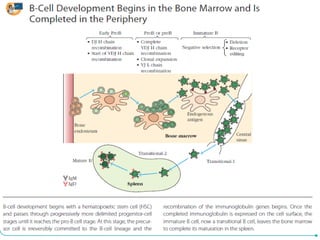
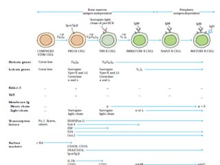
DEVELOPMENT OF B-LYMPHOCYTE
Dr Sitaram Swain
B-cell development begins in the bone marrow and
continues through a series of progressively form common
lymphoid progenitors (CLPs), which can give rise to either
B cells or T cells.
Progenitor cells destined to become T cells migrate to the
thymus where they complete their maturation ; the majority
of those that remain in the bone marrow become B cells.
As differentiation proceeds, the developing B cell expresses
on its cell surface a precisely calibrated sequence of cell-
surface receptor and adhesion molecules. Some of the
signals received from these receptors induce the
differentiation of the developing B cell; others trigger its
proliferation at particular stages of development.
These signals collectively allow differentiation of the CLP
through the early B-cell stages to form immature B cell that
leaves marrow to complete its differentiation in the spleen.
The primary function of mature B cells is to secrete
antibodies that protect the host against pathogens, and
so one major focus of those studying B-cell
differentiation is the analysis of the timing and order
of rearrangement and expression of immunoglobulin
receptor heavy- and light-chain genes.
These rearrangements culminate in the cell-surface
expression of the pre-B-cell receptor during the pre-B-
cell stage, in which the rearranged heavy chain is
expressed in combination with the surrogate light
chain.
Rearrangement of the light chain is initiated after
several rounds of division of cells bearing the pre-
BCR.
Developing B cells must capable of recognizing an extensive
array of antigens, while ensuring that self-reactive B cells are
either eliminated by apoptosis or rendered functionally unreactive
or anergic.
In adult animals, hematopoiesis, the generation of blood cells,
occurs in the bone marrow; the HSCs in the marrow are the
source of all blood cells of the erythroid, myeloid, and lymphoid
lineages .
Various non-hematopoietic cells in the bone marrow express cell-
surface molecules and secrete hormones that guide hematopoietic
cell development. Developing lymphocytes move within the bone
marrow
as they mature, thus interacting with different populations of cells
and signals at various developmental stages.
Hematopoiesis is a complex process in the adult animal, and
Red blood cells must be quickly generated de novo in order to
provide the embryo with sufficient oxygen, and HSCs must
proliferate at a rate sufficient to populate the adult as well as
provide for the hematopoietic needs of the maturing fetus.
Furthermore, since the bone marrow appears relatively la
development, the whole process of blood-cell generation must
shift location several times before moving into its final home.
The gestation period for mice is 19 to 21 days.
Hematopoiesis begins, in the mouse, around 7 days post
fertilization , when precursor cells in the yolk sac begin
differentiating to form primitive, nucleated, erythroid cells that
carry the oxygen the embryo needs for early development.
Fetal HSCs capable of generating all blood-cell types can be
detected in the early aorta-gonad-mesonephros (AGM) region
on day 8, when the fetal heart starts beating.
Hematopoiesis in the Fetal Liver
• Developing B cells in the fetal liver differ in important
ways from their counterparts in adult bone marrow. The
liver is the primary site of B-cell generation in the fetus,
and provides the neonatal animal with the cells it needs to
populate its nascent immune system.
• In order to accomplish this, hematopoietic stem cells and
their progeny must undergo a phase of rapid proliferation,
and fetal liver HSCs, as well as their daughter cells,
undergo several rounds of cell division over a short time.
• In contrast, HSCs derived from the bone marrow of a
healthy adult animal are relatively quiescent.
B cells generated from fetal liver precursors are
predominantly B-1 B cells. Briefly, B-1 B cells are
primarily located in the body (specifically the peritoneal
and pleural) cavities. They are therefore well-positioned
to protect the gut and the lungs, which are the major
ports of entry of microbes in the fetus and neonate.
Antibodies secreted by B-1 B cells are broadly cross-
reactive; many bind to carbohydrate antigens expressed
by a number of microbial species. Since terminal
deoxynucleotidyl transferase (TdT) is minimally
expressed at this point in ontogeny, and the RAG1/2
recombinase proteins appear not to use the full range of
V, D, and J region gene segments at this stage in
embryonic development, the immunoglobulin receptors
of B-1 B cells express minimal receptor diversity.
• In expressing an oligoclonal (few, as opposed to many,
clones) repertoire of B-cell receptors that bind to a
limited number of carbohydrate antigens shared among
many microbes, B-1 B cells occupy a functional niche
that bridges the innate and adaptive immune systems.
• Over a period of 2 to 4 weeks after birth, the process of
hematopoiesis in mice shift s from the fetal liver and
spleen to the bone marrow, where it continues
throughout adulthood.
• The B-1 B-cell population represents an exception to
this general rule, as it is self-renewing in the periphery.
This means that new daughter B-1 B cells are generated
continually from preexisting B-1 B cells in the
peritoneal and pleural cavities, and in those other parts
of the body in which B-1 B cells.
By the third month of pregnancy, these HSCs migrate
from yolk sac to the fetal liver, which then becomes
responsible for the majority of hematopoiesis in the fetus.
By the fourth month of pregnancy, HSCs migrate to the
bone marrow, which gradually assumes the hematopoietic
role from the fetal liver until, by the time of birth, it is the
primary generative organ for blood cells.
Prior to puberty in humans, most of the bones of the
skeleton are hematopoietically active, but by the age of
18 years only the vertebrae, ribs, sternum, skull, pelvis,
and parts of the humerus and femur retain hematopoietic
potential. Just as B-cell development in the fetus and
neonate differs from that in the adult, so does B-cell
hematopoiesis in the aging animal.
B-Cell Development in the Bone Marrow
The bone marrow microenvironment is a complex, three-
dimensional structure with distinctive cellular niches which are
specialized to influence the development of the cell populations
that mature there.
A dense network of fenestrated (leaky) thin-walled blood
vessels— the bone marrow sinusoids—permeates the marrow,
allowing the passage of newly formed blood cells to the
periphery and facilitating blood circulation through the marrow.
In addition to serving as a source of hematopoietic stem cells,
bone marrow also contains stem cells that can differentiate into
adipocytes (fat cells), chondrocytes (cartilage cells), osteocytes
(bone cells), myocytes (muscle cells), and potentially other types
of cells as well. Each of these diff erent classes of stem cells
requires specific sets of factors, secreted by particular bone
marrow stromal cells to enable their proper differentiation.
What are bone marrow stromal cells? The term stroma derives from the
Greek for mattress, and a stromal cell is a general term that describes a
large adherent cell that supports the growth of other cells.
During B-cell development, bone marrow stromal cells fulfill two
functions.
First, by interacting with adhesion molecules on the surfaces of HSCs
and progenitor cells, stromal cells retain the developing cell
populations in the specific bone marrow niches where they can receive
the appropriate molecular signals required for their further
differentiation.
Second, diverse populations of stromal cells express different
cytokines. At various points in their development, progenitor and
precursor B cells must interact with stromal cells secreting particular
cytokines, and thus the developing B cells move in an orderly
progression from location to location within the bone marrow.
This progression is guided by chemokines secreted by particular
stromal cell populations.
Once differentiated to the pre-pro-B-cell stage, the developing B
cells require signals from the chemokine CXCL12, which is
secreted by a specialized set of stromal cells, in order to progress
to the pro-B-cell stage.
Pro-B cells then require signaling from the cytokine IL-7, which
is secreted by yet another stromal cell subset (Figure 10-3). Many
of these stromal cell factors serve to induce the expression of
specialized transcription factors important in B-cell development.
B-Cell Maturation
The generation of mature B cells first occurs in the
embryo and continues throughout life. Before birth, the
yolk sac, fetal liver, and fetal bone marrow are the
major sites of B-cell maturation; after birth, generation
of mature B cells occurs in the bone marrow.
Progenitor B Cells Proliferate in Bone Marrow
B-cell development begins as lymphoid stem cells differentiate
into the earliest distinctive B-lineage cell—the progenitor B cell
(pro-B cell)—which expresses a transmembrane tyrosine
phosphatase called CD45R (sometimes called B220 in mice).
Pro-B cells proliferate within the bone marrow, filling the
extravascular spaces between large sinusoids in the shaft of a
bone. Proliferation and differentiation of pro-B cells into
precursor B cells (pre-B cells) requires the microenvironment
provided by the bone-marrow stromal cells.
If pro-B cells are removed from the bone marrow and cultured in
vitro, they will not progress to more mature B-cell stages unless
stromal cells are present. The stromal cells play two important
roles: they interact directly with pro-B and pre-B cells, and they
secrete various cytokines, notably IL-7, that support the
developmental process.
At the earliest developmental stage, pro-B cells require direct
contact with stromal cells in the bone marrow. This interaction is
mediated by several cell-adhesion molecules, including VLA-4 on
the pro-B cell and its ligand, VCAM-1, on the stromal cell.
After initial contact is made, a receptor on the pro-B cell called c-
Kit interacts with a stromal-cell surface molecule known as stem-
cell factor (SCF). This interaction activates c-Kit, which is a
tyrosine kinase, and the pro-B cell begins to divide and
differentiate into a pre-B cell and begins expressing a receptor for
IL-7.
The IL-7 secreted by the stromal cells drives the maturation
process, eventually inducing down-regulation of the adhesion
molecules on the pre-B cells, so that the proliferating cells can
detach from the stromal cells.
At this stage, pre-B cells no longer require direct contact with
stromal cells but continue to require IL-7 for growth and
maturation.
Ig-Gene Rearrangment Produces Immature B Cells
B-cell maturation depends on rearrangement of the immunoglobulin
DNA in the lymphoid stem cells.
First to occur in the pro-B cell stage is a heavy-chain DH-to-JH gene
rearrangement; this is followed by a VH-to-DHJH rearrangement. If
the first heavy-chain rearrangement is not productive, then VH-DH-
JH rearrangement continues on the other chromosome.
Upon completion of heavy-chain rearrangement, the cell is classified
as a pre-B cell. Continued development of a pre-B cell into an
immature B cell requires a productive light-chain gene
rearrangement.
Because of allelic exclusion, only one light-chain isotype is
expressed on the membrane of a B cell. Immature B cells express
mIgM (membrane IgM) on the cell surface.
The recombinase enzymes RAG-1 and RAG-2 are required for
both heavy-chain and light-chain gene rearrangements, are
expressed during the pro-B and pre-B cell stages.
The enzyme terminal deoxyribonucleotidyl transferase (TdT),
which catalyzes insertion of N-nucleotides at the DH-JH and VH-
DHJH coding joints, is active during the pro-B cell stage and
ceases to be active early in the pre–B-cell stage. Because TdT
expression is turned off during the part of the pre–B-cell stage
when light-chain rearrangement occurs,N-nucleotides are not
usually found in the VL-JL coding joints.
The bone-marrow phase of B-cell development culminates in the
production of an IgM-bearing immature B cell. At this stage of
development the B cell is not fully functional, and antigen
induces death or unresponsiveness (anergy) rather than division
and differentiation. Full maturation is signaled

B-Cell development.pdf

  • 1.
  • 2.
    B-cell development beginsin the bone marrow and continues through a series of progressively form common lymphoid progenitors (CLPs), which can give rise to either B cells or T cells. Progenitor cells destined to become T cells migrate to the thymus where they complete their maturation ; the majority of those that remain in the bone marrow become B cells. As differentiation proceeds, the developing B cell expresses on its cell surface a precisely calibrated sequence of cell- surface receptor and adhesion molecules. Some of the signals received from these receptors induce the differentiation of the developing B cell; others trigger its proliferation at particular stages of development. These signals collectively allow differentiation of the CLP through the early B-cell stages to form immature B cell that leaves marrow to complete its differentiation in the spleen.
  • 3.
    The primary functionof mature B cells is to secrete antibodies that protect the host against pathogens, and so one major focus of those studying B-cell differentiation is the analysis of the timing and order of rearrangement and expression of immunoglobulin receptor heavy- and light-chain genes. These rearrangements culminate in the cell-surface expression of the pre-B-cell receptor during the pre-B- cell stage, in which the rearranged heavy chain is expressed in combination with the surrogate light chain. Rearrangement of the light chain is initiated after several rounds of division of cells bearing the pre- BCR.
  • 6.
    Developing B cellsmust capable of recognizing an extensive array of antigens, while ensuring that self-reactive B cells are either eliminated by apoptosis or rendered functionally unreactive or anergic. In adult animals, hematopoiesis, the generation of blood cells, occurs in the bone marrow; the HSCs in the marrow are the source of all blood cells of the erythroid, myeloid, and lymphoid lineages . Various non-hematopoietic cells in the bone marrow express cell- surface molecules and secrete hormones that guide hematopoietic cell development. Developing lymphocytes move within the bone marrow as they mature, thus interacting with different populations of cells and signals at various developmental stages.
  • 7.
    Hematopoiesis is acomplex process in the adult animal, and Red blood cells must be quickly generated de novo in order to provide the embryo with sufficient oxygen, and HSCs must proliferate at a rate sufficient to populate the adult as well as provide for the hematopoietic needs of the maturing fetus. Furthermore, since the bone marrow appears relatively la development, the whole process of blood-cell generation must shift location several times before moving into its final home. The gestation period for mice is 19 to 21 days. Hematopoiesis begins, in the mouse, around 7 days post fertilization , when precursor cells in the yolk sac begin differentiating to form primitive, nucleated, erythroid cells that carry the oxygen the embryo needs for early development. Fetal HSCs capable of generating all blood-cell types can be detected in the early aorta-gonad-mesonephros (AGM) region on day 8, when the fetal heart starts beating.
  • 9.
    Hematopoiesis in theFetal Liver • Developing B cells in the fetal liver differ in important ways from their counterparts in adult bone marrow. The liver is the primary site of B-cell generation in the fetus, and provides the neonatal animal with the cells it needs to populate its nascent immune system. • In order to accomplish this, hematopoietic stem cells and their progeny must undergo a phase of rapid proliferation, and fetal liver HSCs, as well as their daughter cells, undergo several rounds of cell division over a short time. • In contrast, HSCs derived from the bone marrow of a healthy adult animal are relatively quiescent.
  • 10.
    B cells generatedfrom fetal liver precursors are predominantly B-1 B cells. Briefly, B-1 B cells are primarily located in the body (specifically the peritoneal and pleural) cavities. They are therefore well-positioned to protect the gut and the lungs, which are the major ports of entry of microbes in the fetus and neonate. Antibodies secreted by B-1 B cells are broadly cross- reactive; many bind to carbohydrate antigens expressed by a number of microbial species. Since terminal deoxynucleotidyl transferase (TdT) is minimally expressed at this point in ontogeny, and the RAG1/2 recombinase proteins appear not to use the full range of V, D, and J region gene segments at this stage in embryonic development, the immunoglobulin receptors of B-1 B cells express minimal receptor diversity.
  • 11.
    • In expressingan oligoclonal (few, as opposed to many, clones) repertoire of B-cell receptors that bind to a limited number of carbohydrate antigens shared among many microbes, B-1 B cells occupy a functional niche that bridges the innate and adaptive immune systems. • Over a period of 2 to 4 weeks after birth, the process of hematopoiesis in mice shift s from the fetal liver and spleen to the bone marrow, where it continues throughout adulthood. • The B-1 B-cell population represents an exception to this general rule, as it is self-renewing in the periphery. This means that new daughter B-1 B cells are generated continually from preexisting B-1 B cells in the peritoneal and pleural cavities, and in those other parts of the body in which B-1 B cells.
  • 12.
    By the thirdmonth of pregnancy, these HSCs migrate from yolk sac to the fetal liver, which then becomes responsible for the majority of hematopoiesis in the fetus. By the fourth month of pregnancy, HSCs migrate to the bone marrow, which gradually assumes the hematopoietic role from the fetal liver until, by the time of birth, it is the primary generative organ for blood cells. Prior to puberty in humans, most of the bones of the skeleton are hematopoietically active, but by the age of 18 years only the vertebrae, ribs, sternum, skull, pelvis, and parts of the humerus and femur retain hematopoietic potential. Just as B-cell development in the fetus and neonate differs from that in the adult, so does B-cell hematopoiesis in the aging animal.
  • 13.
    B-Cell Development inthe Bone Marrow The bone marrow microenvironment is a complex, three- dimensional structure with distinctive cellular niches which are specialized to influence the development of the cell populations that mature there. A dense network of fenestrated (leaky) thin-walled blood vessels— the bone marrow sinusoids—permeates the marrow, allowing the passage of newly formed blood cells to the periphery and facilitating blood circulation through the marrow. In addition to serving as a source of hematopoietic stem cells, bone marrow also contains stem cells that can differentiate into adipocytes (fat cells), chondrocytes (cartilage cells), osteocytes (bone cells), myocytes (muscle cells), and potentially other types of cells as well. Each of these diff erent classes of stem cells requires specific sets of factors, secreted by particular bone marrow stromal cells to enable their proper differentiation.
  • 14.
    What are bonemarrow stromal cells? The term stroma derives from the Greek for mattress, and a stromal cell is a general term that describes a large adherent cell that supports the growth of other cells. During B-cell development, bone marrow stromal cells fulfill two functions. First, by interacting with adhesion molecules on the surfaces of HSCs and progenitor cells, stromal cells retain the developing cell populations in the specific bone marrow niches where they can receive the appropriate molecular signals required for their further differentiation. Second, diverse populations of stromal cells express different cytokines. At various points in their development, progenitor and precursor B cells must interact with stromal cells secreting particular cytokines, and thus the developing B cells move in an orderly progression from location to location within the bone marrow. This progression is guided by chemokines secreted by particular stromal cell populations.
  • 15.
    Once differentiated tothe pre-pro-B-cell stage, the developing B cells require signals from the chemokine CXCL12, which is secreted by a specialized set of stromal cells, in order to progress to the pro-B-cell stage. Pro-B cells then require signaling from the cytokine IL-7, which is secreted by yet another stromal cell subset (Figure 10-3). Many of these stromal cell factors serve to induce the expression of specialized transcription factors important in B-cell development.
  • 17.
    B-Cell Maturation The generationof mature B cells first occurs in the embryo and continues throughout life. Before birth, the yolk sac, fetal liver, and fetal bone marrow are the major sites of B-cell maturation; after birth, generation of mature B cells occurs in the bone marrow.
  • 18.
    Progenitor B CellsProliferate in Bone Marrow B-cell development begins as lymphoid stem cells differentiate into the earliest distinctive B-lineage cell—the progenitor B cell (pro-B cell)—which expresses a transmembrane tyrosine phosphatase called CD45R (sometimes called B220 in mice). Pro-B cells proliferate within the bone marrow, filling the extravascular spaces between large sinusoids in the shaft of a bone. Proliferation and differentiation of pro-B cells into precursor B cells (pre-B cells) requires the microenvironment provided by the bone-marrow stromal cells. If pro-B cells are removed from the bone marrow and cultured in vitro, they will not progress to more mature B-cell stages unless stromal cells are present. The stromal cells play two important roles: they interact directly with pro-B and pre-B cells, and they secrete various cytokines, notably IL-7, that support the developmental process.
  • 20.
    At the earliestdevelopmental stage, pro-B cells require direct contact with stromal cells in the bone marrow. This interaction is mediated by several cell-adhesion molecules, including VLA-4 on the pro-B cell and its ligand, VCAM-1, on the stromal cell. After initial contact is made, a receptor on the pro-B cell called c- Kit interacts with a stromal-cell surface molecule known as stem- cell factor (SCF). This interaction activates c-Kit, which is a tyrosine kinase, and the pro-B cell begins to divide and differentiate into a pre-B cell and begins expressing a receptor for IL-7.
  • 21.
    The IL-7 secretedby the stromal cells drives the maturation process, eventually inducing down-regulation of the adhesion molecules on the pre-B cells, so that the proliferating cells can detach from the stromal cells. At this stage, pre-B cells no longer require direct contact with stromal cells but continue to require IL-7 for growth and maturation.
  • 22.
    Ig-Gene Rearrangment ProducesImmature B Cells B-cell maturation depends on rearrangement of the immunoglobulin DNA in the lymphoid stem cells. First to occur in the pro-B cell stage is a heavy-chain DH-to-JH gene rearrangement; this is followed by a VH-to-DHJH rearrangement. If the first heavy-chain rearrangement is not productive, then VH-DH- JH rearrangement continues on the other chromosome. Upon completion of heavy-chain rearrangement, the cell is classified as a pre-B cell. Continued development of a pre-B cell into an immature B cell requires a productive light-chain gene rearrangement. Because of allelic exclusion, only one light-chain isotype is expressed on the membrane of a B cell. Immature B cells express mIgM (membrane IgM) on the cell surface.
  • 24.
    The recombinase enzymesRAG-1 and RAG-2 are required for both heavy-chain and light-chain gene rearrangements, are expressed during the pro-B and pre-B cell stages. The enzyme terminal deoxyribonucleotidyl transferase (TdT), which catalyzes insertion of N-nucleotides at the DH-JH and VH- DHJH coding joints, is active during the pro-B cell stage and ceases to be active early in the pre–B-cell stage. Because TdT expression is turned off during the part of the pre–B-cell stage when light-chain rearrangement occurs,N-nucleotides are not usually found in the VL-JL coding joints. The bone-marrow phase of B-cell development culminates in the production of an IgM-bearing immature B cell. At this stage of development the B cell is not fully functional, and antigen induces death or unresponsiveness (anergy) rather than division and differentiation. Full maturation is signaled