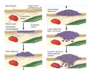
Cancer
Dr Sitaram Swain
Because cancer results from defects in fundamental cell
regulatory mechanisms, it is a disease that ultimately has
to be understood at the molecular and cellular levels.
In fact, many of the proteins that play key roles in cell
signaling, regulation of the cell cycle, and control of
programmed cell death were first identified because
abnormalities in their activities led to the uncontrolled
proliferation of cancer cells.
The study of cancer has thus contributed significantly to
our understanding of normal cell regulation, as well as
vice versa.
The Development and Causes of Cancer
The fundamental abnormality resulting in the
development of cancer is the continual unregulated
proliferation of cancer cells.
Rather than responding appropriately to the signals
that control normal cell behavior, cancer cells grow
and divide in an uncontrolled manner, invading
normal tissues and organs and eventually spreading
throughout the body.
The generalized loss of growth control exhibited by
cancer cells is the net result of accumulated
abnormalities in multiple cell regulatory systems .
Types of Cancer
Cancer can result from abnormal proliferation of any of the
different kinds of cells in the body, so there are more than a
hundred distinct types of cancer, which can vary
substantially in their behavior and response to treatment.
The most important issue in cancer pathology is the
distinction between benign and malignant tumors.
A tumor is any abnormal proliferation of cells, which may
be either benign or malignant. A benign tumor, such as a
common skin wart, remains confined to its original location,
neither invading surrounding normal tissue nor spreading to
distant body sites.
A malignant tumor, however, is capable of both invading
surrounding normal tissue and spreading throughout the
body via the circulatory or lymphatic systems (metastasis).
Only malignant tumors are properly referred to as
cancers, and it is their ability to invade and metastasis
size that makes cancer so dangerous.
Whereas benign tumors can usually be removed
surgically, the spread of malignant tumors to distant
body sites frequently makes them resistant to such
localized treatment.
Both benign and malignant tumors are classified
according to the type of cell from which they arise.
Most cancers fall into one of three main groups:
carcinomas, sarcomas, and leukemias or lymphomas.
Carcinomas, which include approximately 90% of human
cancers, are malignancies of epithelial cells. Sarcomas,
which are rare in humans, are solid tumors of connective
tissues, such as muscle, bone, cartilage, and fibrous tissue.
Leukemias and lymphomas, which account for
approximately 7% of human malignancies, arise from the
blood-forming cells and from cells of the immune system,
respectively.
Tumors are further classified according to tissue of origin
(e.g., lung or breast carcinomas) and the type of cell
involved.
For example, fibrosarcomas arise from fibroblasts, and
erythroid leukemias from precursors of erythrocytes (red
blood cells).
The Development of Cancer
One of the fundamental features of cancer is tumor clonality- the development
of tumors from single cells that begin to proliferate abnormally. The single-
cell origin of many tumors has been demonstrated by analysis of X
chromosome inactivation. One member of the X chromosome pair is
inactivated by being converted to heterochromatin in female cells. X
inactivation occurs randomly during embryonic development, so one X
chromosome is inactivated in some cells, while the other X chromosome is
inactivated in other cells.
Thus, if a female is heterozygous for an X chromosome gene, different alleles
will be expressed in different cells. Normal tissues are composed of mixtures
of cells with different inactive X chromosomes, so expression of both alleles is
detected in normal tissues of heterozygous females.
In contrast, tumor tissues generally express only one allele of a heterozygous
X chromosome gene. The implication is that all of the cells constituting such a
tumor were derived from a single cell of origin in which the pattern of X
inactivation was fixed before the tumor began to develop.
On the contrary, the development of cancer is a
multistep process in which cells gradually
become malignant through a progressive series
of alterations.
One indication of the multistep development of
cancer is that most cancers develop late in life.
The incidence of
At the cellular level, the development of cancer is
viewed as a multistep process involving mutation and
selection for cells with progressively increasing capacity
for proliferation, survival invasion, and metastasis.
The first step in the process, tumor initiation, is thought
to be the result of a genetic alteration leading to
abnormal proliferation of a single cell.
Cell proliferation then leads to the outgrowth of a
population of clonally derived tumor cells. Tumor
progression continues as additional mutations occur
within cells of the tumor population.
Some of these mutations confer a selective
advantage to the cell, such as more rapid growth,
and the descendants of a cell bearing such a
mutation will consequently become dominant within
the tumor population.
The process is called clonal selection, since a new
clone of tumor cells has evolved on the basis of its
increased growth rate or other properties (such as
survival, invasion, or metastasis) that confer a
selective advantage.
Clonal selection continues throughout tumor
development, so tumors continuously become more
rapid-growing and increasingly malignant.
Studies of colon carcinomas have provided a
clear example of tumor progression during the
development of a common human malignancy .
The earliest stage in tumor development is
increased proliferation of colon epithelial cells.
One of the cells within this proliferative cell
population is then thought to give rise to a small
benign neoplasm (an adenoma or polyp).
Further rounds of clonal selection lead to the
growth of adenomas of increasing size and
proliferative potential.
Malignant carcinomas then arise from the benign
adenomas, indicated by invasion of the tumor cells
through the basal lamina into underlying connective
tissue.
The cancer cells then continue to proliferate and
spread through the connective tissues of the colon
wall.
Eventually the cancer cells penetrate the wall of the
colon and invade other abdominal organs, such as the
bladder or small intestine.
In addition, the cancer cells invade blood and
lymphatic vessels, allowing them to metastasize
throughout the body.
Properties of cancer Cells
The uncontrolled growth of cancer cells results from
accumulated abnormalities affecting many of the cell
regulatory mechanisms.
This relationship is reflected in several aspects of cell
behavior that distinguish cancer cells from their
normal counterparts.
Cancer cells typically display abnormalities in the
mechanisms that regulate normal cell proliferation,
differentiation, and survival.
Taken together, these characteristic properties of
cancer cells provide a description of malignancy at
the cellular level.
The uncontrolled proliferation of cancer cells in vivo is
mimicked by their behavior in cell culture.
A primary distinction between cancer cells and normal
cells in culture is that normal cells display density-
dependent inhibition of cell proliferation.
Normal cells proliferate until they reach a finite cell
density, which is determined in part by the availability
of growth factors added to the culture medium (usually
in 'the form of serum).
They then cease proliferating and become quiescent,
arrested in the G0 stage of the cell cycle.
The proliferation of most cancer cells, however,
is not sensitive to density-dependent inhibition.
Rather than responding to the signals that cause
normal cells to cease proliferation and enter G0,
tumor cells generally continue growing to high
cell densities in culture, mimicking their
uncontrolled proliferation in vivo.
A related difference between normal cells and
cancer cells is that many cancer cells have
reduced requirements for extracellular growth
factors.
The proliferation of most cells is controlled by
polypeptide growth factors. Particularly fibroblasts, the
availability of serum growth factors is the principal
determinant of their proliferative capacity in culture.
The growth factor requirements of these cells are
closely related to the phenomenon of density dependent
inhibition, since the density at which normal fibroblasts
become quiescent is proportional to the concentration of
serum growth factors in the culture medium.
The growth factor requirements of many tumor cells are
reduced compared to their normal counterparts,
contributing to the unregulated proliferation of tumor
cells both in vitro and in vivo.
In some cases, cancer cells produce growth factors that
stimulate their own proliferation.
Such abnormal production of a growth factor by a
responsive cell leads to continuous autostimulation of cell
division (autocrine growth stimulation), and the cancer cells
are therefore less dependent on growth factors from other,
physiologically normal sources.
In other cases, the reduced growth factor dependence of
cancer cells results from abnormalities in intracellular
signaling systems,
such as unregulated activity of growth factor receptors or
other proteins (e.g., Ras proteins or protein kinases) that
were elements of signal transduction pathways leading to
cell proliferation.
Cancer cells are also less stringently regulated than normal
cells by cell-cell and cell-matrix interactions.
Most cancer cells are less adhesive than normal cells, often
as a result of reduced expression of cell surface adhesion
molecules.
For example, loss of E-cadherin, the principal adhesion
molecule of epithelial cells, is important in the development
of carcinomas (epithelial cancers).
As a result of reduced expression of cell adhesion molecules,
cancer cells are comparatively unrestrained by interactions
with other cells and tissue components, contributing to the
ability of malignant cells to invade and metastasize.
The reduced adhesiveness of cancer cells also results in
morphological and cytoskeletal alterations: Many tumor cells
are rounder than normal, in part because they are less firmly
attached to either the extracellular matrix or neighboring
cells.
A striking difference in the cell-cell interactions
displayed by normal cells and those of cancer cells is
illustrated by the phenomenon of contact inhibition.
Normal fibroblasts migrate across the surface of a
culture dish until they make contact with a neighboring
cell.
Further cell migration is then inhibited, and normal cells
adhere to each other, forming an orderly array of cells
on the culture dish surface. Tumor cells, in contrast,
continue moving after contact with their neighbors,
migrating over adjacent cells, and growing in
disordered, multilayered patterns.
Not only the movement but also the proliferation of
many normal cells is inhibited by cell-cell contact, and
cancer cells are characteristically insensitive to such
contact inhibition of growth.
Two additional properties of cancer cells affect their
interactions with other tissue components, thereby
playing important roles in invasion and metastasis.
First, malignant cells generally secrete proteases that
digest extracellular matrix components, allowing the
cancer cells to invade adjacent normal tissues. Secretion
of collagenase, for example, appears to be an important
determinant of the ability of carcinomas to digest and
penetrate through basal laminae to invade underlying
connective tissue.
Second, cancer cells secrete growth factors that promote the
formation of new blood vessels (angiogenesis). Angiogenesis is
needed to support the growth of a tumor beyond the size of about a
million cells, at which point new blood vessels are required to
supply oxygen and nutrients to the proliferating tumor cells.
Such blood vessels are formed in response to growth factors,
secreted by the tumor cells, that stimulate proliferation of endothelial
cells in the walls of capillaries in surrounding tissue, resulting in the
outgrowth of new capillaries into the tumor.
The formation of such new blood vessels is important not only in
supporting tumor growth, but also in metastasis. The actively
growing new capillaries formed in response to angiogenic
stimulation are easily penetrated by the tumor cells, providing a
ready opportunity for cancer cells to enter the circulatory system and
begin the metastatic process.
Another general characteristic of most cancer cells is that
they fail to differentiate normally. Such defective
differentiation is closely coupled to abnormal proliferation
since most fully differentiated cells cease cell division.
Rather than carrying out their normal differentiation
program, cancer cells are usually blocked at an early stage
of differentiation, consistent with their continued active
proliferation, of maturation at which they retain their
capacity for proliferation and continue to reproduce.
programmed cell death or apoptosis is an integral part of
the differentiation program of many cell types, including
blood cells. Many cancer cells fail to undergo apoptosis
and therefore exhibit increased life spans compared to
their normal counterparts.
For example, the survival of many normal cells is dependent on
signals from either growth factors or from the extracellular matrix
that prevent apoptosis. In contrast, tumor cells are often able to
survive in the absence of growth factors required by their normal
counterparts. Such a failure of tumor cells to undergo apoptosis
when deprived of normal environmental signals may be important
not only in primary tumor development but also in the survival and
growth of metastatic cells in abnormal tissue sites.
Normal cells also undergo apoptosis following DNA damage, while
many cancer cells fail to do so. In this case, the failure to undergo
apoptosis contributes to the resistance of cancer cells to irradiation
and many chemotherapeutic drugs, which act by damaging DNA. In
addition to evading apoptosis, cancer cells generally acquire the
capacity for unlimited replication as a result of expression of
telomerase, which is required to maintain the ends of eukaryotic
chromosomes.
1.Cancer

1.Cancer

  • 1.
  • 2.
    Because cancer resultsfrom defects in fundamental cell regulatory mechanisms, it is a disease that ultimately has to be understood at the molecular and cellular levels. In fact, many of the proteins that play key roles in cell signaling, regulation of the cell cycle, and control of programmed cell death were first identified because abnormalities in their activities led to the uncontrolled proliferation of cancer cells. The study of cancer has thus contributed significantly to our understanding of normal cell regulation, as well as vice versa.
  • 3.
    The Development andCauses of Cancer The fundamental abnormality resulting in the development of cancer is the continual unregulated proliferation of cancer cells. Rather than responding appropriately to the signals that control normal cell behavior, cancer cells grow and divide in an uncontrolled manner, invading normal tissues and organs and eventually spreading throughout the body. The generalized loss of growth control exhibited by cancer cells is the net result of accumulated abnormalities in multiple cell regulatory systems .
  • 4.
    Types of Cancer Cancercan result from abnormal proliferation of any of the different kinds of cells in the body, so there are more than a hundred distinct types of cancer, which can vary substantially in their behavior and response to treatment. The most important issue in cancer pathology is the distinction between benign and malignant tumors. A tumor is any abnormal proliferation of cells, which may be either benign or malignant. A benign tumor, such as a common skin wart, remains confined to its original location, neither invading surrounding normal tissue nor spreading to distant body sites. A malignant tumor, however, is capable of both invading surrounding normal tissue and spreading throughout the body via the circulatory or lymphatic systems (metastasis).
  • 5.
    Only malignant tumorsare properly referred to as cancers, and it is their ability to invade and metastasis size that makes cancer so dangerous. Whereas benign tumors can usually be removed surgically, the spread of malignant tumors to distant body sites frequently makes them resistant to such localized treatment. Both benign and malignant tumors are classified according to the type of cell from which they arise. Most cancers fall into one of three main groups: carcinomas, sarcomas, and leukemias or lymphomas.
  • 6.
    Carcinomas, which includeapproximately 90% of human cancers, are malignancies of epithelial cells. Sarcomas, which are rare in humans, are solid tumors of connective tissues, such as muscle, bone, cartilage, and fibrous tissue. Leukemias and lymphomas, which account for approximately 7% of human malignancies, arise from the blood-forming cells and from cells of the immune system, respectively. Tumors are further classified according to tissue of origin (e.g., lung or breast carcinomas) and the type of cell involved. For example, fibrosarcomas arise from fibroblasts, and erythroid leukemias from precursors of erythrocytes (red blood cells).
  • 7.
    The Development ofCancer One of the fundamental features of cancer is tumor clonality- the development of tumors from single cells that begin to proliferate abnormally. The single- cell origin of many tumors has been demonstrated by analysis of X chromosome inactivation. One member of the X chromosome pair is inactivated by being converted to heterochromatin in female cells. X inactivation occurs randomly during embryonic development, so one X chromosome is inactivated in some cells, while the other X chromosome is inactivated in other cells. Thus, if a female is heterozygous for an X chromosome gene, different alleles will be expressed in different cells. Normal tissues are composed of mixtures of cells with different inactive X chromosomes, so expression of both alleles is detected in normal tissues of heterozygous females. In contrast, tumor tissues generally express only one allele of a heterozygous X chromosome gene. The implication is that all of the cells constituting such a tumor were derived from a single cell of origin in which the pattern of X inactivation was fixed before the tumor began to develop.
  • 8.
    On the contrary,the development of cancer is a multistep process in which cells gradually become malignant through a progressive series of alterations. One indication of the multistep development of cancer is that most cancers develop late in life. The incidence of
  • 9.
    At the cellularlevel, the development of cancer is viewed as a multistep process involving mutation and selection for cells with progressively increasing capacity for proliferation, survival invasion, and metastasis. The first step in the process, tumor initiation, is thought to be the result of a genetic alteration leading to abnormal proliferation of a single cell. Cell proliferation then leads to the outgrowth of a population of clonally derived tumor cells. Tumor progression continues as additional mutations occur within cells of the tumor population.
  • 11.
    Some of thesemutations confer a selective advantage to the cell, such as more rapid growth, and the descendants of a cell bearing such a mutation will consequently become dominant within the tumor population. The process is called clonal selection, since a new clone of tumor cells has evolved on the basis of its increased growth rate or other properties (such as survival, invasion, or metastasis) that confer a selective advantage. Clonal selection continues throughout tumor development, so tumors continuously become more rapid-growing and increasingly malignant.
  • 13.
    Studies of coloncarcinomas have provided a clear example of tumor progression during the development of a common human malignancy . The earliest stage in tumor development is increased proliferation of colon epithelial cells. One of the cells within this proliferative cell population is then thought to give rise to a small benign neoplasm (an adenoma or polyp). Further rounds of clonal selection lead to the growth of adenomas of increasing size and proliferative potential.
  • 15.
    Malignant carcinomas thenarise from the benign adenomas, indicated by invasion of the tumor cells through the basal lamina into underlying connective tissue. The cancer cells then continue to proliferate and spread through the connective tissues of the colon wall. Eventually the cancer cells penetrate the wall of the colon and invade other abdominal organs, such as the bladder or small intestine. In addition, the cancer cells invade blood and lymphatic vessels, allowing them to metastasize throughout the body.
  • 16.
    Properties of cancerCells The uncontrolled growth of cancer cells results from accumulated abnormalities affecting many of the cell regulatory mechanisms. This relationship is reflected in several aspects of cell behavior that distinguish cancer cells from their normal counterparts. Cancer cells typically display abnormalities in the mechanisms that regulate normal cell proliferation, differentiation, and survival. Taken together, these characteristic properties of cancer cells provide a description of malignancy at the cellular level.
  • 17.
    The uncontrolled proliferationof cancer cells in vivo is mimicked by their behavior in cell culture. A primary distinction between cancer cells and normal cells in culture is that normal cells display density- dependent inhibition of cell proliferation. Normal cells proliferate until they reach a finite cell density, which is determined in part by the availability of growth factors added to the culture medium (usually in 'the form of serum). They then cease proliferating and become quiescent, arrested in the G0 stage of the cell cycle.
  • 18.
    The proliferation ofmost cancer cells, however, is not sensitive to density-dependent inhibition. Rather than responding to the signals that cause normal cells to cease proliferation and enter G0, tumor cells generally continue growing to high cell densities in culture, mimicking their uncontrolled proliferation in vivo. A related difference between normal cells and cancer cells is that many cancer cells have reduced requirements for extracellular growth factors.
  • 19.
    The proliferation ofmost cells is controlled by polypeptide growth factors. Particularly fibroblasts, the availability of serum growth factors is the principal determinant of their proliferative capacity in culture. The growth factor requirements of these cells are closely related to the phenomenon of density dependent inhibition, since the density at which normal fibroblasts become quiescent is proportional to the concentration of serum growth factors in the culture medium. The growth factor requirements of many tumor cells are reduced compared to their normal counterparts, contributing to the unregulated proliferation of tumor cells both in vitro and in vivo.
  • 20.
    In some cases,cancer cells produce growth factors that stimulate their own proliferation. Such abnormal production of a growth factor by a responsive cell leads to continuous autostimulation of cell division (autocrine growth stimulation), and the cancer cells are therefore less dependent on growth factors from other, physiologically normal sources. In other cases, the reduced growth factor dependence of cancer cells results from abnormalities in intracellular signaling systems, such as unregulated activity of growth factor receptors or other proteins (e.g., Ras proteins or protein kinases) that were elements of signal transduction pathways leading to cell proliferation.
  • 21.
    Cancer cells arealso less stringently regulated than normal cells by cell-cell and cell-matrix interactions. Most cancer cells are less adhesive than normal cells, often as a result of reduced expression of cell surface adhesion molecules. For example, loss of E-cadherin, the principal adhesion molecule of epithelial cells, is important in the development of carcinomas (epithelial cancers). As a result of reduced expression of cell adhesion molecules, cancer cells are comparatively unrestrained by interactions with other cells and tissue components, contributing to the ability of malignant cells to invade and metastasize. The reduced adhesiveness of cancer cells also results in morphological and cytoskeletal alterations: Many tumor cells are rounder than normal, in part because they are less firmly attached to either the extracellular matrix or neighboring cells.
  • 22.
    A striking differencein the cell-cell interactions displayed by normal cells and those of cancer cells is illustrated by the phenomenon of contact inhibition. Normal fibroblasts migrate across the surface of a culture dish until they make contact with a neighboring cell. Further cell migration is then inhibited, and normal cells adhere to each other, forming an orderly array of cells on the culture dish surface. Tumor cells, in contrast, continue moving after contact with their neighbors, migrating over adjacent cells, and growing in disordered, multilayered patterns.
  • 23.
    Not only themovement but also the proliferation of many normal cells is inhibited by cell-cell contact, and cancer cells are characteristically insensitive to such contact inhibition of growth. Two additional properties of cancer cells affect their interactions with other tissue components, thereby playing important roles in invasion and metastasis. First, malignant cells generally secrete proteases that digest extracellular matrix components, allowing the cancer cells to invade adjacent normal tissues. Secretion of collagenase, for example, appears to be an important determinant of the ability of carcinomas to digest and penetrate through basal laminae to invade underlying connective tissue.
  • 24.
    Second, cancer cellssecrete growth factors that promote the formation of new blood vessels (angiogenesis). Angiogenesis is needed to support the growth of a tumor beyond the size of about a million cells, at which point new blood vessels are required to supply oxygen and nutrients to the proliferating tumor cells. Such blood vessels are formed in response to growth factors, secreted by the tumor cells, that stimulate proliferation of endothelial cells in the walls of capillaries in surrounding tissue, resulting in the outgrowth of new capillaries into the tumor. The formation of such new blood vessels is important not only in supporting tumor growth, but also in metastasis. The actively growing new capillaries formed in response to angiogenic stimulation are easily penetrated by the tumor cells, providing a ready opportunity for cancer cells to enter the circulatory system and begin the metastatic process.
  • 25.
    Another general characteristicof most cancer cells is that they fail to differentiate normally. Such defective differentiation is closely coupled to abnormal proliferation since most fully differentiated cells cease cell division. Rather than carrying out their normal differentiation program, cancer cells are usually blocked at an early stage of differentiation, consistent with their continued active proliferation, of maturation at which they retain their capacity for proliferation and continue to reproduce. programmed cell death or apoptosis is an integral part of the differentiation program of many cell types, including blood cells. Many cancer cells fail to undergo apoptosis and therefore exhibit increased life spans compared to their normal counterparts.
  • 26.
    For example, thesurvival of many normal cells is dependent on signals from either growth factors or from the extracellular matrix that prevent apoptosis. In contrast, tumor cells are often able to survive in the absence of growth factors required by their normal counterparts. Such a failure of tumor cells to undergo apoptosis when deprived of normal environmental signals may be important not only in primary tumor development but also in the survival and growth of metastatic cells in abnormal tissue sites. Normal cells also undergo apoptosis following DNA damage, while many cancer cells fail to do so. In this case, the failure to undergo apoptosis contributes to the resistance of cancer cells to irradiation and many chemotherapeutic drugs, which act by damaging DNA. In addition to evading apoptosis, cancer cells generally acquire the capacity for unlimited replication as a result of expression of telomerase, which is required to maintain the ends of eukaryotic chromosomes.