ACUTE RESPIRATORY FAILURE MAGDI SASI 2015
•
7 likes•636 views

1. Acute respiratory failure is defined as a severe form of respiratory insufficiency resulting in a PaO2 of less than 60 mmHg or a PaCO2 of more than 50 mmHg. 2. There are two main types - type 1 with low PaO2 and normal or low PaCO2, and type 2 with low PaO2 and high PaCO2. 3. Major causes include diffuse airway obstruction, central airway obstruction, restrictive lung disease, pulmonary vascular disease, pleural and chest wall diseases, and neuromuscular diseases.

Recommended
More Related Content
What's hot
What's hot (20)
Viewers also liked
Viewers also liked (17)
Similar to ACUTE RESPIRATORY FAILURE MAGDI SASI 2015
Similar to ACUTE RESPIRATORY FAILURE MAGDI SASI 2015 (20)
More from cardilogy
More from cardilogy (20)
ACUTE RESPIRATORY FAILURE MAGDI SASI 2015
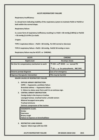
- 1. ACUTE RESPIRATORY FAILURE DR MAGDI AWAD SASI 2015 ACUTE RESPIRATORY FAILURE Respiratory insufficiency: Is a broad term indicating inability of the respiratory system to maintain PaO2 or PaCO2 or both within the normal shape. Respiratory failure: Is a sever form of respiratory insfficiency resulting in a PaO2 < 60 mmHg (( 8KPa)) or PaCO2 > 50 mmHg (( 6.6 KPa )) or both. 2 types: TYPE I respiratory failure ---PaO2 < 60 mmHg , Pa CO2 normal or decrease TYPE II respiratory failure—PaO2 < 60 mmHg , PaCO2 50 mmHg or more Respiratory failure may be ACUTE or CHRONIC. ACUTE CHRONIC Raptd onset Develops slowly No time for compensatory mechanism to work CO2 HCO3 normal PH O2 2ry polycythemia , RBC DPG Deserve prompt diagnosis Less of an emergency Vigorous therapeutic intervention This may be harmful MAJOR CAUSES OF RESPIRATORY FAILURE 1. DIFFUSE AIRWAY OBSTRUCTION: COPD-----hypoxemia ,ventilator failure Bronchial asthma ----hypoxemic failure V. failure iss status sever form and it is an ominous sign. 2. CENTRAL AIRWAY OBSTRUCTION: Foreign body in the larynx or trachea Laryngeal tumour ,tracheal tumor ,cricoids tumors Bilateral laryngeal nerve palsy Tracheal strictures Extrinsic compression of the trachea DIAGNOSTIC CLUES: Stridor Hoarsness Brassy cough Failure to improve on bronchodilator 3. RESTRICTIVE LUNG DISEASE: Hypoxic failure type with low CO2
- 2. ACUTE RESPIRATORY FAILURE DR MAGDI AWAD SASI 2015 Ventilator failure occurs exceptionally with sever cases. Acute pulmonary odema whether cardiac or non cardiac Non cardiac: Shock , High attitude ,Heroin ,Sepsis ,Chemicals ,Neurologic diseases Cardiac : Acute myocardial infarction , mitral or aortic valve disease Alveolar proteinosis Fibrosing alveolitis Pneumocytosis pneumonia Shock lung 4. PULMONARY VASCULAR DISEASE: Acute – pulmonary emboli –fat ,emboli -----hypoxemic failure Preexisting lung disease Pulmonary A/V fistula -----hypoxic failure Chronic ----recurrent thromboembolism ,pulmonary vasculitis 5. PLEURAL AND CHEST WALL DISEASE: ACUTE CHRONIC Flial chest pneumothorax Kyphoscoliosis Obesity Massive pleural effusion Chronic pleural effusion Ankylosing spondylitis 6. NEUROMUSCULAR DISEASE: ventiltory failure A. Muscle disease---polyomyelitis ,muscle dystrophies ,myotonies B. neuromuscular junction –Tetanus ,Mysthenia G. ,Anticholinestrase over dose C. Neurological disease –poliomyelitis ,peripheral neuropathies ,spinal cord injuries ,phrenic nerve paralysis, Amylotrophic lateral sclerosis, Guillian barre syndrome ,Polyneuritis D. Brain disorders: Sedative drugs ,Anesthesia ,Vascular disease ,infections ,Tumours. CLUES: FRC , FEV1/ FVC === NORMAL I.C. ,VC ,ERV ===REDUCED The maximal inspiratory flow rates more depressed then maximal expiratory flow rates more depressed. Central airway obstruction and neuromuscular diseases both lower maximal flow rates but maximal inspireatory and expiratory airway pressure will be normal in the central airway obstruction and decreased in NMD. 7. DEPRESSION OF RESPIRATORY CENTER: The automaticity of ventilation is a function of the respiratory centre in the medulla.
- 3. ACUTE RESPIRATORY FAILURE DR MAGDI AWAD SASI 2015 RESPIRATORY CENTRE CAROTID BODIES RESPIRATORY MUSCLES LUNG RC IN CEREBELLUM RC IN PONS The chemosensitive receptors in the medulla is sensitive to changes in the PH—PaCO2. The peripheral receptors in the carotid and aortic bodies are sensitive to the hypoxic and PH changes. CAUSES OF DEPRESSION OF RESPIRATORY CENTRE: 1. Drugs ----commnest cause ---Sedatives ,Narcotics ,Tranquilizer 2. Chronic hypercapnia 3. Sever hypoxemia 4. Neurological disease : Brain stem tumors/infarction ,Bulbopolio ,M.S., Cerebellar trauma ,Syringomelia 8. OBESITY –HYPOVENTILATION SYNDROME: Many obese patient have arterial hypoxemia due to basilar microatelectasis. Small number of morbidity obese patient manifest the pickwickian syndrome (( Hypersomnia ,Hypoxemia ,Hypercapnia )). 9. SLEEP—APNEA SYNDROME: Central ---obstructive--- mixed. There is transient R.F. during the apnea episodes. Some patient will have chronic day time hypercapnia and hypoxiemia if markedly obese. Most of them have respiratory insufficiency rather than respiratory failure. 10. RESPIRATORY MUSCLE FATIGUE: Is a contributory rather than 1ry cause. Seen in patient with COPD due to : a. Increased work of breathing b. Arterial hypoxemia c. Flattened diaphragm Diagnosis ----Respiatory paradox Chest up & diaphragm down Diagnostic studies in patient with respiratory failure: 1) History and physical examination: Are of major importance Symptoms and signs could be due to underlaying disease process or to blood gases derangements. Signs of hypoxia: Hyperventilation Cyanosis Cerebral hypoxia –headache ,restlessness ,somnolence ,confusion ,coma Pulmonary HTN Cor pulmonale Right side heart failure
- 4. ACUTE RESPIRATORY FAILURE DR MAGDI AWAD SASI 2015 Signs of hypercapnia: Headache Restlessness Confusion and coma Asteriaxis Neurological disease Papilloedema Changes in COP , BP ,and cardiac arrhythmia. Wheezing ---is not completely specific Central airway obstruction Pulmonary embolism Pulmonary oedema Type of breathing : Rapid and shallow breathing in patient with intrapulmonary restrictive lung disease. Absence of dyspnea— Respiratory centre depression Neuromuscular disease of the chest wall Paradoxical movement of the diaphragm Respiratory muscle fatigue Diaphragm paralysis Stridor Extrathoracic trachea or laryngeal obstruction. 2) Standard laboratory test: Is of limited value. Diffuse airway obstruction and peripheral eosinophilia ----bronchila asthma Neuromuscular dysfunction: EMG-----enzyme levels -----serum phosphate CSF examination may help the diagnosis In patient with diffuse pulmonary restrictive disease , RF & ANF & precipitins to organ antigen Urine for fat globules ----serology for fungal disease 3) Chest imaging: CXR will identify blood categories like diffuse histopulmonary opacities---interstitial ,alveolar ,miliary pattern Associated finding like: hilar or mediastinal adenopathy Egg shell calcification of lymph nodes Pleural effusion Cardiomegally Skeletal lesion Pulmonary cavity
- 5. ACUTE RESPIRATORY FAILURE DR MAGDI AWAD SASI 2015 Radioopaque foreign body Localized narrowing of the trachea with shadow endo or paratracheal lesions. Bronchogenic carcinoma with pnemothorax in COPD patient. Pulmonary angiography is available –PE ,A.V .fistula 4) Pulmonary function test: Arterial blood gase , spirometry ,flow volume loops Persistent increased alveolar –arterial O2 gradient after 100% O2 Intracardiac or intrapulmonary ---RT to LT shunt Restrictive or obstructive lung disease Reversibility studies after bronchodilator PFT is an analysis of single forced expiration and most sensitive measure of airway obstruction in obstructive lung disease. PFT is the most sensitive in small airway obstruction assessment. Obstructive lung disease--- FVC (( airway close increase the limit expiration before the patient has breathed out fully )) FEV 1 decreased FEVI /FVC % ----decreased --- increase the airway resistance which slow the rate of expiration. FRC increased and RV increased Restrictive lung disease---- FVC ((limit expansion of lung and chest )) FEV I (( airway resistance normal )) FEV /FVC = normal / increased Decrease RV and decrease lung compliance FEVI : Useful in assessing the efficiency of bronchodilatation therapy and to follow the prognosis of patient with COPD and asthma. Impaired lung function is a frequent association with decreased FEVI Used as apart for reversibility studies after bronchodilatation. Central airway obstruction---- Decrease flow rate ((inspiratory or both )) Relatives normal lung volumes Normal diffusing capacity Normal maximum inspiratory and expiratory airway pressures 5) Bronchoscopy: diagnostic and therapeutic 6) Right heart catheterization : It is of value for diagnosing certain problems e.g,- Teft to right intracardiac shunts Right to left shunts 7) ECHO ,ECG ,contrast ECHO 8) Lung biopsy : Open lung biopsy or trans bronchial lung biopsy
- 6. ACUTE RESPIRATORY FAILURE DR MAGDI AWAD SASI 2015 Conditions that can be diagnosed by lung biopsy and for which effective treatment is available are: CMV pneumonia Pneumocystis pneumonia Bacterial and fungal pneumonia Military TB Chronic eosinophilic pneumonia Lupus pneumonitis Pulmonary alveolar proteinosis Diffuse intra-alveolar haemorrhage Fat embolism Lymphocytic fibrosing alveolitis MANAGEMENT : A. Establishing an airway B. O2 adminstration C. Maintain adequate ventilation D. Identifying and treat the underlaying disease E. Monitor SaO2 ,ECG and vital signs INDICATIONS FOR ENDOTRACHEAL INTUBATION:' Current inability to protect the airway CNS dysfunction: 1. Stroke 2. Head trauma 3. Toxic ---- metabolic central respiratory depression Actual or potential airway compromise: 1. Excessive secretions 2. Airway edema: a. Epiglotitis b. Anaphylaxis c. Facial or neck trauma d. Inhalational injury INDICATIONS FOR VENTILATORY SUPPORT Respiratory rate > 35 (10 – 20)b/min Vital capacity < 15 (65 – 75)ml/kg BWT FEVI < 10 (50 – 60)ml/kg BWT MAX.INSPIRATORY FORCE > 25 ( 75 – 100) cmH2O PaO2 < 50 ( > 500 mmHg ) PaCO2 > 55 ( 35 – 45 mmHg) VD / VT > 0.06 ( 0.25 – 0.40 ) After 10 minutes of 100% O2 except in patients with chronic hypercapnia.
- 7. ACUTE RESPIRATORY FAILURE DR MAGDI AWAD SASI 2015 The final decision remains clinical. INDICATIONS OF CONTROLLED MECHANICAL VENTILATION: 1. Apnea or severe respiratory depression present 2. Patient is unable to maintain adequate spontaneous ventilator activity or assisted ventilation 3. Spontaneous ventilator effort is deleterious –flial chest 4. Application of PEEP is necessary 5. Ventilator is operating near its pressure limit or at high rate ( >25/min) and is unable to deliver T.V. Setting of ventilator: 1. Tidal volume ----start by large tidal volume (( 12 – 15 ml/kg )) 2. Respiratory rate ---- slow R.R. (( 10 – 20 b/min)) Desired F = pressure F X previous PaCO2 Desired PaCO2 3. Inspiratory to expiratory rate I/E (( 1/2 --- 1///4 )) 4. Flow wave form 5. FiO2 ---aim for PaO2 60 – 80mmHg & FiO < 50% 6. Pressure limits 10cm H2O above the peak P. reacting during each cycle 7. PEEP / CPAP: Bypassing the oropharynx by E.T. tube will decrease FRC . The application of PEEP 3 – 5 cmH2O reverse this effect ( physiological CPAP ) . The usual indication of PEEP is the presence of R. hypoxiemia due to lung injury : 1. ARDS 2. Cardiogenic pulmonary oedema 3. Flial chest Start at 5cmH2O and increase progressively every 15—30 min until a satisfactory Oxygenation is reached for FiO2 ideally < 60% Rarely necessary to exceed levels of 20 cmH2o Best way to asses PEEP is by mixed venous SO2. Reduce PEEP if : a. Hypotension b. Step decrement of more than 5% of SV O2 c. > 20 % decrease of C.O. d. Appearance of new cardiac murmur Clinical monitoring of mechanical ventilation: 1. Regular clinical examination of the patient 2. Inspect the ventilator and V. circuit 3. Chest expansion should be symmetrical 4. E.T. check and its pressure < 30 mmHg 5. The humidifier temperature & H2O level checked 6. Pulse oximeter check 7. Ventilator pressure gauge .Peak inspiratory pressure
- 8. ACUTE RESPIRATORY FAILURE DR MAGDI AWAD SASI 2015 8. Compare inspiratory and expiratory tidal volumes for leak in the v. circuit or E.T. tube cuff. Weaning of mechanical ventilation : Negative inspiratory force > -25 cmH2O V.C. > 10 ml/ kg B.W. COMPLICATION OF MECHANICAL VENTILATION: 1. E.T. tube ---displacement and pressure necrosis 2. Nasocomial pneumonia 3. PEEP ventilation : Decrease C.O. Decrease renal perfusion Salt and H2O retention Interstitial lung damage 4. Pneumothorax and mediastinal ---subcutaneous emphysema/pericardial Weaning of mechanical ventilation: CMV SIMV IMV pressure support ventilation PEEP decrease by 5 cmH2O every 10—20 min ABG must be checked for every step of weaning. HISTROY ACUTE HYPOXEMIA CLEAR LUNG FIELDS PHYSICAL EXAMINATION RESPIRATORY (( BLACK LUNG )) CXR FAILURE ABG SHUNT COPD PE DIFFUSE OPACIFICATION (( WHITE LUNG )) INTERSTITIUM ALVEOLAR FILLING VENTILATORY CLEAR LUNG (( BLACK LUNG )) DIFFUSE OPACIFICATION ((WHITE LUNG)) COPD LATE STAGES NEUROGENIC HYPOVENTILATION CHEST WALL RESTRICTION CENTRAL AIRWAY OBSTRUCTION COPD + ADDED PARENCHYMAL DISEASE DIFFUSE PARENCHYMAL DISEASE IN TERMINAL