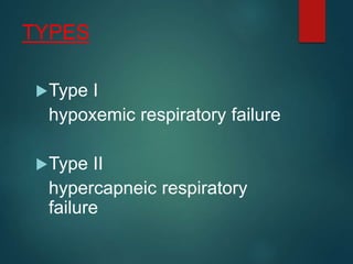
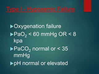
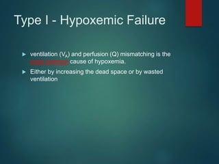
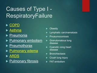
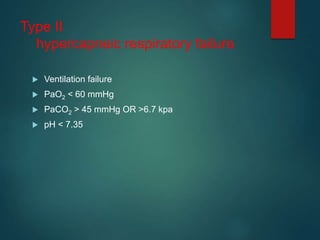
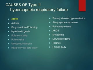
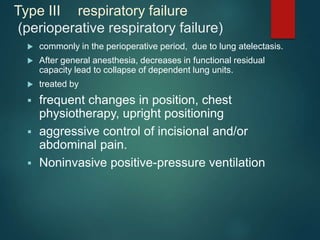
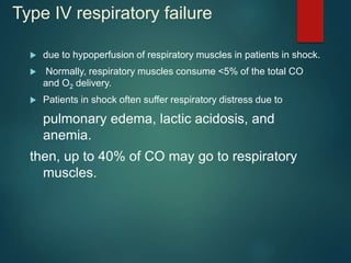
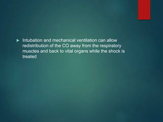
2. DEFINITION
The inability of the respiratory system to adequately
oxygenate the blood with or without a concurrent
alteration in carbon dioxide elimination
3. Acute and Chronic
Depending on underlying cause
Acute
eg: drug overdose, pneumonia, pneumothorax
Chronic
eg: severe COPD
5. Type I - Hypoxemic Failure
Oxygenation failure
PaO2 < 60 mmHg OR < 8
kpa
PaCO2 normal or < 35
mmHg
pH normal or elevated
6. Type I - Hypoxemic Failure
ventilation (VA) and perfusion (Q) mismatching is the
most common cause of hypoxemia.
Either by increasing the dead space or by wasted
ventilation
10. Type III respiratory failure
(perioperative respiratory failure)
commonly in the perioperative period, due to lung atelectasis.
After general anesthesia, decreases in functional residual
capacity lead to collapse of dependent lung units.
treated by
frequent changes in position, chest
physiotherapy, upright positioning
aggressive control of incisional and/or
abdominal pain.
Noninvasive positive-pressure ventilation
11. Type IV respiratory failure
due to hypoperfusion of respiratory muscles in patients in shock.
Normally, respiratory muscles consume <5% of the total CO
and O2 delivery.
Patients in shock often suffer respiratory distress due to
pulmonary edema, lactic acidosis, and
anemia.
then, up to 40% of CO may go to respiratory
muscles.
12. Intubation and mechanical ventilation can allow
redistribution of the CO away from the respiratory
muscles and back to vital organs while the shock is
treated
14. Patient Presentation
Neurological
Hypercapnoea also produce
tremor,myoclonic jerks, asterexis etc.
Increased CNS blood flow causes raised ICT- headache and
papilloedema.
Headache on waking up is common in chronic hypercapnia, may be
due to increased CO2 retention in sleep
20. Immediate determination of upper airway
patency
Examination for central and peripheral
cyanosis
Measurement of the respiratory rate
Observation of the depth and pattern of
respiration
Initial Assessment and Stabilization
of Respiratory Failure
21. Assess the configuration of the chest
wall and its movement during the
respiratory cycle
Palpation and auscultation over each
hemithorax
Signs of respiratory distress including
flaring of nostrils, pursed-lip breathing,
and use of accessory muscles of
respiration
22. Above observations allow an
initial assessment of respiratory
drive, pump function, and delivery
of gas to the lungs
24. PULSE OXIMETRY
There is a relationship between the amount of oxygen dissolved in the
blood and the amount attached to the hemoglobin.
Normal Oxyhemoglobin Dissociation Curve
97% saturation = 97 PaO2 (normal)
90% saturation = 60 PaO2 (danger)
80% saturation = 45 PaO2 (severe hypoxia)
25. MANAGEMENT
Initial therapy be implemented before the specific DIAGNOSIS
Adequate airway protection, oxygenation, and ventilation should be assured
and stabilize the Patient
Hypoxemia and hypercarbia can rapidly lead to circulatory failure and death
THEN, if possible treat the primary condition.
26. Type I respiratory failure
GOAL: to increase the oxygen saturation to 85 to 90 %
by giving oxygen at increasing FiO2.
Maintain adequate cardiac output and correct anemia.
Treat contitions like fever, agitation, overfeeding,
vigorous respiratory activity & sepsis which increase
O2 demand
27. Type I respiratory failure
prolonged exposure to high FiO2(>50%)/prolonged
duration of treatment is avoided, due to pulmonary
toxicity.
28. Type I respiratory failure
Indications of mechanical ventilation are:
1. Inadequate oxygenation despite of high FiO2
2. Increasing PaCO2 associated with altered mental
status or increasing fatigue
3. Failure to control secretions
29. Type II respiratory failure
Most commonly COPD, there is some degree of c/c
resp failure leading to hypercapnea.
Acute on c/c: acidemia and increase in bicarbonate in
ABG
30. Type II respiratory failure
1. Relief of hypoxia
By giving supplemental O2.
by nasal prongs at flow of 1 to 3 L/ min
Or by venturi mask with flow set to 24 to 28 %.
Recheck arterial blood gases/O2 saturation to look for
improvement.
PaO2 of > 50 mmHg is considered as adequate
31. Type II respiratory failure
Avoid sedatives, as they decrease ventilatory drive
To improve acidosis and hypercapnea and
oxygenation respiratory stimulants like doxapram can
be tried, by close monitoring of ABG.
32. Type II respiratory failure
Non invasive positive pressure ventilation.
Advantage : avoiding intubation
Aims at improving ventilation and gas exchange and reduces
work of respiratory muscles
33. Mechanical Ventilation in
respiratory failure Indications
1. Failure to attain PaO2 of 60 mmHg despite of
FiO2 of 0.6
2. Rapidly increasing hypercapnea, producing
uncompensated acidosis
3. Mental confusion either due to
hypoxemia/hypercapnea
4. Tachypnoea(>35/min)
5. Clinical judgement of impending exhuation of
the patient
34. Airway acsess
Nasotracheal/ orotracheal intubation
dis adv: laryngeal/ tracheal trauma,
used only up to 72 hrs
Tracheostomy:
complications; hemorrhage, infection
erosion of tube to esophagus, tracheal
stenosis, tube blockade and
respiratory infection
35. Ventilator settings
Tidal volume of approx 10 ml/kg
RR of 10 to 20/ min
So minute ventilation is 100 to 150 ml/kg
36. In COPD/asthma
In COPD tidal volume is kept at a little lower level(7 – 9 ml/kg) to
avoid auto PEEP and to prevent high inflation pressures to
already over inflated lungs to prevent barotrauma.
I:E ratio is kept at 1:4 or 1:5 to minimise air trapping
Peak inflation pressures are kept under 30 – 35 cm H2O
FiO2 is kept at 35%
37. In COPD/asthma
Regular monitoring of blood gases is needed.
Adjust inspired O2 and level of PEEP to PaO2 and
minute ventilation against PaCO2
38. Auto PEEP/ intrinsic PEEP
Develops in COPD due to decreased elastic recoil, decreased
expiratory flow and expiratory time due to tachypnoea- air
trapping and positive alveolar pressure at end of breathing.
Can impede venous return and decrease cardiac output.
Increase chance of barotrauma
39. In ARDS..
Tidal volume is kept low to prevent barotruama and
pnuemothorax and maintaining CVP at a lower range;
PEEP is maintained at a higher range to minimise FiO2 and
prevent alveolar collapse
I:E ratio is kept >1:1
This results in better survival than conventional ventilation
strategies.
40. Managenment of patient on
ventilator
Monitor ECG, heart rate, BP, oxygenation, ABG, urine output
Do not lower PaCO2 suddenly in a patient whom the resting
level is known to be high.
Better to aim at treating acidosis
Adeqaute sedation
Regular suction of airways
Adequate nutrition : enteral/ parenteral
41. Weaning..
Some times difficult with COPD
Can be tried when underlying condition/
infection has subsided, SaO2>90% with FiO2
< 0.4 and PEEP<5,alveolar ventilation is
adequate with pH normal, cardiovascular
function is stable, upper airway function is
normal,
Weaning index; ratio of beathing frequency to tidal
volume(breaths/min/L) <105 in spont ventilation thru tube,
succesful Extubation is likley
42. Weaning modes
T piece and CPAP Weaning: best tolerated by patients
with mechanical ventilation for brief periods
SIMV & PSV modes
best for intubated for extended periods &require
gradual respiratory-muscle reconditioning
43. complications
Barotrauma(1 – 8%)
more common with ARDS
Assc with high peak airway pressures,
High levels of external and auto PEEP,
High tidal volumes
GI bleeding due to gastric erosions
Nosocomial pneumonia
Cardiac arrhythmias, pulmonary embolism etc
44. Prognosis
Best predictor of mortality in patients with acute on chronic
respiratory failure is degree of acidemia.
pH< 7.26 is assc with higher mortality.
Long term mortality of patients who survive an episode of acute
respiratory failure depends on underlying illness.
Eg: COPD, 50% survival at end of 3 years
45. THANK YOU
“tasmādasaktaḥ satataṃ kāryaṃ karma samācara
asakto hyācarankarma paramāpnoti pūruṣaḥ”
“go on efficiently doing your duty at all times without
attachment.
doing work without attachment man attains the supreme.”