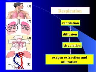
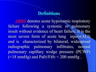
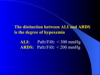
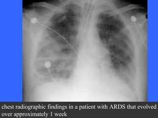
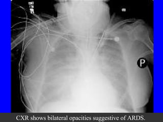
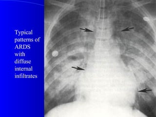
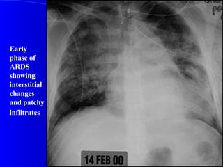
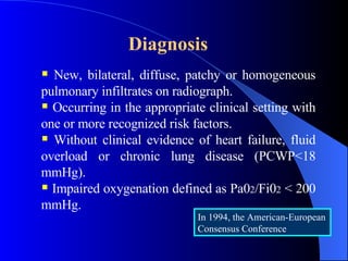
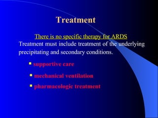
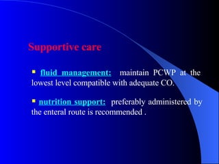
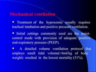
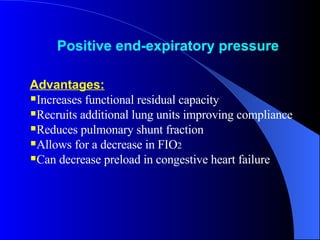
Acute Respiratory Distress Syndrome (ARDS) Yuan  Zhiming Department of Emergency  Medicine  The General Hospital  Tianjin Medical University
●   General Considerations ●   Etiology ●   Pathogenesis  ●   Pathophysiology ●   Pathology ●   Clinical Presentation ●   Diagnosis ●   Treatment ●   Prognosis Contents
 
Respiration   ventilation diffusion circulation oxygen extraction and utilization   (1) (2) (3) (4)
General considerations Definitions Retrospect Incidence
ARDS  denotes acute hypoxemic respiratory failure following a systemic or pulmonary insult without evidence of heart failure. It is the most severe form of acute lung  injury(ALI) and is  characterized by bilateral, widespread radiographic pulmonary infiltrates, normal pulmonary capillary wedge pressure (PCWP) (<18 mmHg) and Pa0 2 /Fi0 2  < 200 mmHg . Definitions
ALI   is a syndrome of inflammation and increased permeability that is associated with a constellation of clinical, radiologic, and physiologic abnormalities that cannot be explained by, but may coexist with, left atrial or pulmonary capillary hypertension.   Exclusion of left atrial hypertension as the primary cause of hypoxemia is critical   to this   definition
The distinction between ALI and ARDS is the degree of hypoxemia   ALI:   Pa0 2 /Fi0 2   < 300 mmHg  ARDS:   Pa0 2 /Fi0 2   < 200 mmHg
Retrospect ARDS was first reported by Ashbaugh in 1967.  Petty and Ashbaugh renamed it as   adult  respiratory distress syndrome in 1971 in order to distinguish them from neonate respiratory distress syndrome. The American-European Consensus Committee on ARDS standardized the definition in 1994 and renamed it acute rather than adult respiratory distress syndrome because it occurs at all ages.
Incidence Incidence  :  12.6 to 18 per 100,000 persons annually  Controversy  still  exists  about  the  correct incidence because of differing criteria used to define ARDS
Risk Factors (common) Direct lung injury Pneumonia Aspiration of gastric contents Inhalation injury  Near drowning  Lung contusion  Fat embolism Indirect lung injury Sepsis  Shock Severe trauma Acute pancreatitis  Massive transfusions Cardiopulmonary bypass Drug overdose
Pathogenesis The pathogenesis of ARDS is not well known  inflammatory cells stimulated  damage of capillary endothelial cells and alveolar epithelial cells vascular permeability   surfactant  interstitial and alveolar pulmonary edema risk factors  pro-inflammatory cytokines and mediators released  hypoxemia alveolar collapse
Pathophysiology
Pathology In ARDS, the injured lung go through three phases:  exudative ,  proliferative , and  fibrotic , but the course of each phase and the overall disease progression is variable, and the three phases don’t have a definite borderline, they overlap partly.
damage to the alveolar epithelium (type I alveolar cells mainly) and vascular endothelium  Exudative phase(<7d): leakage of water, protein, and inflammatory and red blood cells into the interstitium and alveolar lumen; producing hyaline membranes
Proliferative phase (1-3wk): Type II cells proliferate with some epithelial cell regeneration, fibroblastic reaction, and remodeling.  Injury to the surfactant-producing type II cells  alveolar collapse
Fibrotic phase ( > 3wk): Collagen deposition in alveolar, vascular, and interstitial beds with development of microcysts.
Nondescript very heavy and poorly aerated lungs. Each lung in  ARDS usually weighs over 1000 grams. (A normal lung weighs 200-300 grams)
Heavy, red lungs showing congestion and edema.
3-4 weeks after onset of ARDS, extensive interstitial fibrosis developed.
Photomicrograph shows ARDS in the exudative stage. Note the hyaline membranes and loss of alveolar epithelium in this early stage.
Photomicrograph shows ARDS in the early proliferative stage. Note the type 2 pneumocytic proliferation, with widening of the septa and interstitial fibroblast proliferation.
Photomicrograph shows ARDS in the late proliferative stage. Note the extensive fibroblast proliferation, with incorporation of the hyaline membranes.
Diffuse Alveolar Damage Hyaline membranes are lining the alveolar ducts and alveolar septa.
Clinical  Presentation   Rapid onset of profound dyspnea that usually occurs 12-48 h after the initiating event.  Labored breathing, tachypnea, intercostal retractions, and crackles. Marked hypoxemia that is refractory to treatment with supplemental oxygen.  Many patients demonstrate multiple organ failure.
Chest radiography:   In nondirect insults, the initial radiograph may be nonspecific or similar to congestive heart failure with mild effusions (interstitial pulmonary edema with diffuse infiltrates);   As the disease progresses, the characteristic bilateral diffuse alveolar and reticular opacities become evident. In patients with direct pulmonary insults, focal changes may be evident early. Characterized by diffuse or patchy bilateral pulmonary infiltrates
Chest radiograph shows an endotracheal tube, left subclavian central venous catheter into the superior vena cava, and bilateral patchy opacities in mostly the middle and lower lung zones.  The patient had been in respiratory failure for 1 week with the diagnosis of ARDS.
chest radiographic findings in a patient with ARDS that evolved over approximately 1 week
CXR shows bilateral opacities suggestive of ARDS.
Typical patterns of ARDS with diffuse internal infiltrates
Early phase of ARDS showing interstitial changes and patchy infiltrates
Late stage of ARDS showing bilateral and diffuse alveolar and reticular opacification
CT scan of the chest showing diffuse infiltrates, ground glass appearance, and air bronchograms.  ARDS
a small right pleural effusion, consolidation with air-bronchograms, and some ground-glass appearing opacities. The findings indicate an alveolar process, in this case, alveolar damage.  High-resolution computed tomographic (HRCT) image in a patient with ARDS
Diagnosis   New, bilateral, diffuse, patchy or homogeneous pulmonary infiltrates on radiograph.  Occurring in the appropriate clinical setting with one or more recognized risk factors. Without clinical evidence of heart failure, fluid overload or chronic lung disease (PCWP<18 mmHg). Impaired oxygenation defined as Pa0 2 /Fi0 2  < 200 mmHg. In 1994, the American-European Consensus Conference
Treatment   There is no specific therapy for ARDS   Treatment must include treatment of the underlying precipitating and secondary conditions.   supportive care   mechanical ventilation   pharmacologic treatment
Supportive care   fluid management:   maintain PCWP at the lowest level compatible with adequate CO.  nutrition support:   preferably administered by the enteral route is recommended .
Mechanical ventilation  Treatment of  the  hypoxemia  usually  requires tracheal intubation and positive pressure ventilation.  Initial settings commonly used are the assist- control mode with provision of adequate positive end-expiratory pressure (PEEP).  A detailed volume ventilation protocol that employs small tidal volume(<6ml/kg of body  weight)  resulted  in  the lowest mortality (31%).
Positive end-expiratory pressure   Advantages:   Increases functional residual capacity Recruits additional lung units improving compliance Reduces pulmonary shunt fraction Allows for a decrease in FIO 2   Can decrease preload in congestive heart failure
Disadvantages: Increase mean airway pressure leading to reduced venous return Can increase pulmonary vascular resistance and right-heart  dsyfunction Altered renal blood flow with increase in ADH(antidiuretic hormone )  release Barotrauma caused by increased pressure
Pharmacologic treatment   Corticosteroids:  not indicated in the early course, to use in late-phase might be beneficial . Antibiotics  Stress ulcer and thromboembolic prophylaxis  Other drugs:   like prostacyclin, nitric oxide, prostaglandins, and receptor antagonists to various cytokines, etc.
Prognosis   Mortality rate : greater than 50% between 1979 and 1994,  more recent studies show a decline in mortality to be 35%~40%  If  ARDS  is accompanied by sepsis,  the mortality rate may reach 90%. The major causes of death in ARDS are the primary illness and secondary complications such as MODS or sepsis.  Most survivors of ARDS are left with some pulmonary symptoms (cough, dyspnea, sputum production), which tend to improve over time
Summarization   ARDS denotes acute hypoxemic respiratory failure following a systemic or pulmonary insult without evidence of HF,and is the most severe form of ALI. Provision of supplemental oxygen, lung rest, and supportive care are the fundamentals of therapy, endotracheal intubation and mechanical ventilation are frequently required, Timely correction of the inciting clinical condition is essential for preventing further injury.   It is characterized by dyspnea, profound hypoxemia, decreased lung compliance, and diffuse bilateral infiltrates on chest radiography.
Emphases Definition of ARDS Pathophysiology Clinical presentation & diagnosis of ARDS The principles of treatment   of ARDS
THANKS

Ards(En终)

  • 1.
    Acute Respiratory DistressSyndrome (ARDS) Yuan Zhiming Department of Emergency Medicine The General Hospital Tianjin Medical University
  • 2.
    General Considerations ● Etiology ● Pathogenesis ● Pathophysiology ● Pathology ● Clinical Presentation ● Diagnosis ● Treatment ● Prognosis Contents
  • 3.
  • 4.
    Respiration ventilation diffusion circulation oxygen extraction and utilization (1) (2) (3) (4)
  • 5.
  • 6.
    ARDS denotesacute hypoxemic respiratory failure following a systemic or pulmonary insult without evidence of heart failure. It is the most severe form of acute lung injury(ALI) and is characterized by bilateral, widespread radiographic pulmonary infiltrates, normal pulmonary capillary wedge pressure (PCWP) (<18 mmHg) and Pa0 2 /Fi0 2 < 200 mmHg . Definitions
  • 7.
    ALI is a syndrome of inflammation and increased permeability that is associated with a constellation of clinical, radiologic, and physiologic abnormalities that cannot be explained by, but may coexist with, left atrial or pulmonary capillary hypertension. Exclusion of left atrial hypertension as the primary cause of hypoxemia is critical to this definition
  • 8.
    The distinction betweenALI and ARDS is the degree of hypoxemia ALI: Pa0 2 /Fi0 2 < 300 mmHg ARDS: Pa0 2 /Fi0 2 < 200 mmHg
  • 9.
    Retrospect ARDS wasfirst reported by Ashbaugh in 1967. Petty and Ashbaugh renamed it as adult respiratory distress syndrome in 1971 in order to distinguish them from neonate respiratory distress syndrome. The American-European Consensus Committee on ARDS standardized the definition in 1994 and renamed it acute rather than adult respiratory distress syndrome because it occurs at all ages.
  • 10.
    Incidence Incidence : 12.6 to 18 per 100,000 persons annually Controversy still exists about the correct incidence because of differing criteria used to define ARDS
  • 11.
    Risk Factors (common)Direct lung injury Pneumonia Aspiration of gastric contents Inhalation injury Near drowning Lung contusion Fat embolism Indirect lung injury Sepsis Shock Severe trauma Acute pancreatitis Massive transfusions Cardiopulmonary bypass Drug overdose
  • 12.
    Pathogenesis The pathogenesisof ARDS is not well known inflammatory cells stimulated damage of capillary endothelial cells and alveolar epithelial cells vascular permeability surfactant interstitial and alveolar pulmonary edema risk factors pro-inflammatory cytokines and mediators released hypoxemia alveolar collapse
  • 13.
  • 14.
    Pathology In ARDS,the injured lung go through three phases: exudative , proliferative , and fibrotic , but the course of each phase and the overall disease progression is variable, and the three phases don’t have a definite borderline, they overlap partly.
  • 15.
    damage to thealveolar epithelium (type I alveolar cells mainly) and vascular endothelium Exudative phase(<7d): leakage of water, protein, and inflammatory and red blood cells into the interstitium and alveolar lumen; producing hyaline membranes
  • 16.
    Proliferative phase (1-3wk):Type II cells proliferate with some epithelial cell regeneration, fibroblastic reaction, and remodeling. Injury to the surfactant-producing type II cells alveolar collapse
  • 17.
    Fibrotic phase (> 3wk): Collagen deposition in alveolar, vascular, and interstitial beds with development of microcysts.
  • 18.
    Nondescript very heavyand poorly aerated lungs. Each lung in ARDS usually weighs over 1000 grams. (A normal lung weighs 200-300 grams)
  • 19.
    Heavy, red lungsshowing congestion and edema.
  • 20.
    3-4 weeks afteronset of ARDS, extensive interstitial fibrosis developed.
  • 21.
    Photomicrograph shows ARDSin the exudative stage. Note the hyaline membranes and loss of alveolar epithelium in this early stage.
  • 22.
    Photomicrograph shows ARDSin the early proliferative stage. Note the type 2 pneumocytic proliferation, with widening of the septa and interstitial fibroblast proliferation.
  • 23.
    Photomicrograph shows ARDSin the late proliferative stage. Note the extensive fibroblast proliferation, with incorporation of the hyaline membranes.
  • 24.
    Diffuse Alveolar DamageHyaline membranes are lining the alveolar ducts and alveolar septa.
  • 25.
    Clinical Presentation Rapid onset of profound dyspnea that usually occurs 12-48 h after the initiating event. Labored breathing, tachypnea, intercostal retractions, and crackles. Marked hypoxemia that is refractory to treatment with supplemental oxygen. Many patients demonstrate multiple organ failure.
  • 26.
    Chest radiography: In nondirect insults, the initial radiograph may be nonspecific or similar to congestive heart failure with mild effusions (interstitial pulmonary edema with diffuse infiltrates); As the disease progresses, the characteristic bilateral diffuse alveolar and reticular opacities become evident. In patients with direct pulmonary insults, focal changes may be evident early. Characterized by diffuse or patchy bilateral pulmonary infiltrates
  • 27.
    Chest radiograph showsan endotracheal tube, left subclavian central venous catheter into the superior vena cava, and bilateral patchy opacities in mostly the middle and lower lung zones. The patient had been in respiratory failure for 1 week with the diagnosis of ARDS.
  • 28.
    chest radiographic findingsin a patient with ARDS that evolved over approximately 1 week
  • 29.
    chest radiographic findingsin a patient with ARDS that evolved over approximately 1 week
  • 30.
    CXR shows bilateralopacities suggestive of ARDS.
  • 31.
    Typical patterns ofARDS with diffuse internal infiltrates
  • 32.
    Early phase ofARDS showing interstitial changes and patchy infiltrates
  • 33.
    Late stage ofARDS showing bilateral and diffuse alveolar and reticular opacification
  • 34.
    CT scan ofthe chest showing diffuse infiltrates, ground glass appearance, and air bronchograms. ARDS
  • 35.
    a small rightpleural effusion, consolidation with air-bronchograms, and some ground-glass appearing opacities. The findings indicate an alveolar process, in this case, alveolar damage. High-resolution computed tomographic (HRCT) image in a patient with ARDS
  • 36.
    Diagnosis New, bilateral, diffuse, patchy or homogeneous pulmonary infiltrates on radiograph. Occurring in the appropriate clinical setting with one or more recognized risk factors. Without clinical evidence of heart failure, fluid overload or chronic lung disease (PCWP<18 mmHg). Impaired oxygenation defined as Pa0 2 /Fi0 2 < 200 mmHg. In 1994, the American-European Consensus Conference
  • 37.
    Treatment There is no specific therapy for ARDS Treatment must include treatment of the underlying precipitating and secondary conditions. supportive care mechanical ventilation pharmacologic treatment
  • 38.
    Supportive care fluid management: maintain PCWP at the lowest level compatible with adequate CO. nutrition support: preferably administered by the enteral route is recommended .
  • 39.
    Mechanical ventilation Treatment of the hypoxemia usually requires tracheal intubation and positive pressure ventilation. Initial settings commonly used are the assist- control mode with provision of adequate positive end-expiratory pressure (PEEP). A detailed volume ventilation protocol that employs small tidal volume(<6ml/kg of body weight) resulted in the lowest mortality (31%).
  • 40.
    Positive end-expiratory pressure Advantages: Increases functional residual capacity Recruits additional lung units improving compliance Reduces pulmonary shunt fraction Allows for a decrease in FIO 2 Can decrease preload in congestive heart failure
  • 41.
    Disadvantages: Increase meanairway pressure leading to reduced venous return Can increase pulmonary vascular resistance and right-heart  dsyfunction Altered renal blood flow with increase in ADH(antidiuretic hormone ) release Barotrauma caused by increased pressure
  • 42.
    Pharmacologic treatment Corticosteroids: not indicated in the early course, to use in late-phase might be beneficial . Antibiotics Stress ulcer and thromboembolic prophylaxis Other drugs: like prostacyclin, nitric oxide, prostaglandins, and receptor antagonists to various cytokines, etc.
  • 43.
    Prognosis Mortality rate : greater than 50% between 1979 and 1994, more recent studies show a decline in mortality to be 35%~40% If ARDS is accompanied by sepsis, the mortality rate may reach 90%. The major causes of death in ARDS are the primary illness and secondary complications such as MODS or sepsis. Most survivors of ARDS are left with some pulmonary symptoms (cough, dyspnea, sputum production), which tend to improve over time
  • 44.
    Summarization ARDS denotes acute hypoxemic respiratory failure following a systemic or pulmonary insult without evidence of HF,and is the most severe form of ALI. Provision of supplemental oxygen, lung rest, and supportive care are the fundamentals of therapy, endotracheal intubation and mechanical ventilation are frequently required, Timely correction of the inciting clinical condition is essential for preventing further injury. It is characterized by dyspnea, profound hypoxemia, decreased lung compliance, and diffuse bilateral infiltrates on chest radiography.
  • 45.
    Emphases Definition ofARDS Pathophysiology Clinical presentation & diagnosis of ARDS The principles of treatment of ARDS
  • 46.