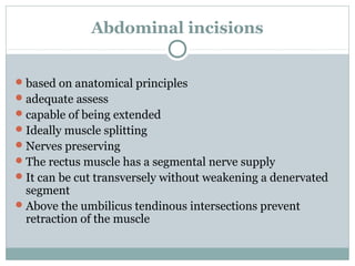
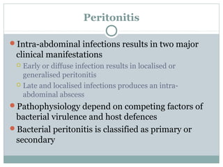
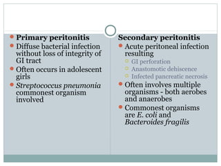
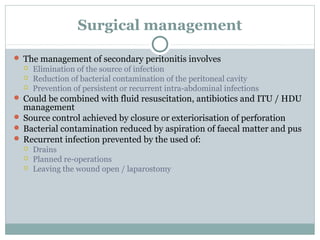
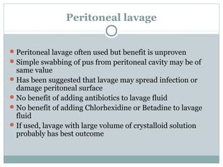
Abdominal access
       & Peritonitis


               S A NAQVI
      CONSULTANT GENERAL SURGEON
MID WESTERN REGIONAL HOSPITALS, LIMERICK,
                IRELAND
Abdominal incisions


 based on anatomical principles
 adequate assess
 capable of being extended
 Ideally muscle splitting
 Nerves preserving
 The rectus muscle has a segmental nerve supply
 It can be cut transversely without weakening a denervated
  segment
 Above the umbilicus tendinous intersections prevent
  retraction of the muscle
Midline incision

Commonest approach
The following structures are divided:
    Skin
    Linea alba
    Transversalis fascia
    Extraperitoneal fat
    Peritoneum
Can be extended around the umbilicus
 Falciform ligament should be
  avoided
 The bladder can be accessed via
  an extraperitoneal approach
  through the space of Retzius
 Mass closure technique
 The most popular sutures are
  either non-absorbable or
  absorbable monofilaments
 At least 1 cm bits should be
  taken 1 cm apart
 Length of sutures, four times
  the wound length
Paramedian incision

Parallel to and approximately 3 cm from the
 midline
The incision Cut
  Skin
  Anterior rectus sheath

  Rectus - retracted laterally

  Posterior rectus sheath (above the arcuate line)

  Transversalis fascia

  Extraperitoneal fat

  Peritoneum
Paramedian incision Cont…
The potential advantages
1. The rectus muscle is not divided
     1.   The incisions in the anterior and
          posterior rectus sheath are
          separated by muscle
2.   Closed in layers
3.   Had a lower incidence of
     incisional hernia (when sutures
     were not so good)

The potential Disadvantages
1. Takes longer to make and close
Peritonitis

Intra-abdominal infections results in two major
 clinical manifestations
  Early or diffuse infection results in localised or
   generalised peritonitis
  Late and localised infections produces an intra-
   abdominal abscess
Pathophysiology depend on competing factors of
 bacterial virulence and host defences
Bacterial peritonitis is classified as primary or
 secondary
 Primary peritonitis            Secondary peritonitis
 Diffuse bacterial infection     Acute peritoneal infection
  without loss of integrity of     resulting
  GI tract                            GI perforation
 Often occurs in adolescent          Anastomotic dehiscence
  girls                               Infected pancreatic necrosis
 Streptococcus pneumonia         Often involves multiple
  commonest organism               organisms - both aerobes
  involved                         and anaerobes
                                  Commonest organisms
                                   are E. coli and
                                   Bacteroides fragilis
Surgical management

 The management of secondary peritonitis involves
     Elimination of the source of infection
     Reduction of bacterial contamination of the peritoneal cavity
     Prevention of persistent or recurrent intra-abdominal infections
 Could be combined with fluid resuscitation, antibiotics and ITU / HDU
  management
 Source control achieved by closure or exteriorisation of perforation
 Bacterial contamination reduced by aspiration of faecal matter and pus
 Recurrent infection prevented by the used of:
     Drains
     Planned re-operations
     Leaving the wound open / laparostomy
Peritoneal lavage


 Peritoneal lavage often used but benefit is unproven
 Simple swabbing of pus from peritoneal cavity may be of
  same value
 Has been suggested that lavage may spread infection or
  damage peritoneal surface
 No benefit of adding antibiotics to lavage fluid
 No benefit of adding Chlorhexidine or Betadine to lavage
  fluid
 If used, lavage with large volume of crystalloid solution
  probably has best outcome
Intra-abdominal abscesses


An intra-abdominal abscess may arise following:
  Localisation of peritonitis
  Gastrointestinal perforation
  Anastomotic leak
  Haematogenous spread
They develop in sites of gravitational drainage
  Pelvis
  Subhepatic spaces
  Subphrenic spaces
  Paracolic gutters
Clinical features

Postoperative abscesses usually present at between
 5 and 10 days after surgery
Suspect if unexplained persistent or swinging
 pyrexia
May also cause abdominal pain and diarrhoea
A mass may be present with overlying erythema
 and tenderness
A pelvic abscess may be palpable only on rectal
 examination
Management

 Ultrasound scanning may reveal the diagnosis
 Contrast-enhanced CT is probably the investigation of
  choice
 May delineate a gastrointestinal or anastomotic leak
 Identifies collection and often allows percutaneous
  drainage
 Operative drainage may be required if:
     Multi-locular abscess
     No safe route for per cutaneous drainage
     Recollection after percutaneous drainage
 Patients should receive antibiotic therapy guided by
  organism sensitivities

Abdominal access

  • 1.
    Abdominal access & Peritonitis S A NAQVI CONSULTANT GENERAL SURGEON MID WESTERN REGIONAL HOSPITALS, LIMERICK, IRELAND
  • 2.
    Abdominal incisions  basedon anatomical principles  adequate assess  capable of being extended  Ideally muscle splitting  Nerves preserving  The rectus muscle has a segmental nerve supply  It can be cut transversely without weakening a denervated segment  Above the umbilicus tendinous intersections prevent retraction of the muscle
  • 4.
    Midline incision Commonest approach Thefollowing structures are divided:  Skin  Linea alba  Transversalis fascia  Extraperitoneal fat  Peritoneum Can be extended around the umbilicus
  • 5.
     Falciform ligamentshould be avoided  The bladder can be accessed via an extraperitoneal approach through the space of Retzius  Mass closure technique  The most popular sutures are either non-absorbable or absorbable monofilaments  At least 1 cm bits should be taken 1 cm apart  Length of sutures, four times the wound length
  • 6.
    Paramedian incision Parallel toand approximately 3 cm from the midline The incision Cut  Skin  Anterior rectus sheath  Rectus - retracted laterally  Posterior rectus sheath (above the arcuate line)  Transversalis fascia  Extraperitoneal fat  Peritoneum
  • 7.
    Paramedian incision Cont… Thepotential advantages 1. The rectus muscle is not divided 1. The incisions in the anterior and posterior rectus sheath are separated by muscle 2. Closed in layers 3. Had a lower incidence of incisional hernia (when sutures were not so good) The potential Disadvantages 1. Takes longer to make and close
  • 8.
    Peritonitis Intra-abdominal infections resultsin two major clinical manifestations  Early or diffuse infection results in localised or generalised peritonitis  Late and localised infections produces an intra- abdominal abscess Pathophysiology depend on competing factors of bacterial virulence and host defences Bacterial peritonitis is classified as primary or secondary
  • 9.
     Primary peritonitis Secondary peritonitis  Diffuse bacterial infection  Acute peritoneal infection without loss of integrity of resulting GI tract  GI perforation  Often occurs in adolescent  Anastomotic dehiscence girls  Infected pancreatic necrosis  Streptococcus pneumonia  Often involves multiple commonest organism organisms - both aerobes involved and anaerobes  Commonest organisms are E. coli and Bacteroides fragilis
  • 10.
    Surgical management  Themanagement of secondary peritonitis involves  Elimination of the source of infection  Reduction of bacterial contamination of the peritoneal cavity  Prevention of persistent or recurrent intra-abdominal infections  Could be combined with fluid resuscitation, antibiotics and ITU / HDU management  Source control achieved by closure or exteriorisation of perforation  Bacterial contamination reduced by aspiration of faecal matter and pus  Recurrent infection prevented by the used of:  Drains  Planned re-operations  Leaving the wound open / laparostomy
  • 11.
    Peritoneal lavage  Peritoneallavage often used but benefit is unproven  Simple swabbing of pus from peritoneal cavity may be of same value  Has been suggested that lavage may spread infection or damage peritoneal surface  No benefit of adding antibiotics to lavage fluid  No benefit of adding Chlorhexidine or Betadine to lavage fluid  If used, lavage with large volume of crystalloid solution probably has best outcome
  • 12.
    Intra-abdominal abscesses An intra-abdominalabscess may arise following:  Localisation of peritonitis  Gastrointestinal perforation  Anastomotic leak  Haematogenous spread They develop in sites of gravitational drainage  Pelvis  Subhepatic spaces  Subphrenic spaces  Paracolic gutters
  • 13.
    Clinical features Postoperative abscessesusually present at between 5 and 10 days after surgery Suspect if unexplained persistent or swinging pyrexia May also cause abdominal pain and diarrhoea A mass may be present with overlying erythema and tenderness A pelvic abscess may be palpable only on rectal examination
  • 14.
    Management  Ultrasound scanningmay reveal the diagnosis  Contrast-enhanced CT is probably the investigation of choice  May delineate a gastrointestinal or anastomotic leak  Identifies collection and often allows percutaneous drainage  Operative drainage may be required if:  Multi-locular abscess  No safe route for per cutaneous drainage  Recollection after percutaneous drainage  Patients should receive antibiotic therapy guided by organism sensitivities