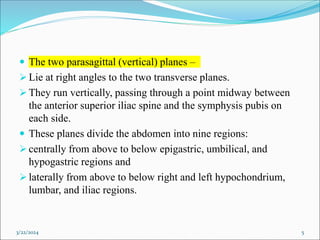
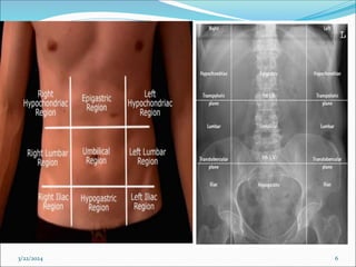
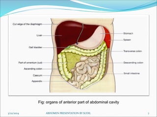
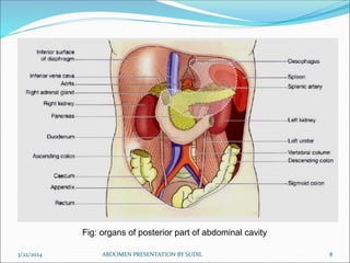
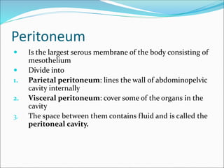
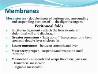
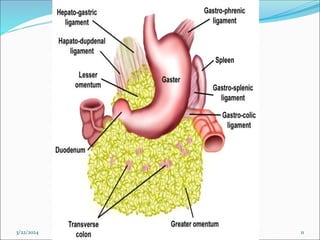
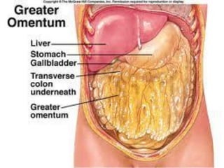
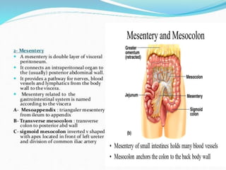
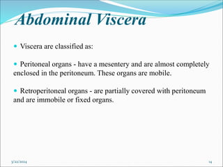
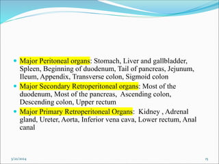
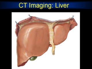
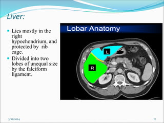
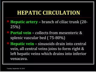
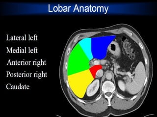
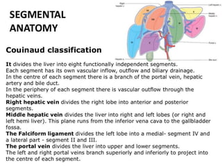
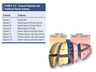
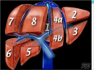
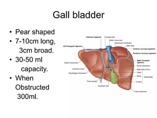
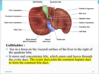
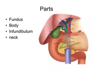
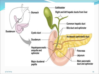
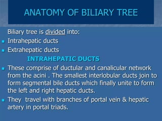
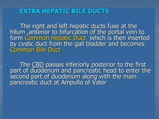
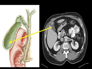
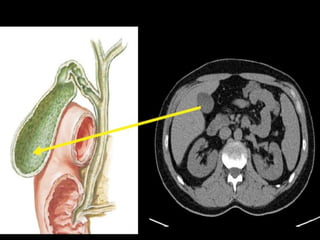
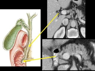
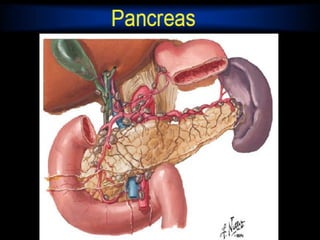
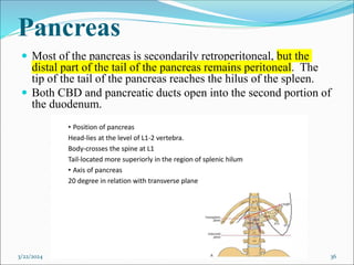
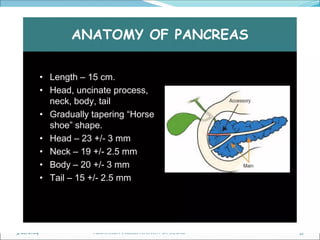
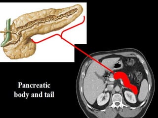
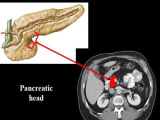
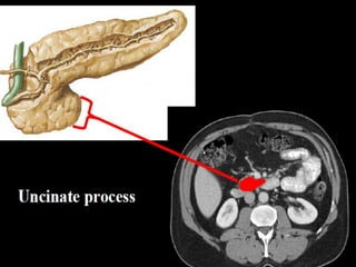
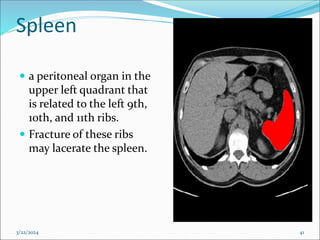
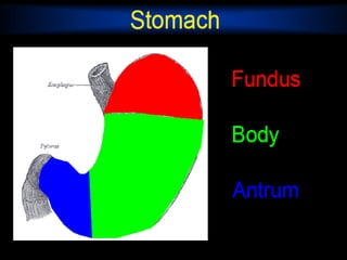
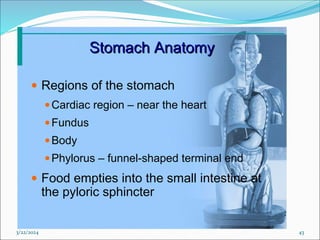
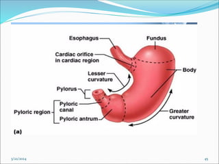
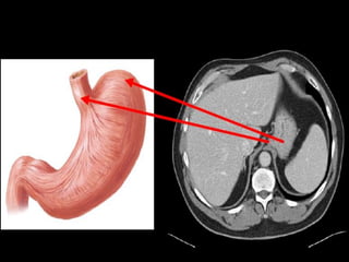
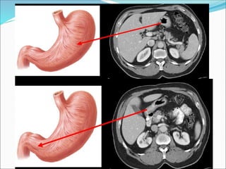
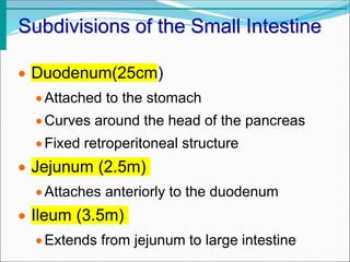
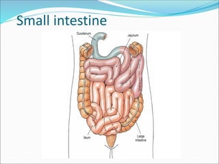
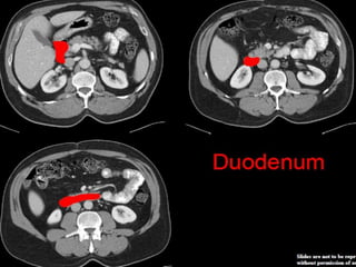
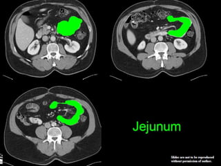
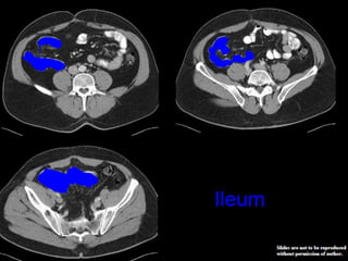
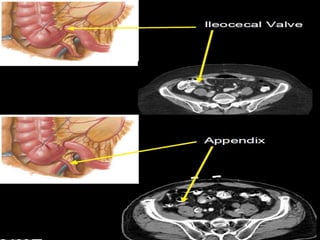
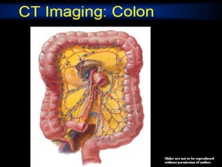
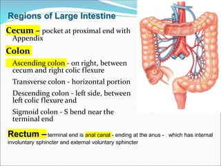
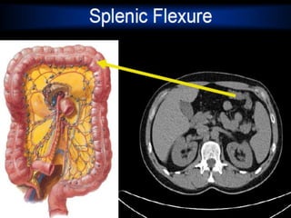
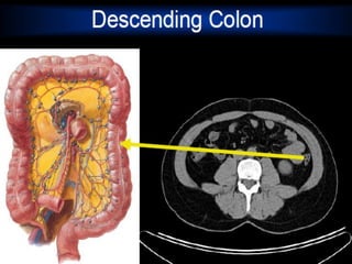
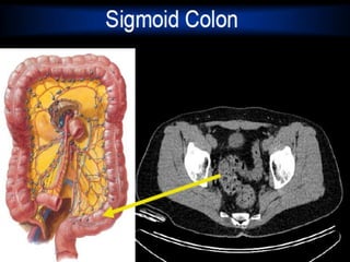
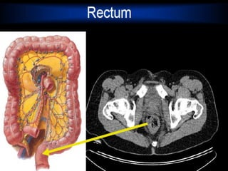
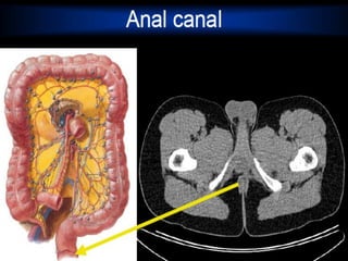
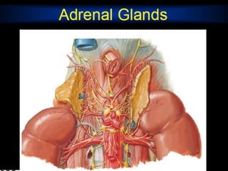
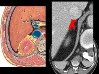
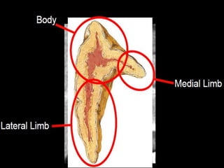
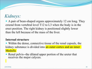
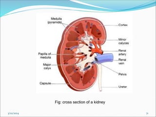
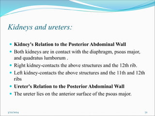
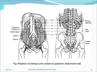
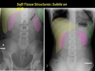
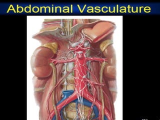
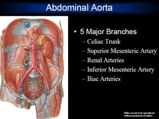
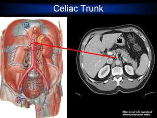
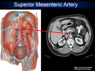
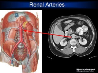
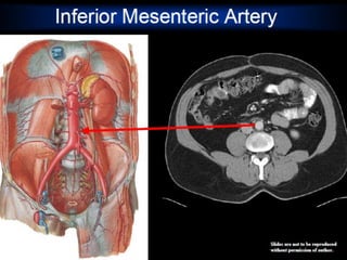
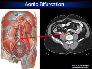
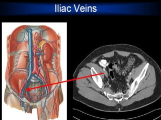
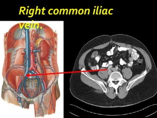
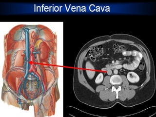
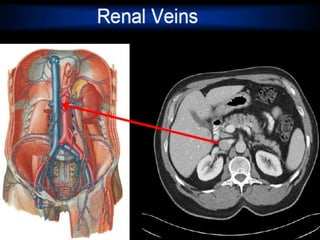
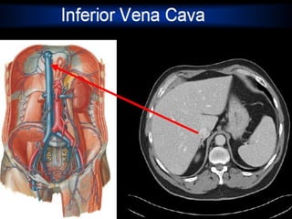
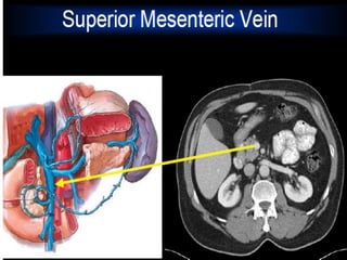
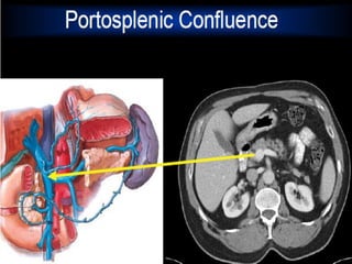
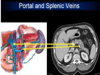
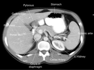
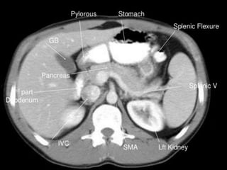
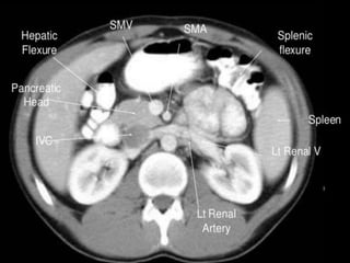
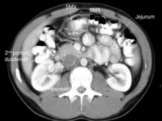
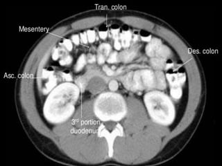
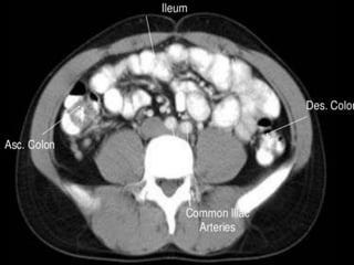
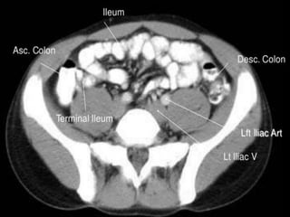
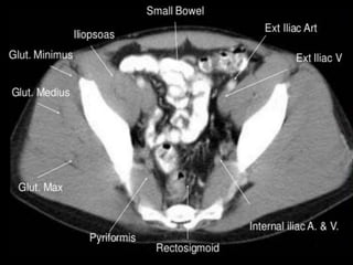
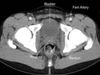
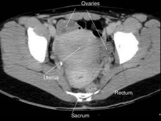
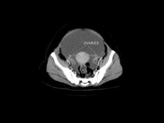
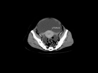
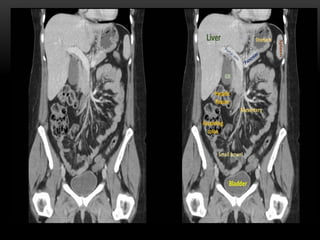
General Anatomy:
The largest cavity of the body,
Bounded
Anteriorly - by abdominal wall muscles
Posteriorly - by the vertebral column and posterior wall
muscles
Laterally - by lower ribs and parts of muscles of abdominal
wall
Superiorly - by the diaphragm
Inferiorly - by pelvic cavity
3/22/2024 1
Abdominal walls:
 Five pairs of muscles form the anterior wall:
 Rectus abdominis
 External oblique
 Internal oblique
 Transversus abdominis
 Three pairs form the posterior wall:
 Quadratus lumborum
 Psoas major
 Iliacus
Linea alba: A very strong midline tendinous cord, that extends from the
xiphoid process to the symphysis pubis. Divides the anterior abdominal wall
longitudinally into two identical halves.
3/22/2024 2
3/22/2024 3
Fig: Anterior abdominal wall muscles Fig: Posterior abdominal wall muscles
Planes and regions:
 Divided into nine regions by two transverse and two vertical
planes
 The upper transverse plane - The Transpyloric Plane,
 approximately midway between the upper border of
xiphisternum and umbilicus. Posteriorly, passes through the
body of the first lumbar vertebra; Anteriorly, passes through the
tips of the right and left ninth costal cartilages.
 The lower transverse plane - The Transtubercular Plane,
Lies at the level of tubercles of the iliac crest anteriorly, and
near the upper border of the fifth lumbar vertebra posteriorly.
3/22/2024 4
 The two parasagittal (vertical) planes –
Lie at right angles to the two transverse planes.
They run vertically, passing through a point midway between
the anterior superior iliac spine and the symphysis pubis on
each side.
 These planes divide the abdomen into nine regions:
centrally from above to below epigastric, umbilical, and
hypogastric regions and
laterally from above to below right and left hypochondrium,
lumbar, and iliac regions.
3/22/2024 5
3/22/2024 6
Fig: organs of anterior part of abdominal cavity
3/22/2024 7
ABDOMEN PRESENTATION BY SUDIL
3/22/2024 ABDOMEN PRESENTATION BY SUDIL 8
Fig: organs of posterior part of abdominal cavity
Peritoneum
 Is the largest serous membrane of the body consisting of
mesothelium
 Divide into
1. Parietal peritoneum: lines the wall of abdominopelvic
cavity internally
2. Visceral peritoneum: cover some of the organs in the
cavity
3. The space between them contains fluid and is called the
peritoneal cavity.
Membranes
Mesenteries - double sheets of peritoneum, surrounding
and suspending portions of the digestive organs
Peritoneal folds
1. falciform ligament:- attach the liver to anterior
abdominal wall and diaphragm
2. Greater omentum - "fatty apron", hangs anteriorly from
stomach, double layer encloses fat
3. Lesser omentum - between stomach and liver
4. Mesentery proper - suspends and wraps the small
intestine
5. Mesocolon - suspends and wraps the colon, parts are
i. transverse mesocolon
ii. sigmoid mesocolon
3/22/2024 ABDOMEN PRESENTATION BY SUDIL 11
Abdominal Viscera
 Viscera are classified as:
 Peritoneal organs - have a mesentery and are almost completely
enclosed in the peritoneum. These organs are mobile.
 Retroperitoneal organs - are partially covered with peritoneum
and are immobile or fixed organs.
3/22/2024 14
 Major Peritoneal organs: Stomach, Liver and gallbladder,
Spleen, Beginning of duodenum, Tail of pancreas, Jejunum,
Ileum, Appendix, Transverse colon, Sigmoid colon
 Major Secondary Retroperitoneal organs: Most of the
duodenum, Most of the pancreas, Ascending colon,
Descending colon, Upper rectum
 Major Primary Retroperitoneal Organs: Kidney , Adrenal
gland, Ureter, Aorta, Inferior vena cava, Lower rectum, Anal
canal
3/22/2024 15
Liver:
 Lies mostly in the
right
hypochondrium, and
protected by rib
cage.
 Divided into two
lobes of unequal size
by the falciform
ligament.
3/22/2024 17
3/22/2024 ABDOMEN PRESENTATION BY SUDIL 18
3/22/2024 ABDOMEN PRESENTATION BY SUDIL 26
Gallbladder :
 lies in a fossa on the visceral surface of the liver to the right of
the quadrate lobe.
 It stores and concentrates bile, which enters and leaves through
the cystic duct. The cystic duct joins the common hepatic duct
to form the common bile duct.
3/22/2024 27
3/22/2024 ABDOMEN PRESENTATION BY SUDIL 28
3/22/2024 29
3/22/2024 ABDOMEN PRESENTATION BY SUDIL 30
3/22/2024 ABDOMEN PRESENTATION BY SUDIL 31
Pancreas
 Most of the pancreas is secondarily retroperitoneal, but the
distal part of the tail of the pancreas remains peritoneal. The
tip of the tail of the pancreas reaches the hilus of the spleen.
 Both CBD and pancreatic ducts open into the second portion of
the duodenum.
3/22/2024 36
3/22/2024 ABDOMEN PRESENTATION BY SUDIL 37
Spleen
 a peritoneal organ in the
upper left quadrant that
is related to the left 9th,
10th, and 11th ribs.
 Fracture of these ribs
may lacerate the spleen.
3/22/2024 41
3/22/2024 43
Stomach:
 has a lesser curvature, which is connected to the porta hepatis
of the liver by the lesser omentum, and a greater curvature
from which the greater omentum is suspended.
 The cardiac region receives the esophagus.
 The dome-shaped upper portion of the stomach, which is
normally filled with air, is the fundus.
 The main center portion of the stomach is the body.
 The pyloric portion of the stomach has a thick muscular wall
and a narrow lumen that leads to the duodenum.
3/22/2024 44
3/22/2024 45
Small Intestine
 The body’s major digestive organ
 Site of nutrient absorption into the blood
 Muscular tube extending from the
pyloric sphincter to the ileocecal valve
 Suspended from the posterior
abdominal wall by the mesentery
Subdivisions of the Small Intestine
 Duodenum(25cm)
Attached to the stomach
Curves around the head of the pancreas
Fixed retroperitoneal structure
 Jejunum (2.5m)
Attaches anteriorly to the duodenum
 Ileum (3.5m)
Extends from jejunum to large intestine
Small intestine
Cecum – pocket at proximal end with
Appendix
Colon
Ascending colon - on right, between
cecum and right colic flexure
Transverse colon - horizontal portion
Descending colon - left side, between
left colic flexure and
Sigmoid colon - S bend near the
terminal end
Regions of Large Intestine
Rectum –terminal end is anal canal - ending at the anus - which has internal
involuntary sphincter and external voluntary sphincter
Kidneys:
 A pair of bean-shaped organs approximately 12 cm long. They
extend from vertebral level T12 to L3 when the body is in the
erect position. The right kidney is positioned slightly lower
than the left because of the mass of the liver.
Internal structure
 Within the dense, connective tissue of the renal capsule, the
kidney substance is divided into an outer cortex and an inner
Medulla
 Renal pelvis--the dilated upper portion of the ureter that
receives the major calyces.
3/22/2024 70
3/22/2024 71
Fig: cross section of a kidney
Kidneys and ureters:
 Kidney's Relation to the Posterior Abdominal Wall
 Both kidneys are in contact with the diaphragm, psoas major,
and quadratus lumborum .
 Right kidney-contacts the above structures and the 12th rib.
 Left kidney-contacts the above structures and the 11th and 12th
ribs
 Ureter's Relation to the Posterior Abdominal Wall
 The ureter lies on the anterior surface of the psoas major.
3/22/2024 72
3/22/2024 ABDOMEN PRESENTATION BY SUDIL 73
Fig: Relation of kidneys and ureters to posterior abdominal wall
Ureters : are fibro-muscular tubes that connect the kidneys to
the urinary bladder in the pelvis.
Urinary Bladder:
 The urinary bladder is covered superiorly by peritoneum.
 The body is a hollow muscular cavity.
 The neck is continuous with the urethra.
 The trigone is a smooth triangular area of mucosa located
internally at the base of the bladder.
 The base of the triangle is superior and bounded by the two
openings of the ureters.
 The apex of the trigone points inferiorly and is the opening for
the urethra.
3/22/2024 ABDOMEN PRESENTATION BY SUDIL 74
3/22/2024 75
ABDOMEN PRESENTATION BY SUDIL
Small Bowel.
Central position
in the abdomen
Valvulae
conniventes -
mucosal folds
that cross the
full width of the
bowel
(arrowheads)
• Colon has sacculations
called haustra as teniae coli
are shorter than the colonic
wall
• Colon is relatively peripheral
SoftTissue Structures: Subtle on
KUB.
Right common iliac
vein.
CT cross sectional
anatomy.
Ct abdominal ………………………………………………………...pdf

Ct abdominal ………………………………………………………...pdf

  • 1.
    General Anatomy: The largestcavity of the body, Bounded Anteriorly - by abdominal wall muscles Posteriorly - by the vertebral column and posterior wall muscles Laterally - by lower ribs and parts of muscles of abdominal wall Superiorly - by the diaphragm Inferiorly - by pelvic cavity 3/22/2024 1
  • 2.
    Abdominal walls:  Fivepairs of muscles form the anterior wall:  Rectus abdominis  External oblique  Internal oblique  Transversus abdominis  Three pairs form the posterior wall:  Quadratus lumborum  Psoas major  Iliacus Linea alba: A very strong midline tendinous cord, that extends from the xiphoid process to the symphysis pubis. Divides the anterior abdominal wall longitudinally into two identical halves. 3/22/2024 2
  • 3.
    3/22/2024 3 Fig: Anteriorabdominal wall muscles Fig: Posterior abdominal wall muscles
  • 4.
    Planes and regions: Divided into nine regions by two transverse and two vertical planes  The upper transverse plane - The Transpyloric Plane,  approximately midway between the upper border of xiphisternum and umbilicus. Posteriorly, passes through the body of the first lumbar vertebra; Anteriorly, passes through the tips of the right and left ninth costal cartilages.  The lower transverse plane - The Transtubercular Plane, Lies at the level of tubercles of the iliac crest anteriorly, and near the upper border of the fifth lumbar vertebra posteriorly. 3/22/2024 4
  • 5.
     The twoparasagittal (vertical) planes – Lie at right angles to the two transverse planes. They run vertically, passing through a point midway between the anterior superior iliac spine and the symphysis pubis on each side.  These planes divide the abdomen into nine regions: centrally from above to below epigastric, umbilical, and hypogastric regions and laterally from above to below right and left hypochondrium, lumbar, and iliac regions. 3/22/2024 5
  • 6.
  • 7.
    Fig: organs ofanterior part of abdominal cavity 3/22/2024 7 ABDOMEN PRESENTATION BY SUDIL
  • 8.
    3/22/2024 ABDOMEN PRESENTATIONBY SUDIL 8 Fig: organs of posterior part of abdominal cavity
  • 9.
    Peritoneum  Is thelargest serous membrane of the body consisting of mesothelium  Divide into 1. Parietal peritoneum: lines the wall of abdominopelvic cavity internally 2. Visceral peritoneum: cover some of the organs in the cavity 3. The space between them contains fluid and is called the peritoneal cavity.
  • 10.
    Membranes Mesenteries - doublesheets of peritoneum, surrounding and suspending portions of the digestive organs Peritoneal folds 1. falciform ligament:- attach the liver to anterior abdominal wall and diaphragm 2. Greater omentum - "fatty apron", hangs anteriorly from stomach, double layer encloses fat 3. Lesser omentum - between stomach and liver 4. Mesentery proper - suspends and wraps the small intestine 5. Mesocolon - suspends and wraps the colon, parts are i. transverse mesocolon ii. sigmoid mesocolon
  • 11.
  • 14.
    Abdominal Viscera  Visceraare classified as:  Peritoneal organs - have a mesentery and are almost completely enclosed in the peritoneum. These organs are mobile.  Retroperitoneal organs - are partially covered with peritoneum and are immobile or fixed organs. 3/22/2024 14
  • 15.
     Major Peritonealorgans: Stomach, Liver and gallbladder, Spleen, Beginning of duodenum, Tail of pancreas, Jejunum, Ileum, Appendix, Transverse colon, Sigmoid colon  Major Secondary Retroperitoneal organs: Most of the duodenum, Most of the pancreas, Ascending colon, Descending colon, Upper rectum  Major Primary Retroperitoneal Organs: Kidney , Adrenal gland, Ureter, Aorta, Inferior vena cava, Lower rectum, Anal canal 3/22/2024 15
  • 17.
    Liver:  Lies mostlyin the right hypochondrium, and protected by rib cage.  Divided into two lobes of unequal size by the falciform ligament. 3/22/2024 17
  • 18.
  • 26.
  • 27.
    Gallbladder :  liesin a fossa on the visceral surface of the liver to the right of the quadrate lobe.  It stores and concentrates bile, which enters and leaves through the cystic duct. The cystic duct joins the common hepatic duct to form the common bile duct. 3/22/2024 27
  • 28.
  • 29.
  • 30.
  • 31.
  • 36.
    Pancreas  Most ofthe pancreas is secondarily retroperitoneal, but the distal part of the tail of the pancreas remains peritoneal. The tip of the tail of the pancreas reaches the hilus of the spleen.  Both CBD and pancreatic ducts open into the second portion of the duodenum. 3/22/2024 36
  • 37.
  • 41.
    Spleen  a peritonealorgan in the upper left quadrant that is related to the left 9th, 10th, and 11th ribs.  Fracture of these ribs may lacerate the spleen. 3/22/2024 41
  • 43.
  • 44.
    Stomach:  has alesser curvature, which is connected to the porta hepatis of the liver by the lesser omentum, and a greater curvature from which the greater omentum is suspended.  The cardiac region receives the esophagus.  The dome-shaped upper portion of the stomach, which is normally filled with air, is the fundus.  The main center portion of the stomach is the body.  The pyloric portion of the stomach has a thick muscular wall and a narrow lumen that leads to the duodenum. 3/22/2024 44
  • 45.
  • 48.
    Small Intestine  Thebody’s major digestive organ  Site of nutrient absorption into the blood  Muscular tube extending from the pyloric sphincter to the ileocecal valve  Suspended from the posterior abdominal wall by the mesentery
  • 49.
    Subdivisions of theSmall Intestine  Duodenum(25cm) Attached to the stomach Curves around the head of the pancreas Fixed retroperitoneal structure  Jejunum (2.5m) Attaches anteriorly to the duodenum  Ileum (3.5m) Extends from jejunum to large intestine
  • 50.
  • 56.
    Cecum – pocketat proximal end with Appendix Colon Ascending colon - on right, between cecum and right colic flexure Transverse colon - horizontal portion Descending colon - left side, between left colic flexure and Sigmoid colon - S bend near the terminal end Regions of Large Intestine Rectum –terminal end is anal canal - ending at the anus - which has internal involuntary sphincter and external voluntary sphincter
  • 70.
    Kidneys:  A pairof bean-shaped organs approximately 12 cm long. They extend from vertebral level T12 to L3 when the body is in the erect position. The right kidney is positioned slightly lower than the left because of the mass of the liver. Internal structure  Within the dense, connective tissue of the renal capsule, the kidney substance is divided into an outer cortex and an inner Medulla  Renal pelvis--the dilated upper portion of the ureter that receives the major calyces. 3/22/2024 70
  • 71.
    3/22/2024 71 Fig: crosssection of a kidney
  • 72.
    Kidneys and ureters: Kidney's Relation to the Posterior Abdominal Wall  Both kidneys are in contact with the diaphragm, psoas major, and quadratus lumborum .  Right kidney-contacts the above structures and the 12th rib.  Left kidney-contacts the above structures and the 11th and 12th ribs  Ureter's Relation to the Posterior Abdominal Wall  The ureter lies on the anterior surface of the psoas major. 3/22/2024 72
  • 73.
    3/22/2024 ABDOMEN PRESENTATIONBY SUDIL 73 Fig: Relation of kidneys and ureters to posterior abdominal wall
  • 74.
    Ureters : arefibro-muscular tubes that connect the kidneys to the urinary bladder in the pelvis. Urinary Bladder:  The urinary bladder is covered superiorly by peritoneum.  The body is a hollow muscular cavity.  The neck is continuous with the urethra.  The trigone is a smooth triangular area of mucosa located internally at the base of the bladder.  The base of the triangle is superior and bounded by the two openings of the ureters.  The apex of the trigone points inferiorly and is the opening for the urethra. 3/22/2024 ABDOMEN PRESENTATION BY SUDIL 74
  • 75.
  • 76.
    Small Bowel. Central position inthe abdomen Valvulae conniventes - mucosal folds that cross the full width of the bowel (arrowheads)
  • 77.
    • Colon hassacculations called haustra as teniae coli are shorter than the colonic wall • Colon is relatively peripheral
  • 78.
  • 91.
  • 102.