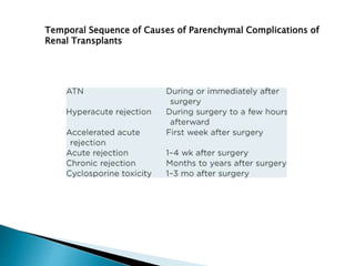
2. Incidence of ESRD is rising.
the most desirable treatment Renal transplant.
Sources of donor kidneys
1. Cadavers
2. Living donors.
3. Depends on
1. Immunosuppression.
2. HLA matching.
3. Transplant team.
4. Ideal recipient age ( 5- 50 years).
90-95% of recipients survive the first year of
surgery with at least 80 % of them functioning at
that time.
4. Despite careful selection and management of patients only a
minority of kidneys retain function useful than 10 years.
Patients may receive subsequent grafts after a first one has
failed ,and the duration of function of the first graft is a
useful predictor of the longevity of the next transplantation
5. Selection of donors and recipients.
Detection and management of post-
transplant complications.
8. 20 %.
HLA matching Radiological evaluation.
To rule out morphological/vascular
abnormalities of donor kidney.
Remaining kidney is sufficiently normal –
donor should not be at risk for subsequent
renal insufficiency.
9. Laparoscopic nephrectomy.
CT is the preferred imaging due to better spatial resolution
and sensitivity to vascular calcifications and renal stones.
CTA >> MRA
1. Distinguishing single/two main renal arteries.
2. Anatomy and to identify the point at which first branch
occurs.
3. Renal venous anatomy,
4. Tiny polar accessory arteries.
10. RENAL ARTERY
Origin : from the abdominal aorta at the L1-2 vertebral body
level, inferior to the origin of the superior mesenteric artery.
Course
The right renal artery courses inferiorly and passes posterior
to the IVC and the right renal vein to reach the renal hilum.
The left renal artery is much shorter and arises slightly more
superior to the right main renal artery. The left renal artery
courses more horizontally, posterior to the left renal vein to
enter the renal hilum.
Renal arteries are 4-6 cm in length and 5-6 mm in diameter.
11. Branches
Each renal artery gives off small branches in its proximal course,
prior to dividing into dorsal and ventral rami.
◦ inferior adrenal artery
◦ ureteric artery
◦ capsular artery
The dorsal and ventral rami segmental branches within the renal
hilum before entering the parenchyma {apical, anterior superior,
anterior inferior (middle), inferior and posterior segmental renal
arteries}
lobar branches interlobar, arcuate, and interlobular arteries.
The afferent arterioles, which supply the glomeruli, originate from
the interlobular arteries.
12. Doppler ultrasound
◦ normal PSV 150-180 cm/s and beyond this ,indicate a renal
artery stenosis of >60% .
◦ Normal RI ≈ 0.60
Variant anatomy
◦ accessory renal arteries:
◦ aberrant renal arteries: enter via the renal capsule rather than the
hilum
◦ early branching (or prehilar branching):
occurs within 1.5-2.0 cm of origin in the left renal artery or in
the retrocaval segment of the right renal artery
important to recognize in renal transplant for successful
anastomoses
25. I. Assessment of Renal Volume:-
◦ Main predictor of renal function.
◦ Larger kidneys :-
Remaining kidney:- better renal function.
Donor kidney :- Fewer complication in recipient.
Contraindication
◦ Tumors
◦ Post inflammatory fibrosis
◦ Hydronephrosis.
◦ Any medical condition affecting health of recipient/donor.
◦ Small renal stones/cortical cyst – not really a
contraindication
26. 1. Atherosclerosis.
2. FMD
3. Accessory renal artery.
4. Duplicated renal artery.
5. Triplicated renal artery.
6. Occluded small lower polar arteries.
7. Short distance between the origin and first
bifurcation of all major renal arteries.
27.
28.
29. Renal venous anatomy
◦ Retro-aortic/circum-aortic left renal vein.
◦ Duplicated right renal veins
◦ Distance between junction of vein with IVC and first
bifurcation
◦ Renal venous tributaries: adrenal/lumbar/gonadal
veins.
30. 1) Left kidney is preferred for donation.
Left renal vein is longer and surgical resection is easier.
2) Single renal artery is preferred.
3) Donor kidney with lower pole accessory artery is
avoided.
4) Peri-renal fat is usually removed before
transplantation.
33. Heterotopic renal transplantation.
1. Donor kidney is placed in an extra peritoneal location in
iliac fossa.
2. Renal artery and vein is anastomosed to external iliac
vessels
3. Urinary tract is reconstructed by an anti-refluxing
urterneocystostomy.
34.
35. USG/CT/MRI/Radionuclide studies.
Specific imaging – Voiding Cystourethrography.
Evaluation of vessels is by NECT
Assess the degree of calcification = Degree of
difficulty in performing the anastomosis.
ESRD , usually undergo biopsy AVF/pseudo-
aneurysm.
39. immediately after transplantation.
Due to ischemia of the transplanted kidney before vascular
anastomosis.
Occur in cadaveric donors during the agonal period of the donor
and in any donor kidney during the delay between the harvesting
of the kidney and the completion of vascular anastomoses in the
recipient.
Duration of the ischemia is directly related to the likelihood of
ATN.
resolve spontaneously and have little adverse effect on ultimate
graft survival
40. Manifest as anuria, rising creatinine, and mild enlargement and
tenderness of the graft.
Graft tenderness and fever are less likely to be prominent signs
of ATN than of cases of acute rejection, but the clinical picture
seldom allows confident differentiation between the two
conditions.
ATN usually appears within ½ days after transplantation and
usually resolves within a few days to a few weeks of initial onset;
Patients who experience ATN after transplantation may require
dialysis for a week until renal function returns.
ATN is much more common in cadaveric transplants than living
related donors
41. Cyclosporine,+ prednisone,
Nephrotoxic & hepatotoxic.
Newer drugs, sirolimus, tacrolimus, and mycophenolate
mofetil ,permit reduction in doses of cyclosporine and
diminished nephrotoxicity at the cost of increased risk of
hyperlipidemia and diabetes.
Cyclosporine nephrotoxicity acute, subacute, or chronic.
Acute toxicity potentiate and prolong the graft dysfunction
due to iscahemia.
42. (1) hyperacute rejection,
(2) accelerated acute rejection,
(3) acute rejection
(4) chronic rejection.
Differentiation of graft rejection from other causes of intrinsic renal
dysfunction is crucial because rejection may require increasing the
dose of immunosuppressive therapy
44. Hyperacute rejection
Doppler usg shows little or nil cortical blood flow
Radionuclide studies show complete absence of perfusion or
tubular accumulation
Angiography : total or near total lack of filling of vessels.
45.
46. Accelerated acute rejection
demonstrates imaging
findings consistent with acute
rejection, but occurs within
the first week after
transplantation.
Rare cases of cortical
nephrocalcinosis (Fig.9.12)
have been described in
patients whose transplants
underwent severe immediate
rejection but were left insitu
for several years.
47. On a grey scale :
With acute transplant rejection may show an
1. increase in the volume of the kidney
2. Swelling
3. Altered echogenicity of the renal pyramids and the
cortex.
The high echogenicity of the sinus of a normal kidney may be diminished.
These findings are thought to reflect edema of the parenchyma and of the
renal sinus fat. Edema of the collecting system walls may make them appear
thickened.
The sensitivity of these findings to diagnose rejection, however, and the
ability to distinguish rejection from cyclosporine nephrotoxicity and ATN
when the findings are not clearly present are poor
48.
49. Diminution in blood flow
1. Rejection.
2. Cyclosporine nephrotoxicity.
3. ATN.
If RI > 0.9 Acute rejection.
If PI > 1.5 rejection.
In ATN = higher RI/PI indicates longer period of recovery.
50. RI and PI have prognostic significance also.
1. If they remain elevated in first transplant month
increased risk for chronic allograft nephropathy.
2. Elevated RI increased risk for subsequent development or
worsening of general CVD. And also indicates decreased
compliance of systemic arteries in the body.
51.
52.
53.
54.
55.
56.
57.
58. Graft recipient >>> donor…
Incidence and severity rely on
1. dose of immunotherapy
2. DM
3. Co-existant graft dysfunction.
Organisms:- Gram negative pathogens/CMV/HSV.
59.
60.
61.
62.
63. Unusual complication.
First 2 weeks of transplant surgery.
Predisposing causes :- Acute rejection. ATN, vascular occlusion.
Imaging
1. Renal parenchymal laceration
2. Intra-renal hematoma
3. Peri-renal hematoma.
1. USG:- hypoechoic fluid collection represents a hematoma within the
laceration/peri-nephric space.
2. CT:- dense clot within the laceration/peri-nephric space.
64.
65. 1.Thrombosis.
◦ Immediate post-operative period.
◦ Due to complete/partial occlusion at the
anastamotic site or result of an intimal flap.
66.
67. No flow in the inferior portion
of the transplanted kidney
Little isotope in the same region
68.
69. 2.Renal artery stenosis.
Mc vascular complication
reduced renal function; htn; a bruit.
@ host artery proximal to anastomosis related to
atherosclerosis or trauma at time of surgery.
@ anastomotic site Surgical technique , suture material ,
perfusion injury of vessels.
Post anastomotic site :- refection , abnormal local
hemodynamics , extrinsic compression.
Require arterial imaging for accurate complication either with
USG doppler , CTA/MRA.
Percutaneous Transluminal angioplasty is the preferred mode of
treatment
87. Increased risk of malignancy in post RT patients.
Related to the dose and duration of the immunosuppressive
drugs.
1. Lymphoprolefeative disorders(PTLD):- esp in patients with
EBV.
2. Renal cell carcinoma (2 %):- 1oo times increased risk and
most common cell type is Papillary carcinoma.
3. Lymphoma.