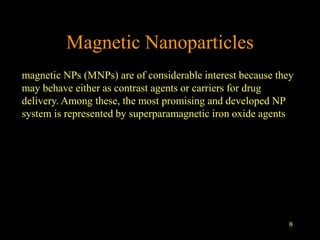
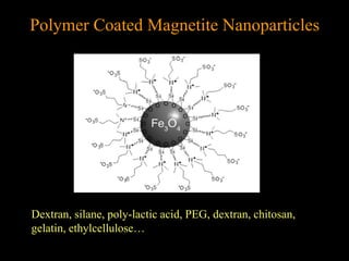
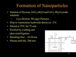
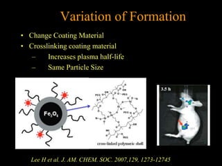
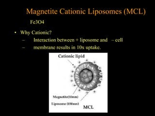
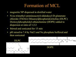
1
Multifunctional NP for Imaging
Imaging techniques and related contrast agents
X-ray  Iodinated contrast materials
Au
MRI  Gadolinium-based
Fe-based
19F-based
PET  Radioactively labelled agents (11C, 18F)
X-ray CT
CT is ubiquitous in the clinical setting as. The increasing use and
development of micro-CT and hybrid systems that with PET,
MRI.
The most investigated NPs in this field are gold NPs, since they
have large absorption coefficients against the x-ray source used
for CT imaging and may increase the signal-to-noise ratio of the
technique.
To date, different types of gold NPs have been tested in a
preclinical setting as contrast agents for molecular imaging:
nanospheres, nanocages, nanorods and nanoshells. Gold NPs
formulations as an injectable imaging agent have been utilized to
study the distribution in rodent brain ex vivo
nanocage
nanosphere
nanorod
nanoshell
Size 4-40 nm
PET
The strategy utilized is consisting in incorporating PET emitters
within the components of the NP, or entrapping them within the
core.
brain cancer
Oku et al (2011), Int. J. Pharm. 403 :170–177
MRI
MRI relies upon the enhancement of local water proton relaxation in the
presence of a contrast agent (CA). CA are compounds that catalytically shorten
the relaxation times of bulk water protons.
T1 (longitudinal relaxation– in simple terms, the time taken for the protons to
realign with the external magnetic field) Positive CA (Gd)
T2 (transverse relaxation –in simple terms, the time taken for the protons to
exchange energy with other nuclei) Negative CA (Iron oxide agents )
Gd-based NP
several nanotechnological approaches have been devised, based on
2 ideas:
-carrying many Gd chelates;
-slow down the rotation of the complex
Examples include liposomes micelles dendrimers fullerenes.
However, this approach has not yet achieved clinical applications.
8
Magnetic Nanoparticles
magnetic NPs (MNPs) are of considerable interest because they
may behave either as contrast agents or carriers for drug
delivery. Among these, the most promising and developed NP
system is represented by superparamagnetic iron oxide agents
9
Types of Magnets
• Ferromagnetic materials: the magnetic moments of
neighboring atoms align resulting in a net magnetic moment.
• Paramagnetic materials are randomly oriented due to Brownian
motion, except in the presence of external magnetic field.
• Superparamagnetic Combination of paramagnetic and ferromagnetic
properties. Made of nano-sized ferrous magnetic particles, but
affected by Brownian Motion. They will align in the presence of an
external magnetic field.
The most promising and developed NP system is
represented by superparamagnetic iron oxide
agents, consisting of a magnetite (Fe3O4) and/or
maghemite (Fe2O3) crystalline core surrounded by a
low molecular weight carbohydrate (usually dextran
or carboxydextran) or polymer coat.
. Iron oxide NPs can be classified according to their
core structure, such as Monocrystalline (MION;
10–30 nm diameter), or according to their size as
ultra-small superparamagnetic (USPIO) (20–50 nm
diameter), superparamagnetic (SPIO) (60–250 nm).
J Lodhia et al. Biomed Imaging Interv J 2010; 6(2):e12
Polymer Coated Magnetite Nanoparticles
Dextran, silane, poly-lactic acid, PEG, dextran, chitosan,
gelatin, ethylcellulose…
13
Formation of Nanoparticles
• Solution of Dextran, FeCl3.6H2O and FeCl2.4H2O (acidic
solution)
– Less Dextran Larger Particles
• Drip in Ammonium hydroxide (basic) at ~2oC
• Stirred at 75oC for 75 min.
• Purified by washing and
ultra-centrifugation
• Resulting Size ~ 10-20 nm
• Plasma half-life: 200 min
Variation of Formation
• Change Coating Material
• Crosslinking coating material
– Increases plasma half-life
– Same Particle Size
Lee H et al. J. AM. CHEM. SOC. 2007,129, 1273-12745
• Why Cationic?
– Interaction between + liposome and – cell
– membrane results in 10x uptake.
Magnetite Cationic Liposomes (MCL)
Fe3O4
Formation of MCL
• magnetite NP dispersed in distilled water
• N-(a-trimethyl-amminoacetyl)-didodecyl-D-glutamate
chloride (TMAG) Dilauroylphosphatidylcholine (DLPC)
Dioleoylphosphatidyl-ethanolamine (DOPE) added to
dispersion at ratio of 1:2:2
• Stirred and sonicated for 15 min
• pH raised to 7.4 by NaCl and Na phosphate buffered and
then sonicated
DLPC
DOPE

7a mri

  • 1.
  • 2.
    Imaging techniques andrelated contrast agents X-ray  Iodinated contrast materials Au MRI  Gadolinium-based Fe-based 19F-based PET  Radioactively labelled agents (11C, 18F)
  • 3.
    X-ray CT CT isubiquitous in the clinical setting as. The increasing use and development of micro-CT and hybrid systems that with PET, MRI. The most investigated NPs in this field are gold NPs, since they have large absorption coefficients against the x-ray source used for CT imaging and may increase the signal-to-noise ratio of the technique. To date, different types of gold NPs have been tested in a preclinical setting as contrast agents for molecular imaging: nanospheres, nanocages, nanorods and nanoshells. Gold NPs formulations as an injectable imaging agent have been utilized to study the distribution in rodent brain ex vivo
  • 4.
  • 5.
    PET The strategy utilizedis consisting in incorporating PET emitters within the components of the NP, or entrapping them within the core. brain cancer Oku et al (2011), Int. J. Pharm. 403 :170–177
  • 6.
    MRI MRI relies uponthe enhancement of local water proton relaxation in the presence of a contrast agent (CA). CA are compounds that catalytically shorten the relaxation times of bulk water protons. T1 (longitudinal relaxation– in simple terms, the time taken for the protons to realign with the external magnetic field) Positive CA (Gd) T2 (transverse relaxation –in simple terms, the time taken for the protons to exchange energy with other nuclei) Negative CA (Iron oxide agents )
  • 7.
    Gd-based NP several nanotechnologicalapproaches have been devised, based on 2 ideas: -carrying many Gd chelates; -slow down the rotation of the complex Examples include liposomes micelles dendrimers fullerenes. However, this approach has not yet achieved clinical applications.
  • 8.
    8 Magnetic Nanoparticles magnetic NPs(MNPs) are of considerable interest because they may behave either as contrast agents or carriers for drug delivery. Among these, the most promising and developed NP system is represented by superparamagnetic iron oxide agents
  • 9.
    9 Types of Magnets •Ferromagnetic materials: the magnetic moments of neighboring atoms align resulting in a net magnetic moment. • Paramagnetic materials are randomly oriented due to Brownian motion, except in the presence of external magnetic field. • Superparamagnetic Combination of paramagnetic and ferromagnetic properties. Made of nano-sized ferrous magnetic particles, but affected by Brownian Motion. They will align in the presence of an external magnetic field.
  • 10.
    The most promisingand developed NP system is represented by superparamagnetic iron oxide agents, consisting of a magnetite (Fe3O4) and/or maghemite (Fe2O3) crystalline core surrounded by a low molecular weight carbohydrate (usually dextran or carboxydextran) or polymer coat. . Iron oxide NPs can be classified according to their core structure, such as Monocrystalline (MION; 10–30 nm diameter), or according to their size as ultra-small superparamagnetic (USPIO) (20–50 nm diameter), superparamagnetic (SPIO) (60–250 nm).
  • 11.
    J Lodhia etal. Biomed Imaging Interv J 2010; 6(2):e12
  • 12.
    Polymer Coated MagnetiteNanoparticles Dextran, silane, poly-lactic acid, PEG, dextran, chitosan, gelatin, ethylcellulose…
  • 13.
    13 Formation of Nanoparticles •Solution of Dextran, FeCl3.6H2O and FeCl2.4H2O (acidic solution) – Less Dextran Larger Particles • Drip in Ammonium hydroxide (basic) at ~2oC • Stirred at 75oC for 75 min. • Purified by washing and ultra-centrifugation • Resulting Size ~ 10-20 nm • Plasma half-life: 200 min
  • 14.
    Variation of Formation •Change Coating Material • Crosslinking coating material – Increases plasma half-life – Same Particle Size Lee H et al. J. AM. CHEM. SOC. 2007,129, 1273-12745
  • 15.
    • Why Cationic? –Interaction between + liposome and – cell – membrane results in 10x uptake. Magnetite Cationic Liposomes (MCL) Fe3O4
  • 16.
    Formation of MCL •magnetite NP dispersed in distilled water • N-(a-trimethyl-amminoacetyl)-didodecyl-D-glutamate chloride (TMAG) Dilauroylphosphatidylcholine (DLPC) Dioleoylphosphatidyl-ethanolamine (DOPE) added to dispersion at ratio of 1:2:2 • Stirred and sonicated for 15 min • pH raised to 7.4 by NaCl and Na phosphate buffered and then sonicated DLPC DOPE