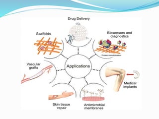
This document discusses the biomedical applications of nanoparticles. It begins by defining nanoparticles as particles between 1-100 nanometers in size. It then outlines several types of nanoparticles that have biomedical applications, including gold nanoparticles, quantum dots, iron oxide nanoparticles, carbon nanotubes, dendrimers, and lipid-based nanoparticles. For each type of nanoparticle, it provides examples of their biomedical uses such as drug delivery, cancer treatment, biomedical imaging, and diagnosis. It also discusses considerations for the toxicity of nanoparticles and their potential effects on cells and animals. In closing, it covers antimicrobial nanoparticles and their use against bacteria, fungi, and viruses.
Nanomaterials in biomedical applicationsumeet sharma
An introduction to emerging technology in medicinal science, "nanodrugs" a fruitful combination of nano-science and medical science. In this presentation, use of nano shells for delivery of drugs to targeted cancer cells has been explained. along with In Vivo and In Vitro studies on use of nanomaterials for biomedical application. For any information please feel free to contact me or refer to the references.
here you can find the most rare topics in detail
all fields of chemistry are deeply understood here for presenting the lectures
stay blessed and keep supporting
Different types of methods can be used for the preparation of Magnetic Nanoparticles, their advantages and disadvantages and applications of the materials in various fields are given in the presentation
Introduction
Nanoparticle characterization techniques
Electron Microscope
Scanning electron microscope
Transmission electron Microscope
X-ray powder diffraction
Nuclear Magnetic Resonance
Review paper on the applications and challenges of gold nanoparticles in medicine and dentistry.
Gold nanoparticles is a game-changer in delivering patient care. Its versatility can be put to use in diagnosis, imaging and treatment of various conditions. It relatively recent innovation although gold is a metal that has had a lot of meaning in human civilisation.With a lot of potential left unexplored one has to what and watch the miracles this breakthrough has in store for medical science.
The following presentation is only for quick reference. I would advise you to read the theoretical aspects of the respective topic and then use this presentation for your last minute revision. I hope it helps you..!!
Mayur D. Chauhan
NANOTECHNOLOGY comprises technological developments on the nanometer scale, usually 0.1 to 100 nm. Nanotechnology, the science of the small. Nano is Greek for dwarf, and nanoscience deals with the study of molecular and atomic particles.
Nanotechnology and its Application in Cancer TreatmentHasnat Tariq
Nanotechnology
Nanomaterials
Nanostructures
Nanoparticles
Unexpected Optical Properties of Nanoparticles
Synthesis of Nanoparticles
Nanotechnology in Cancer Treatment
Role of Sulfur NPs in Cancer Treatment
Human Tumour Cell Lines Used in Research
Ehrlich ascites carcinoma (EAC)
Sulfur Nanoparticles Preparation
MTT Assay
Sulphorhodamine-B (SRB) Assay
Median lethal dose (LD 50)
Experimental design
FT-IR Characterization of Sulfur Nanoparticles
SEM Characterization of Sulfur Nanoparticles
EDS Characterization of Sulfur Nanoparticles
XRD Characterization of Sulfur Nanoparticles
Chemical Studies on Sulfur Nanoparticles In Vitro
Biochemical investigations
Conclusion
Applications of Nanoparticles in cancer treatment
Nanoshells
Nano X-Ray therapy
Drug Delivery by Nanoparticles
'Nano', a Greek word that means 'dwarf’.
The word 'nano' is used to refer to 10-9 or a billionth part of one meter.
The term 'Nanotechnology' was first defined by Taniguchi of the Tokyo Science University in 1974.
It is generally used for materials of size between 1 to 100 nm.• They are also referred to as Nanoparticles.
In Nanotechnology, a particle is a small object that behaves as a unit with respect to its transport and properties.
Abstract
In the last decade, developments in nanotechnology have provided a new field in medicine called “Nanomedicine”. Nanomedicine has provided new tools for photodynamic therapy. Quantum dots (QDs) are approximately spherical nanoparticles that have attracted broad attention and have been used in nanomedicine applications. QDs have high molar extinction coefficients and photoluminescence quantum yield, narrow emission spectra, broad absorption, large effective stokes shifts. QDs are more photostable and resistant to metabolic degradation. These photosensitizing properties can be used as photosensitizers for Photodynamic Therapy (PDT). PDT has been recommended for its unique characteristic, such as low side effect and more efficiency. Therefore, nanomedicine leads a promising future for targeted therapy in cancer tumor. Furthermore, QDs have recently been applied in PDT, which will be addressed in this review letter. Also this review letter evaluates key aspects of nano-particulate design and engineering, including the advantage of the nanometer scale size range, biological behavior, and safety profile.
Toxic effects of heavy metals : Lead and Arsenicsanjana502982
Heavy metals are naturally occuring metallic chemical elements that have relatively high density, and are toxic at even low concentrations. All toxic metals are termed as heavy metals irrespective of their atomic mass and density, eg. arsenic, lead, mercury, cadmium, thallium, chromium, etc.
Slide 1: Title Slide
Extrachromosomal Inheritance
Slide 2: Introduction to Extrachromosomal Inheritance
Definition: Extrachromosomal inheritance refers to the transmission of genetic material that is not found within the nucleus.
Key Components: Involves genes located in mitochondria, chloroplasts, and plasmids.
Slide 3: Mitochondrial Inheritance
Mitochondria: Organelles responsible for energy production.
Mitochondrial DNA (mtDNA): Circular DNA molecule found in mitochondria.
Inheritance Pattern: Maternally inherited, meaning it is passed from mothers to all their offspring.
Diseases: Examples include Leber’s hereditary optic neuropathy (LHON) and mitochondrial myopathy.
Slide 4: Chloroplast Inheritance
Chloroplasts: Organelles responsible for photosynthesis in plants.
Chloroplast DNA (cpDNA): Circular DNA molecule found in chloroplasts.
Inheritance Pattern: Often maternally inherited in most plants, but can vary in some species.
Examples: Variegation in plants, where leaf color patterns are determined by chloroplast DNA.
Slide 5: Plasmid Inheritance
Plasmids: Small, circular DNA molecules found in bacteria and some eukaryotes.
Features: Can carry antibiotic resistance genes and can be transferred between cells through processes like conjugation.
Significance: Important in biotechnology for gene cloning and genetic engineering.
Slide 6: Mechanisms of Extrachromosomal Inheritance
Non-Mendelian Patterns: Do not follow Mendel’s laws of inheritance.
Cytoplasmic Segregation: During cell division, organelles like mitochondria and chloroplasts are randomly distributed to daughter cells.
Heteroplasmy: Presence of more than one type of organellar genome within a cell, leading to variation in expression.
Slide 7: Examples of Extrachromosomal Inheritance
Four O’clock Plant (Mirabilis jalapa): Shows variegated leaves due to different cpDNA in leaf cells.
Petite Mutants in Yeast: Result from mutations in mitochondrial DNA affecting respiration.
Slide 8: Importance of Extrachromosomal Inheritance
Evolution: Provides insight into the evolution of eukaryotic cells.
Medicine: Understanding mitochondrial inheritance helps in diagnosing and treating mitochondrial diseases.
Agriculture: Chloroplast inheritance can be used in plant breeding and genetic modification.
Slide 9: Recent Research and Advances
Gene Editing: Techniques like CRISPR-Cas9 are being used to edit mitochondrial and chloroplast DNA.
Therapies: Development of mitochondrial replacement therapy (MRT) for preventing mitochondrial diseases.
Slide 10: Conclusion
Summary: Extrachromosomal inheritance involves the transmission of genetic material outside the nucleus and plays a crucial role in genetics, medicine, and biotechnology.
Future Directions: Continued research and technological advancements hold promise for new treatments and applications.
Slide 11: Questions and Discussion
Invite Audience: Open the floor for any questions or further discussion on the topic.
Seminar of U.V. Spectroscopy by SAMIR PANDASAMIR PANDA
Spectroscopy is a branch of science dealing the study of interaction of electromagnetic radiation with matter.
Ultraviolet-visible spectroscopy refers to absorption spectroscopy or reflect spectroscopy in the UV-VIS spectral region.
Ultraviolet-visible spectroscopy is an analytical method that can measure the amount of light received by the analyte.
(May 29th, 2024) Advancements in Intravital Microscopy- Insights for Preclini...Scintica Instrumentation
Intravital microscopy (IVM) is a powerful tool utilized to study cellular behavior over time and space in vivo. Much of our understanding of cell biology has been accomplished using various in vitro and ex vivo methods; however, these studies do not necessarily reflect the natural dynamics of biological processes. Unlike traditional cell culture or fixed tissue imaging, IVM allows for the ultra-fast high-resolution imaging of cellular processes over time and space and were studied in its natural environment. Real-time visualization of biological processes in the context of an intact organism helps maintain physiological relevance and provide insights into the progression of disease, response to treatments or developmental processes.
In this webinar we give an overview of advanced applications of the IVM system in preclinical research. IVIM technology is a provider of all-in-one intravital microscopy systems and solutions optimized for in vivo imaging of live animal models at sub-micron resolution. The system’s unique features and user-friendly software enables researchers to probe fast dynamic biological processes such as immune cell tracking, cell-cell interaction as well as vascularization and tumor metastasis with exceptional detail. This webinar will also give an overview of IVM being utilized in drug development, offering a view into the intricate interaction between drugs/nanoparticles and tissues in vivo and allows for the evaluation of therapeutic intervention in a variety of tissues and organs. This interdisciplinary collaboration continues to drive the advancements of novel therapeutic strategies.
Deep Behavioral Phenotyping in Systems Neuroscience for Functional Atlasing a...Ana Luísa Pinho
Functional Magnetic Resonance Imaging (fMRI) provides means to characterize brain activations in response to behavior. However, cognitive neuroscience has been limited to group-level effects referring to the performance of specific tasks. To obtain the functional profile of elementary cognitive mechanisms, the combination of brain responses to many tasks is required. Yet, to date, both structural atlases and parcellation-based activations do not fully account for cognitive function and still present several limitations. Further, they do not adapt overall to individual characteristics. In this talk, I will give an account of deep-behavioral phenotyping strategies, namely data-driven methods in large task-fMRI datasets, to optimize functional brain-data collection and improve inference of effects-of-interest related to mental processes. Key to this approach is the employment of fast multi-functional paradigms rich on features that can be well parametrized and, consequently, facilitate the creation of psycho-physiological constructs to be modelled with imaging data. Particular emphasis will be given to music stimuli when studying high-order cognitive mechanisms, due to their ecological nature and quality to enable complex behavior compounded by discrete entities. I will also discuss how deep-behavioral phenotyping and individualized models applied to neuroimaging data can better account for the subject-specific organization of domain-general cognitive systems in the human brain. Finally, the accumulation of functional brain signatures brings the possibility to clarify relationships among tasks and create a univocal link between brain systems and mental functions through: (1) the development of ontologies proposing an organization of cognitive processes; and (2) brain-network taxonomies describing functional specialization. To this end, tools to improve commensurability in cognitive science are necessary, such as public repositories, ontology-based platforms and automated meta-analysis tools. I will thus discuss some brain-atlasing resources currently under development, and their applicability in cognitive as well as clinical neuroscience.
Comparing Evolved Extractive Text Summary Scores of Bidirectional Encoder Rep...University of Maribor
Slides from:
11th International Conference on Electrical, Electronics and Computer Engineering (IcETRAN), Niš, 3-6 June 2024
Track: Artificial Intelligence
https://www.etran.rs/2024/en/home-english/
Salas, V. (2024) "John of St. Thomas (Poinsot) on the Science of Sacred Theol...Studia Poinsotiana
I Introduction
II Subalternation and Theology
III Theology and Dogmatic Declarations
IV The Mixed Principles of Theology
V Virtual Revelation: The Unity of Theology
VI Theology as a Natural Science
VII Theology’s Certitude
VIII Conclusion
Notes
Bibliography
All the contents are fully attributable to the author, Doctor Victor Salas. Should you wish to get this text republished, get in touch with the author or the editorial committee of the Studia Poinsotiana. Insofar as possible, we will be happy to broker your contact.
2. NANOPARTICLES
Nanoparticles are particles between 1 and 100
nanometers in size.
In nanotechnology , a particle is defined as a small
object that behaves as a whole unit with respect to
its transport and properties.
Nanomaterials hold immense promise for
significantly improving existing diagnosis , therapy
and designing novel approaches to treat a variety
of human ailments.
3. NANOPARTICLES While some of the applications of nanotechnology
have been translated into clinical settings, many
more potential uses of nanomedicines have been
demonstrated in experimental systems.
Since a variety of materials can be nanosized, the
scope of nanomedicine is also large and may even
become larger.
At the same time, the impact of nanomaterials on
cellular and animal models will need to be carefully
evaluated under both acute and chronic exposures
at toxicological and pharmacological doses.
5. GOLD
Inorganic nanoparticles are emerging as versatile
tools in imaging as a result of their unique
chemical, physical and optical properties .
Gold nanoparticles were discovered > 100 years ago
and owing to their surface chemistry, projected
biocompatibility, relatively low short-term toxicity,
high atomic number and high X-ray absorption
coefficient.
Gold nanoparticles have received significant
interest recently for use in multiple imaging
technologies.
6. APPICATIONS OF GOLD
NANOPARTICLES
Gold nanoparticles may be used in different domains,
one of most important being the biomedical field.
They have suitable properties for :
controlled drug delivery
cancer treatment
biomedical imaging
diagnosis and many others due to their excellent
compatibility with the humans, low toxicity and
tunable stability, small dimensions, and possibility to
interact with a variety of substances.
7. APPICATIONS OF GOLD
NANOPARTICLES
Gold nanoparticles are intensively studied in
biomedicine, and recent studies revealed the fact
that they can cross the blood-brain barrier, may
interact with the DNA and produce genotoxic
effects.
Because of their ability of producing heat, they can
target and kill the tumors, being used very often in
photodynamic therapy.
Gold nanoparticles have also emerged in
diagnostics as colorimetric biosensors
8. Quantum dots
Quantum dots (QDs) are fluorescent semiconductor
nanocrystals (∼ 1 – 100 nm) with unique optical and
electrical properties .
Quantum dots are increasingly used as fluorophores
for in vivo fluorescence imaging.
Fluorescence imaging has several advantages compared
with other imaging modalities because this method has
good sensitivity and is non-invasive in nature, using
readily available and relatively inexpensive instruments.
Being an optical technique, it is limited in terms of
tissue penetration depth. A wide variety of in vivo studies
have validated the potency of QDs.
9. Iron oxide nanoparticles
Magnetic nanoparticles have received considerable
attention because of their potential use in optical,
magnetic and electronic devices.
Magnetic iron oxide nanoparticles have been
functionalized with antibodies, nucleosides, proteins and
enzymes for directing them to diseased tissues such as
tumors.
MRI is a non-invasive medical imaging technique that is
commonly used in clinical medicine to visualize the
structure and function of tissues.
MRI is based on the behavior, alignment and interaction
of protons in the presence of an applied magnetic field.
10. Carbon nanotubes
CNT’s are used as field emission devices, tips for
scanning microscopy, nanoscale transistors, or
components for composite materials.
A few bioimaging applications using CNTs have
been developed for cancer cell destruction,
detection and dynamic imaging.
SWNTs(single walled carbon nanotubes)
covalently linked to visible-wavelength
fluorophores have been imaged in cells using
confocal microscopy .
11. Dendrimers
Dendrimers are well-defined, highly branched
molecules that are synthesized with precise structural
control and low polydispersity .
Magnevist, a gadolinium(III)-
diethylenetriaminepentaacetic acid (Gd-DTPA)
complex, is a commonly used contrast agent for MRI.
12. Polyelectrolyte complex
nanoparticles
Polyelectrolyte complex nanoparticles have attractive
properties for imaging and drug delivery.
PECs have been established as potential materials for
gene delivery because of their relatively efficient
transfection efficiency and simple formulation .
More recently, these nanocarriers have proved
effective for entrapping and delivering small
molecules, peptides, RNAs and even large proteins .
Few reports are also available on their use in
biomedical imaging applications and also as
therapeutic agents.
13. Calcium phosphate nanoparticles
Calcium phosphate nanoparticles have received
attention because of reports of low toxicity,
biocompatibility and solubility in cells.
Calcium phosphate has been used as a delivery
vehicle for DNA and has been studied as a
nanoparticle for gene delivery.
Also, these particles have been used for the
encapsulation of organic molecules, fluorescent
dyes and chemotherapeutic drugs, suggesting the
potential for in vivo biomedical imaging and drug
delivery applications.
14. Perfluorocarbon nanoparticles
Perfluorocarbon nanoparticles (PFCNPs) have
received considerable attention for their applications
in molecular imaging and targeted drug delivery
applications.
PFCNPs act as a platform to carry contrast-enhancing
agents or chemotherapeutic drugs.
Functionalized PFCNPs are compatible with several
molecular imaging modalities, including MRI and CT.
Functionalized PFCNP contrast agents have been used
as molecular imaging agents in MRI assessments of
tumor angiogenesis, cellular tracking and
atherosclerosis .
15. Lipid-based nanoparticles
Lipid-based nanoparticles such as liposomes and micelles
have been developed for pharmaceuticals and aim to
address the traditional boundaries to drug delivery .
Mulder et al. pioneered the use of lipid-based
nanoparticles for contrast-enhanced MRI and molecular
imaging applications .
Koole et al. recently developed paramagnetic lipid-coated
silica nanoparticles containing a quantum dot core as a
contrast agent for multimodal imaging (fluorescence and
MRI) of αvβ3-integrin expression on cultured endothelial
cells.
16. Lipid-based nanoparticles
Also, Cressman et al. recently developed an RGD-
labeled lipid incorporated into liposomal
nanoparticles and studied trafficking in cultured
endothelial cells.
Senarath-Yapa et al. furthered this work by
reporting the use of poly(lipid)-coated,
fluorophore-doped silica nanoparticles for
biolabeling and cellular imaging applications .
17.
18.
19. Toxicity considerations
Owing to the rapid growth of nanoparticle use in biomedical
research, the toxicity of these materials should be considered
in detail .
Complete characterization of size , shape, charge , surface
chemistry and material properties is important when
correlating toxicity.
Nanomaterials may agglomerate in vitro or in vivo and may
chemically degrade, making it difficult to relate systematically
nanoparticle toxicity to such a diverse set of materials.
Detailed studies of biodistribution, pharmacokinetics and
local and systemic toxicity for each type of nanoparticle, will
be important in overall toxicity evaluations.
20. ANTIMICROBIAL NANOPARTICLES
Despite numerous existing potent antibiotics and
other antimicrobial means, bacterial infections are
still a major cause of morbidity and mortality.
Moreover, the need to develop additional
bactericidal means has significantly increased due
to the growing concern regarding multidrug-
resistant bacterial strains and biofilm associated
infections.
Consequently, attention has been especially
devoted to new and emerging nanoparticle-based
materials in the field of antimicrobial
chemotherapy.
21. ANTIMICROBIAL NANOPARTICLES
Metals and metal oxides have been widely studied
for their antimicrobial activities.
Metal oxide nanoparticles, well known for their
highly potent antibacterial effect, include silver
(Ag), iron oxide (Fe3O4), titanium oxide (TiO2),
copper oxide (CuO), and zinc oxide (ZnO).
Most metal oxide nanoparticles exhibit
bactericidal properties through reactive oxygen
species (ROS) generation although some are
effective due to their physical structure and metal
ion release.
22. Silver
Of the metal nanoparticles, silver nanoparticles have
been widely used as an effective antimicrobial agent
against bacteria, fungi, and viruses .
Ag and its compounds have long been used for the
disinfection of medical devices and water purification.
In medicine, Ag compounds are commonly applied to
treat burns, wounds, and a variety of infectious
diseases .
Although the Ag nanoparticle mechanism of action is
still not clear, small diameter Ag nanoparticles have a
superior antimicrobial effect to those of a larger
diameter .
23. Titanium Oxide
Titanium dioxide (TiO2) is another metal oxide
that has been extensively studied for its
antimicrobial activities .
TiO2 has long been known for its ability to kill
both Gram-positive and Gram-negative bacteria .
More recent reports have shown its efficiency
against various viral species and parasites.
24. Titanium Oxide
Titanium dioxide (TiO2) NM as antibacterial compounds
have been on the market for quite some time .
They are photocatalytic, their toxicity induced by visible
light, near-UV or UV , stimulates ROS burst.
The ROS damage the membrane, DNA, and many other
macromolecules and functions of the bacterial cell .
TiO2 is effective against many bacteria including spores
of Bacillus , which is the most resistant organism known.
As with other NM, combinations of Ti or TiO2 with
other NM such as Ag were found to have a synergistic
effect and to enhance their activity.
25. Zinc Oxide
Additional broad spectrum bactericidal NM are
ZnO-based nanoparticles .
ZnO nanoparticles were shown to have a wide
range of antimicrobial activity against various
microorganisms, which is significantly dependent
on the chosen concentration and particle size
26. Zinc Oxide
ZnO nanoparticles were shown to inhibit the
growth of methicillin-sensitive S. aureus (MSSA),
methicillin-resistant S. aureus (MRSA), and
methicillin-resistant S. epidermidis (MRSE) strains
and proved to be effective bactericidal agents that
were not affected by the drug-resistant
mechanisms of MRSA and MRSE .
27. Zinc Oxide
Zinc oxide (ZnO) NM are of relatively low cost and
effective in size dependency against a wide range
of bacteria .
These include pathogens such as Klebsiella
pneumonia , Listeria monocytogenes, Salmonella
enteritidis , Streptococcus mutans,
Lactobacillus and E. coli with low toxicity to
human cells.
28. Zinc Oxide
Their white color, UV-blocking, and ability to prevent
biofilm formation makes them suitable for fabric and
glass industries as coating materials designated for
medical and other devices.
Furthermore, treatment using zinc was approved by
the FDA and nowadays Zn is available as a food
additive .
ZnO NM affect bacterial cells by binding to
membranes, disrupting their potential and integrity,
and by inducting ROS production .
In addition and as a result, Zn NM are also weak
mutagens.
29. Iron Oxide and Gold
Fe3O4 nanoparticles and gold (Au) represent an
additional class of antimicrobial materials that are
being researched for their use in health care .
Microbiological assays have proved that surfaces
modified using Fe3O4 nanoparticles demonstrate
antiadherent properties and significantly reduce
both Gram-negative and Gram-positive bacterial
colonization .
Au nanoparticles and nanorods have been
reported to be bactericidal when photothermally
functionalized.
30. Magnesium Oxide
Nano-magnesium oxides (MgO) are additional
antibacterial metal oxide NM that have been shown to
exhibit bactericidal activity.
Nano-MgO particles were reported to exhibit efficient
antimicrobial activity against bacteria (both Gram-
positive and Gram-negative), spores, and viruses.
Magnesium (Mg) can be used in various NM in the
form of MgO or MgX2 (e.g., MgF2) .
In addition to inducing ROS, Mg-containing NM may
directly inhibit essential enzymes of the bacteria .
MgF2 NM were found to prevent biofilm formation
of E. coli and S. aureus .
31. Superparamagnetic Iron Oxide
Superparamagnetic iron oxide (SPION) represents
a relatively new approach using magnetic particles
that cause local hyperthermia in the presence of a
magnetic field or alternatively, they can be coated
by other NM such as Ag and Au and their
magnetic effect can be utilized to penetrate and
destroy biofilms.
32. Nitric Oxide
Nitric oxide (NO) NM differ from other metal NM
by specifically affecting reactive nitrogen species
(RNS), rather than ROS.
NO NM were found to effectively kill methicillin-
resistant S. aureus (MRSA) in skin infections and
to enhance wound healing of normal and diabetic
mice.
NO NM are also effective in biofilm eradication of
multiple bacterial species.
33. Aluminum Oxide
It is not clear if aluminum oxide (Al2O3) nanoparticles are
suitable for antibacterial treatment.
First, their bactericidal effect is relatively mild and they work
only at high concentrations unless in combination with other
NM such as Ag.
Second and more disturbing is their ability to promote
horizontal transfer of multiresistance genes mediated by
plasmids across genera.
The mechanism of action of aluminum NM, as recently
shown for E. coli, is by diffusion and accumulation inside the
cells, causing pit formation, perforation, and membrane
disorganization, leading to cell death .