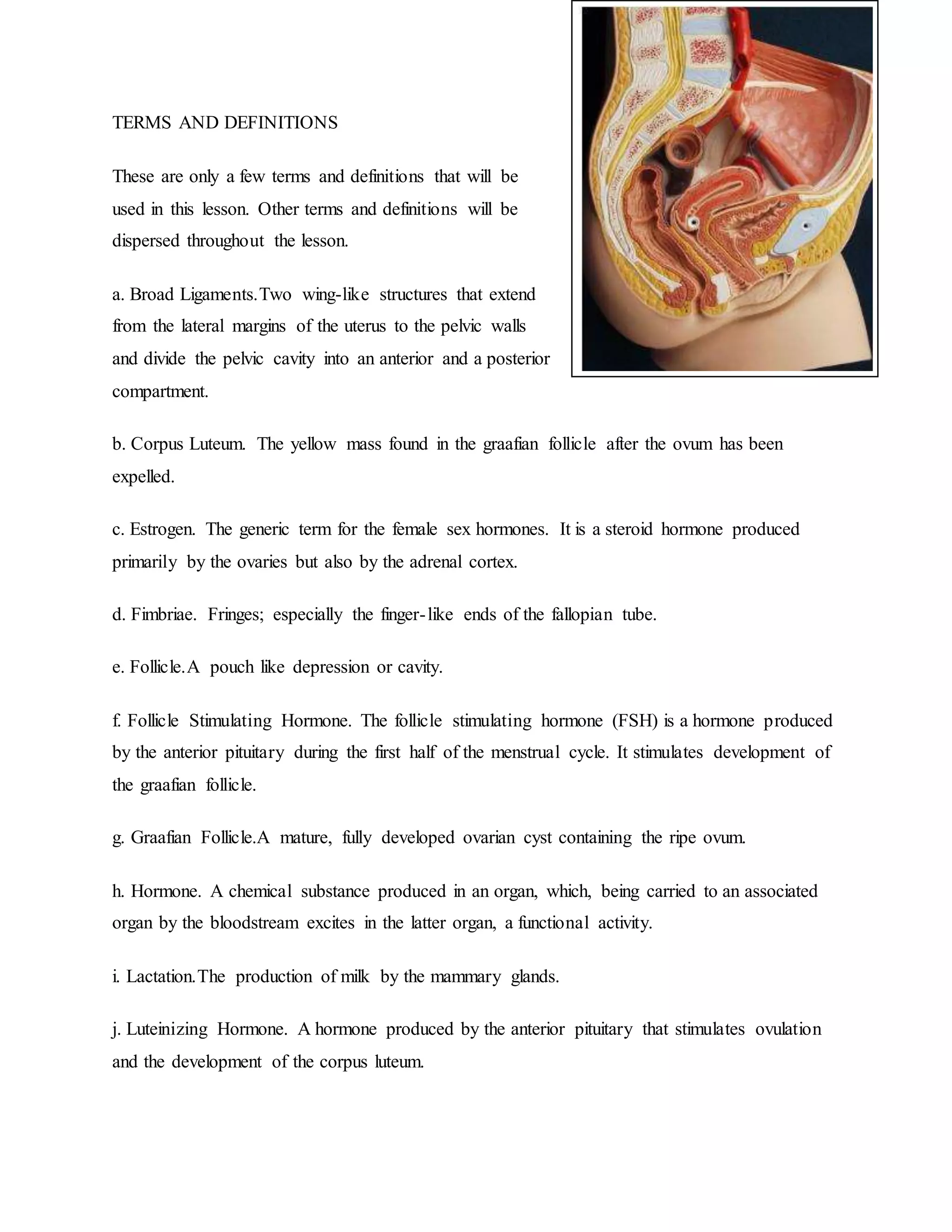
I. INTRODUCTION:
Dysfunctional uterine bleeding (DUB) is heavy or irregular menstrual bleeding that is
not caused by an underlying anatomical abnormality, such as a fibroid, lesion, or tumor. DUB is
the most common type of abnormal uterine bleeding.</p>
Most cases of DUB are associated with anovulatory bleeding (menstruation that occurs without
ovulation). Anovulatory bleeding is common in women who have just started menstruating and
during the several years preceding menopause. When ovulation does not occur, the level of
estrogen and progesterone in the uterus is disturbed, leading to DUB. Anovulation, however,
does not always lead to DUB and there are other causes as well. Women with ovulatory cycles
(cycles that involve ovulation) may also experience DUB.
Menstrual cycles vary in duration, frequency, and intensity, making abnormalities difficult to
determine. Women who have DUB may experience a variety of patterns of bleeding. A woman
who bleeds for longer than a week, bleeds more than every 3 weeks or so, bleeds between
periods, or bleeds excessively should see a doctor or other health care provider.
DUB is usually painless. Diagnosis involves ruling out other causes of abnormal
bleeding. Treatment depends on the intensity and timing of the bleeding, the patient's age, and if
she is trying to conceive.
DUB and Anovulation
Anatomy of the endometrium
The endometrium is the mucous surface that lines the inside of the uterus. It is responsive to
hormonal changes and contains several layers of cells that vary in appearance and number
throughout the menstrual cycle. During the luteal phase (i.e., 2 weeks prior to menstruation), the
endometrium is thick, its epithelial cells and glands are enlarged, and the arteries are swollen. At
menstruation, the endometrium sheds. Following menstruation, the endometrium regenerates.
The normal menstrual cycle
Menstruation is triggered by a sudden decrease in progesterone and estrogen secretions. The
menstrual flow is made up of endometrial cells and tissue, blood, and cervical and vaginal mucus
and cells.
After menstruation, the increased secretion of estrogen causes cellular growth and the
regeneration of the endometrium. This first half of the menstrual cycle is known as the follicular
phase.
Ovulation (the release of an egg from the ovary) normally occurs 2 weeks after the first day of
the last menstrual cycle. After ovulation, the secretion of progesterone stops the growth of the
endometrium, balancing out the effects of the estrogen. If conception does not occur,
progesterone production declines, and menstrual bleeding begins again.
Anovulatory bleeding
Normally during the menstrual cycle, the production of progesterone in the latter 2 weeks of the
cycle balances out the regenerative effects of estrogen, halting further endometrial growth. In
anovulation, the level of estrogen does not decline, and progesterone is not secreted to balance
out the effects of estrogen.
Anemia
Anemia is a condition in which the number of red blood cells or the amount of hemoglobin (the
protein that carries oxygen in them) is low.
Red blood cells contain hemoglobin, a protein that enables them to carry oxygen from the lungs
and deliver it to all parts of the body. When the number of red blood cells is reduced or the
amount of hemoglobin in them is low, the blood cannot carry an adequate supply of oxygen. An
inadequate supply of oxygen in the tissues produces the symptoms of anemia.
Anemia may be caused by excessive bleeding. Bleeding may be sudden, as may occur in
an injury or during surgery. Often, bleeding is gradual and repetitive, typically from
abnormalities in the digestive or urinary tract or heavy menstrual periods. Chronic bleeding
typically leads to low levels of iron, which leads to worsening anemia.
Anemia may also result when the body does not produce enough red blood cells. Many
nutrients are needed for red blood cell production. The most critical are iron, vitamin B12, and
folate (folic acid), but the body also needs trace amounts of vitamin C, riboflavin, and copper, as
well as a proper balance of hormones, especially erythropoietin (a hormone that stimulates red
blood cell production). Without these nutrients and hormones, production of red blood cells is
slow and inadequate, or the red blood cells may be deformed and unable to carry oxygen
adequately. Chronic disease also may affect red blood cell production. In some circumstances,
the bone marrow space may be invaded and replaced (for example, by leukemia, lymphoma, or
metastatic cancer), resulting in decreased production of red blood cells.
Anemia may also result when too many red blood cells are destroyed. Normally, red blood cells
live about 120 days. Scavenger cells in the bone marrow, spleen, and liver detect and destroy red
blood cells that are near or beyond their usual life span. If red blood cells are destroyed
prematurely (hemolysis), the bone marrow tries to compensate by producing new cells faster.
When destruction of red blood cells exceeds their production, hemolytic anemia results.
Hemolytic anemia is relatively uncommon compared with the anemia caused by excessive
bleeding and decreased red blood cell production.
II. OBJECTIVES
GENERAL OBJECTIVES
Our primary objectives in nursing are adequate understanding by the patient of
certain details of the condition, adequate and comfortable daily elimination, a certain
amount of rest, a balanced diet, and participation in specific items of self-care.
SPECIFIC OBJECTIVES
- Conduct an effective plan of management for a patient with anemia.
- Control the bleeding, reduce morbidity, and prevent complications
- Select patients in need of vitamin defiency anemia
- Conduct counseling and education of patients with anemia caused by nutritional
defeciencies and hemoglobinopathies.
- Should be to arrest bleeding , replace iron to avoid anemia, and prevent future
bleeding.
- Determine the presence of anemina in clinical context; all 3 laboratory indices of
anemia are concentration measurements (blood loss will decrease hemoglobin,
hematocrit or red blood cell count only after self transfusion or extracelluar fluid the
following day.
- Interpret the signs and symptoms of anemia with the understanding that they are
dependent on the rapidity with which anemia developed.
- Determine the presence of anemia; differentiate between causes according to the
patient’s age.
III. PATIENTS PROFILE
A. Biographical and Demographic Information
1. Name :maria veronica camildo
2. Address: malolos bulacan
3. Gender: female
4. Date of birth:
5. Place of birth: malolos bulacan
6. Marital status: single
7. Religion: catholic
8. Occupation: N/A
9. Educational attainment: highschool level
10. Ethnic origin: tagalog
11. Date of admission:
12. Time of admission:
B. Resource and Reliability of Information
Researchers obtained subjective and objective data. Most of the patient’s biographical data
was achieved from relying on the chart of the patient. Subjective data from the patient achieved
by the researchers was used in the physical examination. Interview of the patient with the mother
was done and details were used in the Gordon’s Typology and NCP.
C. Health History
1. Chief Complaint
 Heavy Bleeding
2. History of Present Illness
 2 days PTA, the patient experienced dysmenorrhea and heavy bleeding during her
menarche.
.
3. Past Medical History
- Pediatric/childhood Illness: none
- Adult Illness: none
- Injuries or accidents: none
- Hospitalizations: none
- Allergies: allergic to seafoods
4. Family Health History:
 The client has a family history of DM and HPN.
5. Obstetric History:
 (-) pregnancies
6. Personal and Social History:
 The pt. is active in participating in school activities; she's not fond of having a regular
exercise daily. She client is always concern with her hygiene.
7. Socio-economic History:
 She is living with her auntie and cousins, because her family is residing in Manila. The
pt.'s status of living is average class.
D. GORDON’S FUNCTIONAL HEALTH PATTERN
HEALTH PERCEPTION
During the interview with the patient, she rate her health 8/10 since hospitalization helped her to
get well. She told that before she was admitted in the hospital she had a heavy bleeding due to
her menstruation. Since it is her first year of her menstruation, she thinks that it should be not
heavy and irregular. The patient’s daily activities are affected because she needs bed rest.
The pt also has dysmenorrhea so she takes pain relievers and she always stand to ease the pain.
For her bleeding, she does bed rest.
NURITIONAL METABOLIC PATTERN
The patients meal on a typical day would be;
Breakfast: noodles and milk
Lunch: rice and vegetables
Dinner: rice, chicken and vegetables
The patient usually drinks water two glasses during the day and another two glasses during the
evening. The patient doesn’t experience nausea and vomiting nor abdominal pains,
She always take a bath everyday before hospitalization. She doesn’t put oils or any lotions. Her
skin doesn’t have itches but it is quite dry as well as her hair.
ELIMINATION PATTERN
The patient has one bowel movement a day. Her stool is color black and it’s soft. She urinates
twice a day. It has a yellowish color and has a pungent odor. The patient doesn’t have any
problems urinating. She also doesn’t have any bladder surgeries nor urinary catheter,
ACTIVITY EXERCISE PATTERN
The patient stated her activities on a normal day. She takes a bath everyday and she go to school,
she eat three to four times a day and she do some household chores. Right after she had a health
problem her usual daily activities was interrupted since she needs bed rest and hospitalization.
Her leisure activities are watching television and eating. She believe that she has an exercise and
that is playing badminton two to three times a week.
SLEEP REST PATTERN
She always watch television before going to sleep. It takes about 10-30 mins before she get
falling asleep. She has a good quality of sleep. She usually sleep at 9pm and wake up at 4am. She
also sleep in the afternoon that last for about three hours.
COGNITIVE PERCEPTUAL
The patient understand the doctor’s and nurse’s instructions. She told that she’s scared about
her health problem but now that she’s getting better, she feels relieved. The patient doesn’t have
any difficulties expressing herself. She was able to remember the recent events and events long
ago. She can also make her own decisions but it is her mother that guides her.
SELF-PERCEPTION-SELF-CONCEPT
When we interviewed her we observed that she is shy and nervous because she cannot look to
as directly and she keeps on playing her cellphone while we are asking her a question.She was
wearing a simple white t-shirt and a blue jogging pants.She said that she take a bath two times
a day.She rated her level of anxiety of 8.Her face was so white.Her voice volume changes
from loud to soft because when she answered our question sometimes we can hear her but
sometimes we can’t.She answer our questions readily without hesitation.Her usual view of
self is somewhat negative but her mother encouraged her to be strong and always think
positive.Her current illness changed her body structure and function. She cannot do the
activities that she usually do because it is already limited.
ROLE-RELATIONSHIP
She lived with her Aunt and study in Bulacan.Her parents are with her brother and
sister in Valenzuela,Manila because they were studying there.She is the youngest child in the
family.Her mother give support and take care of her while she is in the hospital.She said that
she has a good interaction with her family because they were calling and texting each other
and sometimes she is chatting with her brother and sister.When she was admitted to the
hospital she cannot go to school already and had many absences.
SEXUALITY-REPRODUCTIVE
She said that she had her first menstruation last April 2010 at the age of 13 years
old.She has no history of sexually transmitted disease.She is now more concerned with proper
hygiene and limit to take a bath two times a day because she already know that bathing at
night can cause Anemia.
COPING-STRESS TOLERANCE
She has no signs of stress like that of crying because when we saw her she is smiling.
She decrease and relieve her stress by doing something like playing her cellphone, listening
music and most of the time eating. When she experience a heavy menstruation she just think
that it was just normal because it was her first time to have a menstruation.
VALUE-BELIEF
She is a Catholic. She pray every night before go to bed. She also attend the mass every
Sunday with her Aunt. When she was admitted to the hospital she already cannot attend the
mass every Sunday but still she keeps on praying every night before go to bed to ask God’s
blessings and help her make her feelings better.
E. REVIEW OF SYSTEMS AND PHYSICAL EXAMINATION
1. General Survey
In first contact with the patient, she is asleep. Her general appearance are just normal. Her facial
grimace doesn’t show any problem with pain that time. Her gait wasn’t observed but the patient
told us that she can walk normally. Her clothes were just appropriate as well as her appearance.
There is no deformity visible from the patient. She has a normal size for her age and she has a
good posture. The patient has no noted body odor.
3. Head to toe Assessment and general appearance
RR:
PR:
TEMP:
BP:
BODY PARTS NORMAL FINDINGS ACTUAL FINDINGS ANALYSIS
General
Appearance
 Body built
 Posture
 Grooming:
Appropriately
dressed
 odor
 medium in size
 good posture
 Grooming:
Appropriately
dressed
 odorless
Normal
Mental Status  LOC: conscious
and oriented to
time, place and
person
 Conscious and
oriented to time,
place and Person
Normal
Skin  Color: pinkish  Color: pinkish Normal
brown in color
 Temperature:
uniform; with
normal range,
warm
 Moisture: dry,
clammy, sweaty,
oily
 Texture: smooth
 Skin Turgor:
when
pinched,skin
springs back to
previous state
 Hair distribution:
equal
brown in color
 Temperature:
normal range;
warm
 moisture: dry
 Texture: smooth
 Skin Turgor: when
pinched,skin
springs back to
previous state
 Hair distribution:
equal
Nails  Nail bed color:
pink
 Plate shape:
convex
 Condition:
Smooth texture
 Blanch test:
Promt return to
pink (less than 3
seconds)
 Nail bed color:
pink
 Plate shape: convex
 Condition: Smooth
texture
 Blanch test:
Promt return to
pink (less than 3
seconds)
Normal
Head  Size: head
circumference
smaller than the
body
 Size: head
circumference
smaller than the
body
Normal
 Shape: round and
smooth
 Symmetry:
symmetrical
 Scalp: White
 Shape: round and
smooth
 Symmetry:
symmetrical
 Scalp: White
Hair  Color: Black
 Texture: Straight
and silky hair
 Growth patterned
toward head and
neck
 Color: Black
 Texture: Straight
and silky hair
 Growth patterned
toward head and
neck
Normal
Face  Skin color:
brown
 Texture: smooth
 Symmetry:
symmetrical
 Facial
movements:
symmetrical
 Skin color:
 brown
 Texture: smooth
 Symmetry:
symmetrical
 Facial movements:
symmetrical
Normal
Eyes  Eyebrows:
evenly
distributed
 Eyelashes:
evenly
distributed,
curved outward
 Eyelids:
Effective closure
 Conjunctiva:
pink conjunctiva
 Sclera: White in
 Eyebrows: evenly
distributed
 Eyelashes: evenly
distributed, curved
outward
 Eyelids: Effective
closure
 Conjunctiva: pink
conjunctiva
 Sclera: White in
color; clear; no
yellowish
Normal
color; clear; no
yellowish
discoloration
 Cornea: no
irregularities on
the surface;
looks smooth
and clear or
transparent
 Iris: Anterior
chamber is
transparent; no
noted visible
materials; color
depends on th
person’s race
 Pupil: ranges
from 3-7mm and
are equal in size;
equally round;
constrict when
light is directed
to the eye, both
directly and
consensual
 Reflex: blink
consensually
 discoloration
 Cornea: no
irregularities on the
surface; looks
smooth and clear or
transparent
 Iris: Anterior
chamber is
transparent; no
noted visible
materials; color
depends on th
person’s race
 Pupil: ranges from
3-7mm and are
equal in size;
equally round;
constrict when light
is directed to the
eye, both directly
and consensual
 Reflex: blink
consensually
Ears  Color: pinkish
brown
 Symmetry:
symmetrical
 Auricle: elastic
and firm
 Pinna: recoils
when folded
 Hearing acquity:
responds to
normal voice
 Color: pinkish
brown
 Symmetry:
symmetrical
 Auricle: elastic and
firm
 Pinna: recoils when
folded
 Hearing acquity:
responds to normal
voice
Normal
Nose  Color: Pinkish
Brown
 Symmetry:
Symmetrical
 Nares: Patent;
not flaring
 Sinuses: non-
tender
 Color: Pinkish
Brown
 Symmetry:
Symmetrical
 Nares: Patent; not
flaring
 Sinuses: non-tender
Normal
Lips  Symmetry:
symmetrical
 Color: pink
 Symmetry:
symmetrical
 Color: pink
Normal
Teeth  Color and
number: varies
depending on
age; color is
Epstein pearl
 Color and number:
Has 28 tooth and
has a color of
Epstein pearl
Normal
Tongue  Position: central
position
 Color: pink
 Texture: soft
and rough
 Reflex: gag
 Position: central
position
 Color: pink
 Texture: soft and
rough
 Reflex: gag
Normal
Gums  Color: pink  Color: pink Normal
Pharynx  Uvula: midline
 Mucosa: pink
 Tonsils: not
inflamed
 Gag reflex:
present
 Uvula: midline
 Mucosa: pink
 Tonsils: not
inflamed
 Gag reflex: present
Normal
Neck and lymph
nodes
 Length: short,
thick, surrounded
 Length: short,
thick, surrounded
Normal
by skin folds
 Mobility: head
held midline,
free movement
from side to side.
 Lymph nodes:
not palpable
by skin folds
 Mobility: head held
midline, free
movement from
side to side.
 Lymph nodes: not
palpable
Thorax  Chest: not
protruding
 Breast contour:
round,
 Skin Character:
smooth
 Anterior chest:
(+)
bronchovescicula
r and vescicular
breath sounds
 Spinal
Alignment:
vertically aligned
 Chest: not
protruding
 Breast contour:
round,
 Skin Character:
smooth
 Anterior chest: (+)
bronchovescicular
and vescicular
breath sounds
 Spinal Alignment:
vertically aligned
Normal
Heart  Rate: normal
(regular rhythm)
pulmonic, aortic
 Rate: normal
(regular rhythm)
pulmonic, aortic
Normal
Abdomen  Contour and
symmetry:
round, non
protruding
 Color: brown
 Bowel sounds:
normal
 Contour and
symmetry: round,
non protruding
 Color: brown
 Bowel sounds:
normal
 Palpation: non-
Normal
 Palpation: non-
tender
 Liver: not
palpable
tender
 Liver: not palpable
Upper and lower
extremities
 Arms and hands
( length: equal
length , arms
longer than the
legs
Movement:
Spontaneous
muscle tone:
generally
flexed)
 Legs and feet
(Appearance:
bowed legs,
length: legs are
shorter than arms
Mobility: full
ROM
muscle tone:
Flexion
 Arms and hands (
length: equal length
, arms longer than
the legs
Movement:
Spontaneous
muscle tone:
generally flexed)
 Legs and feet
(Appearance:
bowed legs, length:
legs are shorter
than arms
Mobility: full ROM
muscle tone:
Flexion
Normal
4. Review of Systems
 General: Fatigue; change in weight
 Head, eyes, ears, nose, throat:
reddish eyes, Has no Nose bleeding, colds, obstruction and discharge.
No presence of dental problems, Neck stiffness, pain, tenderness and masses in thyroid or
other areas
 Cardiovascular: no palpitation nor tachycardia but has anemia
 Respiratory: has no dyspnea, wheezing, stridor and cough
 Gastrointestinal: decreased appetite, has no dysphagia, indigestion, food idiosyncrasy,
heartburn, eructation, hematemesis, jaundice, constipation, or diarrhea. Presense of black
stool due to ferrous sulfate
 Genitourinary: Dysuria; vaginal discharge; dysmenorrhea
 Hematologic: Easy bleeding or bruising
 Neurologic: Headaches; blurring of vision
 Skin: quite dry and has frizzy hair
 Breast: Nipple discharge, breast tenderness
F. Anatomy and Pathophysiology
*PATHOPYSIOLOGY OF ANEMIA*
*Anatomy and Physiology*
The Circulatory System
The circulatory system is responsible for the transport of water and dissolved materials
throughout the body, including oxygen, carbon dioxide, nutrients, and waste. The circulatory
system transports oxygen from the lungs and nutrients from the digestive tract to every cell in the
body, allowing for the continuation of cell metabolism. The circulatory system also transports
the waste products of cell metabolism to the lungs and kidneys where they can be expelled from
the body. Without this important function toxic substances would quickly build up in the body.
Anatomy of the Circulatory System
The human circulatory system is organized into two major circulations. Each has its own pump
with both pumps being incorporated into a single organ—the heart. The two sides of the human
heart are separated by partitions, the interatrial septum and the interventricular septum. Both
septa are complete so that the two sides are anatomically and functionally separate pumping
units. The right side of the heart pumps blood through the pulmonary circulation (the lungs)while
the left side of the heart pumps blood through the systemic circulation (the body).
The human heart is a specialized, four-chambered muscle that maintains the blood flow in the
circulatory system. It lies immediately behind the sternum, or breastbone, and between the lungs.
The apex, or bottom of the heart, is tilted to the left side. At rest, the heart pumps about 59 cc (2
oz) of blood per beat and 5 l (5 qt) per minute. During exercise it pumps 120-220 cc (4-7.3 oz) of
blood per beat and 20-30 l (21-32 qt) per minute. The adult human heart is about the size of a fist
and weighs about 250-350 gm (9 oz).
The human heart begins beating early in fetal life and continues regular beating throughout the
life span of the individual. If the heart stops beating for more than 3 or 4 minutes permanent
brain damage may occur. Blood flow to the heart muscle itself also depends on the continued
beating of the heart and if this flow is stopped for more than a few minutes, as in a heart attack,
the heart muscle may be damaged to such a great extent that it may be irreversibly stopped.
The heart is made up of two muscle masses. One of these forms the two atria (the upper
chambers) of the heart, and the other forms the two ventricles (the lower chambers). Both atria
contract or relax at the same time, as do both ventricles.
An electrical impulse called an action potential is generated at regular intervals in a specialized
region of the right atrium called the sinoauricular (or sinoatrial, or SA) node. Since the two atria
form a single muscular unit, the action potential will spread over the atria. A fraction of a second
later, having been triggered by the action potential,the atrial muscle contracts.
The ventricles form a single muscle mass separate from the atria. When the atrial action potential
reaches the juncture of the atria and the ventricles, the atrioventricular or AV node (another
specialized region for conduction) conducts the impulse. After a slight delay, the impulse is
passed by way of yet another bundle of muscle fibers (the Bundle of His and the Purkinje
system.) Contraction of the ventricle quickly follows the onset of its action potential. From this
pattern it can be seen that both atria will contract simultaneously and that both ventricles will
contract simultaneously, with a brief delay between the contraction of the two parts of the heart.
The electrical stimulus that leads to contraction of the heart muscle thus originates in the heart
itself, in the sinoatrial node (SA node), which is also known as the heart's pacemaker. This node,
which lies just in front of the opening of the superior vena cava, measures no more than a few
millimeters. It consists of heart cells that emit regular impulses. Because of this spontaneous
discharge of the sinoatrial node, the heart muscle is automated. A completely isolated heart can
contract on its own as long as its metabolic processes remain intact.
The rate at which the cells of the SA node discharge is externally influenced through the
autonomic nervous system, which sends nerve branches to the heart. Through their stimulatory
and inhibitory influences they determine the resultant heart rate. In adults at rest this is between
60 and 74 beats a minute. In infants and young children it may be between 100 and 120 beats a
minute. Tension, exertion, or fever may cause the rate of the heart to vary between 55 and 200
beats a minute.The heart's pacemaker is the Sinoatrial (SA) Node.
The Blood
The blood transports life-supporting food and oxygen to every cell of the body and removes their
waste products. It also helps to maintain body temperature, transports hormones, and fights
infections. The brain cells in particular are very dependent on a constant supply of oxygen. If the
circulation to the brain is stopped, death shortly follows.
Blood has two main constituents. The cells, or corpuscles, comprise about 45 percent, and the
liquid portion, or plasma, in which the cells are suspended comprises 55 percent. The blood cells
comprise three main types: red blood cells, or erythrocytes; white blood cells, or leukocytes,
which in turn are of many different types; and platelets, or thrombocytes. Each type of cell has
its own individual functions in the body. The plasma is a complex colorless solution, about 90
percent water, that carries different ions and molecules including proteins, enzymes, hormones,
nutrients, waste materials such as urea, and fibrinogen, the protein that aids in clotting.
Red Blood Cells
The red blood cells are tiny, round, biconcave disks, averaging about 7.5 microns (0.003 in) in
diameter. A normal-sized man has about 5 l (5.3 qt) of blood in his body, containing more than
25 trillion red cells. Because the normal life span of red cells in the circulation is only about 120
days, more than 200 billion cells are normally destroyed each day by the spleen and must be
replaced. Red blood cells, as well as most white cells and platelets, are made by the bone
marrow.The main function of the red blood cells is to transport oxygen from the lungs to the
tissues and to transport carbon dioxide, one of the chief waste products, it to the lungs for release
from the body.
The substance in the red blood cells that is largely responsible for their ability to carry oxygen
and carbon dioxide is hemoglobin, the material that gives the cells their red color. It is a protein
complex comprising many linked amino acids, and occupies almost the entire volume of a red
blood cell. Essential to its structure and function is the mineral iron.The blood cell type
responsible for the transport of oxygen and carbon dioxide is the red blood cell (erythrocytes.)
White Blood Cells
The leukocytes, or white blood cells, are of three types; granulocytes, lymphocytes, and
monocytes. All are involved in defending the body against foreign organisms.
There are three types of granulocytes: neutrophils, eosinophils, and basophils, with neutrophils
the most abundant. Neutrophils seek out bacteria and phagocytize, or engulf, them.
The lymphocytes' chief function is to migrate into the connective tissue and build antibodies
against bacteria and viruses. Leukocytes are almost colorless, considerably larger than red cells,
have a nucleus, and are much less numerous; only one or two exist for every 1,000 red cells. The
number increases in the presence of infection.
Monocytes, representing only 4 to 8 percent of white cells, attack organisms not destroyed by
granulocytes and leukocytes.
The granulocytes, accounting for about 70 percent of all white blood cells, are formed in the
bone marrow. The lymphocytes on the other hand are produced primarily by the lymphoid
tissues of the body—the spleen and lymph nodes. They are usually smaller than the granulocytes.
Monocytes are believed to originate from lymphocytes. Just as the oxygen-carrying function of
red cells is necessary for our survival, so are normal numbers of leukocytes, which protect us
against infection.Fighting infection and foreign invaders is one of the primary functions of the
Fighting leucocytes.
Platelets
Platelets, or thrombocytes, are much smaller than the red blood cells. They are round or
biconcave disks and are normally about 30 to 40 times more numerous than the white blood
cells. The platelets' primary function is to stop bleeding. When tissue is damaged, the platelets
aggregate in clumps to obstruct blood flow.
Plasma
The plasma is more than 90 percent water and contains a large number of substances, many
essential to life. Its major solute is a mixture of proteins. The most abundant plasma protein is
albumin. The globulins are even larger protein molecules than albumin and are of many chemical
structures and functions. The antibodies, produced by lymphocytes, are globulins and are carried
throughout the body, where many of them fight bacteria and viruses.
An important function of plasma is to transport nutrients to the tissues. glucose, for example,
absorbed from the intestines, constitutes a major source of body energy. Some of the plasma
proteins and fats, or lipids, are also used by the tissues for cell growth and energy. Minerals
essential to body function, although present only in trace amounts, are other important elements
of the plasma. The calcium ion, for example, is essential to the building of bone, as is
phosphorus. Calcium is also essential to the clotting of blood. Copper is another necessary
component of the plasma. a major source of body energy, transported to the cells by the plasma
is glucose.
THE BLOOD VESSEL
The central opening of a blood vessel, the lumen, is surrounded by a wall consisting of three
layers:
 The tunica intima is the inner layer facing the blood. It is composed of an innermost layer
of endothelium (simple squamous epithelium) surrounded by variable amounts of
connective tissues.
 The tunica media, the middle layer, is composed of smooth muscle with variable amounts
of elastic fibers.
 The tunica adventitia, the outer layer, is composed of connective tissue.
The cardiovascular system consists of three kinds of blood vessels that form a closed system of
passageways:
 Arteries carry blood away from the heart. The three kinds of arteries are categorized by
size and function:
 Elastic arteries (conducting arteries) are the largest arteries and include the aorta
and other nearby branches. The tunica media of elastic arteries contains a large
amount of elastic connective tissue, which enables the artery to expand as blood
enters the lumen from the contracting heart. During relaxation of the heart, the
elastic wall of the artery recoils to its original position, forcing blood forward and
smoothing the jerky discharge of blood from the heart.
 Muscular arteries (conducting arteries) branch from elastic arteries and distribute
blood the various body regions. Abundant smooth muscle in the thick tunica
media allows these arteries to regulate blood flow by vasoconstriction (narrowing
of the lumen) or vasodilation (widening of the lumen). Most named arteries of the
body are muscular arteries.
 Arterioles are small, nearly microscopic, blood vessels that branch from muscular
arteries. Most arterioles have all three tunics present in their walls, with
considerable smooth muscle in the tunica media. The smallest arterioles consist of
endothelium surrounded by a single layer of smooth muscle. Arterioles regulate
the flow of blood into capillaries by vasoconstriction and vasodilation.
Capillaries are microscopic blood vessels with extremely thin walls. Only the tunica
intima is present in these walls, and some walls consist exclusively of a single layer of
endothelium. Capillaries penetrate most body tissues with dense interweaving networks
called capillary beds. The thing walls of capillaries allow the diffusion of oxygen and
nutrients out of the capillaries, while allowing carbon dioxide and wastes into the
capillaries.
 Metarterioles (precapillaries) are the blood vessels between arterioles and venules.
Although metarterioles pass through capillary beds with capillaries, they are not
true capillaries because metarterioles, like arterioles, have smooth muscle present
in the tunica media. The smooth muscle of a metarteriole allows it to acts as a
shunt to regulate blood flow into the true capillaries that branch from it. The
thoroughfare channel, the tail end of the metarteriole that connects to the venule,
lacks smooth muscle.
 True capillaries form the bulk of the capillary bed. They branch away from a
metarteriole at its arteriole end and return to merge with the metarteriole at its
venule end (thoroughfare channel).
 Some true capillaries connect directly from an arteriole to a metarteriole or
venule. Although the walls of true capillaries lack muscle fibers, they possess a
ring of smooth muscle called a precapillary sphincter where they emerge from the
metarteriole. The precapillary sphincter regulates blood flow through the
capillary. There are three types of true capillaries:
 Continuous capillaries have continuous, unbroken walls consisting of cells that
are connected by tight junctions. Most capillaries are of this type.
 Fenestrated capillaries have continuous walls between endothelial cells, but the
cells have numerous pores (fenestrations) that increase their permeability. These
capillaries are found in the kidneys, lining the small intestine, and in other areas
where a high transfer rate of substances into or out of the capillary is required.
 Sinusoidal capillaries (sinusoids) have large gaps between endothelial cells that
permit the passage of blood cells. These capillaries are found in the bone marrow,
spleen, and liver.
Veins carry blood toward the heart. The three kinds of veins are listed here in the order
that they merge to form increasingly larger blood vessels:
 Postcapillary venules, the smallest veins, form when capillaries merge as they exit
a capillary bed. Much like capillaries, they are very porous, but with scattered
smooth muscle fibers in the tunica media.
 Venules form when postcapillary venules join. Although the walls of larger
venules contain all three layers, they are still porous enough to allow white blood
cells to pass.
 Veins have walls with all three layers, but the tunica intima and tunica media are
much thinner than in similarly sized arteries. Few elastic or muscle fibers are
present. The wall consists of primarily of a well-developed tunica adventitia.
Many veins, especially those in the limbs, have valves, formed from folds of the
tunica intima, that prevent the backflow of blood.
Many regions of the body receive blood supplies from two or more arteries. The points where
these arteries merge are called arterial anastomoses. Arterial anastomoses allow tissues to receive
blood even after one of the arteries supplying blood has been blocked.
THE FEMALE REPRODUCTIVE SYSTEM
GENERAL
The organs of the reproductive systems are concerned with the general process of reproduction,
and each is adapted for specialized tasks. These organs are unique in that their functions are not
necessary for the survival of each individual. Instead, their functions are vital to the continuation
of the human species. In providing maternity gynecologic health care to women, you will find
that it is vital to your career as a practical nurse and to the patient that you will require a greater
depth and breadth of knowledge of the female anatomy and physiology than usual. The female
reproductive system consists of internal organs and external organs. The internal organs are
located in the pelvic cavity and are supported by the pelvic floor. The external organs are located
from the lower margin of the pubis to the perineum. The appearance of the external genitals
varies greatly from woman to woman, since age, heredity, race, and the number of children a
woman has borne determine the size, shape, and color. See figure 1-1 for the female reproductive
organs.
TERMS AND DEFINITIONS
These are only a few terms and definitions that will be
used in this lesson. Other terms and definitions will be
dispersed throughout the lesson.
a. Broad Ligaments.Two wing-like structures that extend
from the lateral margins of the uterus to the pelvic walls
and divide the pelvic cavity into an anterior and a posterior
compartment.
b. Corpus Luteum. The yellow mass found in the graafian follicle after the ovum has been
expelled.
c. Estrogen. The generic term for the female sex hormones. It is a steroid hormone produced
primarily by the ovaries but also by the adrenal cortex.
d. Fimbriae. Fringes; especially the finger-like ends of the fallopian tube.
e. Follicle.A pouch like depression or cavity.
f. Follicle Stimulating Hormone. The follicle stimulating hormone (FSH) is a hormone produced
by the anterior pituitary during the first half of the menstrual cycle. It stimulates development of
the graafian follicle.
g. Graafian Follicle.A mature, fully developed ovarian cyst containing the ripe ovum.
h. Hormone. A chemical substance produced in an organ, which, being carried to an associated
organ by the bloodstream excites in the latter organ, a functional activity.
i. Lactation.The production of milk by the mammary glands.
j. Luteinizing Hormone. A hormone produced by the anterior pituitary that stimulates ovulation
and the development of the corpus luteum.
k. Oocyte.A developing egg in one of two stages.
l. Ovum.The female reproductive cell.
m. Progesterone. The pure hormone contained in the corpora lutea whose function is to prepare
the endometrium for the reception and development of the fertilized ovum.
n. Reproduction.The process by which an offspring is formed.
1-3. INTERNAL FEMALE ORGANS
The internal organs of the female consists of the uterus, vagina, fallopian tubes, and the ovaries
a. The uterus is a hollow organ about the size and shape of a pear. It serves two important
functions: it is the organ of menstruation and during pregnancy it receives the fertilized ovum,
retains and nourishes it until it expels the fetus during labor.
(1) Location. The uterus is located between the urinary bladder and the rectum. It is suspended in
the pelvis by broad ligaments.
(2) Divisions of the uterus. The uterus consists of the body or corpus, fundus, cervix, and the
isthmus. The major portion of the uterus is called the body or corpus. The fundus is the superior,
rounded region above the entrance of the fallopian tubes. The cervix is the narrow, inferior outlet
that protrudes into the vagina. The isthmus is the slightly constricted portion that joins the corpus
to the cervix.
(3) Walls of the uterus (see figure 1-3). The walls are thick and are composed of three layers: the
endometrium, the myometrium, and the perimetrium. The endometrium is the inner layer or
mucosa. A fertilized egg burrows into the endometrium (implantation) and resides there for the
rest of its development.When the female is not pregnant, the endometrial lining sloughs off about
every 28 days in response to changes in levels of hormones in the blood. This process is called
menses. The myometrium is the smooth muscle component of the wall. These smooth muscle
fibers are arranged. In longitudinal, circular, and spiral patterns, and are interlaced with
connective tissues. During the monthly female cycles and during pregnancy, these layers
undergo extensive changes. The perimetrium is a strong, serous membrane that coats the entire
uterine corpus except the lower one fourth and anterior surface where the bladder is attached.
b. Vagina.
(1) Location. The vagina is the thin in walled muscular tube about 6 inches long leading from the
uterus to the external genitalia. It is located between the bladder and the rectum.
(2) Function. The vagina provides the passageway for childbirth and menstrual flow; it receives
the penis and semen during sexual intercourse.
c. Fallopian Tubes (Two).
(1) Location. Each tube is about 4 inches long and extends medially from each ovary to empty
into the superior region of the uterus.
(2) Function. The fallopian tubes transport ovum from the ovaries to the uterus. There is no
contact of fallopian tubes with the ovaries.
(3) Description. The distal end of each fallopian tube is expanded and has finger-like projections
called fimbriae, which partially surround each ovary. When an oocyte is expelled from the ovary,
fimbriae create fluid currents that act to carry the oocyte into the fallopian tube. Oocyte is carried
toward the uterus by combination of tube peristalsis and cilia, which propel the oocyte forward.
The most desirable place for fertilization is the fallopian tube.
d. Ovaries
(1) Functions. The ovaries are for oogenesis-the production of eggs (female sex cells) and for
hormone production (estrogen and progesterone).
(2) Location and gross anatomy. The ovaries are about the size and shape of almonds. They lie
against the lateral walls of the pelvis, one on each side. They are enclosed and held in place by
the broad ligament. There are compact like tissues on the ovaries, which are called ovarian
follicles. The follicles are tiny sac-like structures that consist of an immature egg surrounded by
one or more layers of follicle cells. As the developing egg begins to ripen or mature, follicle
enlarges and develops a fluid filled central region. When the egg is matured, it is called a
graafian follicle, and is ready to be ejected from the ovary.
(3) Process of egg production--oogenesis
(a) The total supply of eggs that a female can release has been determined by the time she is
born. The eggs are referred to as "oogonia" in the developing fetus. At the time the female is
born, oogonia have divided into primary oocytes, which contain 46 chromosomes and are
surrounded by a layer of follicle cells.
(b) Primary oocytes remain in the state of suspended animation through childhood until the
female reaches puberty (ages 10 to 14 years). At puberty, the anterior pituitary gland secretes
follicle-stimulating hormone (FSH), which stimulates a small number of primary follicles to
mature each month.
(c) As a primary oocyte begins dividing, two different cells are produced, each containing 23
unpaired chromosomes. One of the cells is called a secondary oocyte and the other is called the
first polar body. The secondary oocyte is the larger cell and is capable of being fertilized. The
first polar body is very small, is nonfunctional, and incapable of being fertilized.
(d) By the time follicles have matured to the graafian follicle stage, they contain secondary
oocytes and can be seen bulging from the surface of the ovary. Follicle development to this stage
takes about 14 days. Ovulation (ejection of the mature egg from the ovary) occurs at this 14-day
point in response to the luteinizing hormone (LH), which is released by the anterior pituitary
gland.
(e) The follicle at the proper stage of maturity when the LH is secreted will rupture and release
its oocyte into the peritoneal cavity. The motion of the fimbriae draws the oocyte into the
fallopian tube. The luteinizing hormone also causes the ruptured follicle to change into a
granular structure called corpus luteum, which secretes estrogen and progesterone.
(f) If the secondary oocyte is penetrated by a sperm, a secondary division occurs that produces
another polar body and an ovum, which combines its 23 chromosomes with those of the sperm to
form the fertilized egg, which contains 46 chromosomes.
(4) Process of hormone production by the ovaries.
(a) Estrogen is produced by the follicle cells, which are responsible secondary sex characteristics
and for the maintenance of these traits. These secondary sex characteristics include the
enlargement of fallopian tubes, uterus, vagina, and external genitals; breast development;
increased deposits of fat in hips and breasts; widening of the pelvis; and onset of menses or
menstrual cycle.
(b) Progesterone is produced by the corpus luteum in presence of in the blood. It works with
estrogen to produce a normal menstrual cycle. Progesterone is important during pregnancy and in
preparing the breasts for milk production.
1-4. EXTERNAL FEMALE GENITALIA
the external organs of the female reproductive system include the mons pubis, labia majora, labia
minora, vestibule, perineum, and the Bartholin's glands. As a group, these structures that
surround the openings of the urethra and vagina compose the vulva, from the Latin word
meaning covering.
a. Mons Pubis. This is the fatty rounded area overlying the symphysis pubis and covered with
thick coarse hair.
b. Labia Majora. The labia majora run posteriorly from the mons pubis. They are the 2 elongated
hair covered skin folds. They enclose and protect other external reproductive organs.
c. Labia Minora. The labia minora are 2 smaller folds enclosed by the labia majora. They protect
the opening of the vagina and urethra.
d. Vestibule. The vestibule consists of the clitoris, urethral meatus, and the vaginal introitus.
(1) The clitoris is a short erectile organ at the top of the vaginal vestibule whose function is
sexual excitation.
(2) The urethral meatus is the mouth or opening of the urethra. The urethra is a small tubular
structure that drains urine from the bladder.
(3) The vaginal introitus is the vaginal entrance.
e. Perineum. This is the skin covered muscular area between the vaginal opening (introitus) and
the anus. It aids in constricting the urinary, vaginal, and anal opening. It also helps support the
pelvic contents.
f. Bartholin's Glands (Vulvovaginal or Vestibular Glands). The Bartholin's glands lie on either
side of the vaginal opening. They produce a mucoid substance, which provides lubrication for
intercourse.
1-5. BLOOD SUPPLY
The blood supply is derived from the uterine and ovarian arteries that extend from the internal
iliac arteries and the aorta. The increased demands of pregnancy necessitate a rich supply of
blood to the uterus. New, larger blood vessels develop to accommodate the need of the growing
uterus. The venous circulation is accomplished via the internal iliac and common iliac vein.
1-6. FACTS ABOUT THE MENSTRUAL CYCLE
Menstruation is the periodic discharge of blood, mucus, and epithelial cells from the uterus. It
usually occurs at monthly intervals throughout the reproductive period, except during pregnancy
and lactation, when it is usually suppressed.
a. The menstrual cycle is controlled by the cyclic activity of follicle stimulating hormone (FSH)
and LH from the anterior pituitary and progesterone and estrogen from the ovaries. In other
words, FSH acts upon the ovary to stimulate the maturation of a follicle, and during this
development, the follicular cells secrete increasing amounts of estrogen
b. Hormonal interaction of the female cycle are as follows:
(1) Days 1-5. This is known as the menses phase. A lack of signal from a fertilized egg
influences the drop in estrogen and progesterone production. A drop in progesterone results in
the sloughing off of the thick endometrial lining which is the menstrual flow. This occurs for 3 to
5 days.
(2) Days 6-14. This is known as the proliferative phase. A drop in progesterone and estrogen
stimulates the release of FSH from the anterior pituitary. FSH stimulates the maturation of an
ovum with graafian follicle. Near the end of this phase, the release of LH increases causing a
sudden burst like release of the ovum, which is known as ovulation.
(3) Days 15-28. This is known as the secretory phase. High levels of LH cause the empty
graafian follicle to develop into the corpus luteum. The corpus luteum releases progesterone,
which increases the endometrial blood supply. Endometrial arrival of the fertilized egg. If the
egg is fertilized, the embryo produces human chorionic gonadotropin (HCG). Thehuman
chorionic gonadotropin signals the corpus luteum to continue to supply progesterone to maintain
the uterine lining. Continuous levels of progesterone prevent the release of FSH and ovulation
ceases.
c. Additional Information.
(1) The length of the menstrual cycle is highly variable. It may be as short as 21 days or as long
as 39 days.
(2) Only one interval is fairly constant in all females, the time from ovulation to the beginning of
menses, which is almost always 14-15 days.
(3) The menstrual cycle usually ends when or before a woman reaches her fifties. This is known
as menopause.
1-7. OVULATION
Ovulation is the release of an egg cell from a mature ovarian follicle. Ovulation is stimulated by
hormones from the anterior pituitary gland, which apparently causes the mature follicle to swell
rapidly and eventually rupture. When this happens, the follicular fluid, accompanied by the egg
cell, oozes outward from the surface of the ovary and enters the peritoneal cavity. After it is
expelled from the ovary, the egg cell and one or two layers of follicular cells surrounding it are
usually propelled to the opening of a nearby uterine tube. If the cell is not fertilized by union of a
sperm cell within a relatively short time, it will degenerate.
1-8. MENOPAUSE
As mentioned in paragraph 1-6c(3), menopause is the cessation of menstruation. This usually
occurs in women between the ages of 45 and 50. Some women may reach menopause before the
age of 45 and some after the age of 50. In common use, menopause generally means cessation of
regular menstruation. Ovulation may occur sporadically or may cease abruptly. Periods may end
suddenly, may become scanty or irregular, or may be intermittently heavy before ceasing
altogether. Markedly diminished ovarian activity, that is, significantly decreased estrogen
production and cessation of ovulation, causes menopause.
*PATHOPYSIOLOGY OF DUB
Research Paper help
https://www.homeworkping.com/

59174242 case-study-bmc

  • 1.
    I. INTRODUCTION: Dysfunctional uterinebleeding (DUB) is heavy or irregular menstrual bleeding that is not caused by an underlying anatomical abnormality, such as a fibroid, lesion, or tumor. DUB is the most common type of abnormal uterine bleeding.</p> Most cases of DUB are associated with anovulatory bleeding (menstruation that occurs without ovulation). Anovulatory bleeding is common in women who have just started menstruating and during the several years preceding menopause. When ovulation does not occur, the level of estrogen and progesterone in the uterus is disturbed, leading to DUB. Anovulation, however, does not always lead to DUB and there are other causes as well. Women with ovulatory cycles (cycles that involve ovulation) may also experience DUB. Menstrual cycles vary in duration, frequency, and intensity, making abnormalities difficult to determine. Women who have DUB may experience a variety of patterns of bleeding. A woman who bleeds for longer than a week, bleeds more than every 3 weeks or so, bleeds between periods, or bleeds excessively should see a doctor or other health care provider. DUB is usually painless. Diagnosis involves ruling out other causes of abnormal bleeding. Treatment depends on the intensity and timing of the bleeding, the patient's age, and if she is trying to conceive. DUB and Anovulation Anatomy of the endometrium The endometrium is the mucous surface that lines the inside of the uterus. It is responsive to hormonal changes and contains several layers of cells that vary in appearance and number throughout the menstrual cycle. During the luteal phase (i.e., 2 weeks prior to menstruation), the endometrium is thick, its epithelial cells and glands are enlarged, and the arteries are swollen. At menstruation, the endometrium sheds. Following menstruation, the endometrium regenerates. The normal menstrual cycle Menstruation is triggered by a sudden decrease in progesterone and estrogen secretions. The
  • 2.
    menstrual flow ismade up of endometrial cells and tissue, blood, and cervical and vaginal mucus and cells. After menstruation, the increased secretion of estrogen causes cellular growth and the regeneration of the endometrium. This first half of the menstrual cycle is known as the follicular phase. Ovulation (the release of an egg from the ovary) normally occurs 2 weeks after the first day of the last menstrual cycle. After ovulation, the secretion of progesterone stops the growth of the endometrium, balancing out the effects of the estrogen. If conception does not occur, progesterone production declines, and menstrual bleeding begins again. Anovulatory bleeding Normally during the menstrual cycle, the production of progesterone in the latter 2 weeks of the cycle balances out the regenerative effects of estrogen, halting further endometrial growth. In anovulation, the level of estrogen does not decline, and progesterone is not secreted to balance out the effects of estrogen. Anemia Anemia is a condition in which the number of red blood cells or the amount of hemoglobin (the protein that carries oxygen in them) is low. Red blood cells contain hemoglobin, a protein that enables them to carry oxygen from the lungs and deliver it to all parts of the body. When the number of red blood cells is reduced or the amount of hemoglobin in them is low, the blood cannot carry an adequate supply of oxygen. An inadequate supply of oxygen in the tissues produces the symptoms of anemia. Anemia may be caused by excessive bleeding. Bleeding may be sudden, as may occur in an injury or during surgery. Often, bleeding is gradual and repetitive, typically from abnormalities in the digestive or urinary tract or heavy menstrual periods. Chronic bleeding typically leads to low levels of iron, which leads to worsening anemia.
  • 3.
    Anemia may alsoresult when the body does not produce enough red blood cells. Many nutrients are needed for red blood cell production. The most critical are iron, vitamin B12, and folate (folic acid), but the body also needs trace amounts of vitamin C, riboflavin, and copper, as well as a proper balance of hormones, especially erythropoietin (a hormone that stimulates red blood cell production). Without these nutrients and hormones, production of red blood cells is slow and inadequate, or the red blood cells may be deformed and unable to carry oxygen adequately. Chronic disease also may affect red blood cell production. In some circumstances, the bone marrow space may be invaded and replaced (for example, by leukemia, lymphoma, or metastatic cancer), resulting in decreased production of red blood cells. Anemia may also result when too many red blood cells are destroyed. Normally, red blood cells live about 120 days. Scavenger cells in the bone marrow, spleen, and liver detect and destroy red blood cells that are near or beyond their usual life span. If red blood cells are destroyed prematurely (hemolysis), the bone marrow tries to compensate by producing new cells faster. When destruction of red blood cells exceeds their production, hemolytic anemia results. Hemolytic anemia is relatively uncommon compared with the anemia caused by excessive bleeding and decreased red blood cell production.
  • 4.
    II. OBJECTIVES GENERAL OBJECTIVES Ourprimary objectives in nursing are adequate understanding by the patient of certain details of the condition, adequate and comfortable daily elimination, a certain amount of rest, a balanced diet, and participation in specific items of self-care. SPECIFIC OBJECTIVES - Conduct an effective plan of management for a patient with anemia. - Control the bleeding, reduce morbidity, and prevent complications - Select patients in need of vitamin defiency anemia - Conduct counseling and education of patients with anemia caused by nutritional defeciencies and hemoglobinopathies. - Should be to arrest bleeding , replace iron to avoid anemia, and prevent future bleeding. - Determine the presence of anemina in clinical context; all 3 laboratory indices of anemia are concentration measurements (blood loss will decrease hemoglobin, hematocrit or red blood cell count only after self transfusion or extracelluar fluid the following day. - Interpret the signs and symptoms of anemia with the understanding that they are dependent on the rapidity with which anemia developed. - Determine the presence of anemia; differentiate between causes according to the patient’s age.
  • 5.
    III. PATIENTS PROFILE A.Biographical and Demographic Information 1. Name :maria veronica camildo 2. Address: malolos bulacan 3. Gender: female 4. Date of birth: 5. Place of birth: malolos bulacan 6. Marital status: single 7. Religion: catholic 8. Occupation: N/A 9. Educational attainment: highschool level 10. Ethnic origin: tagalog 11. Date of admission: 12. Time of admission: B. Resource and Reliability of Information Researchers obtained subjective and objective data. Most of the patient’s biographical data was achieved from relying on the chart of the patient. Subjective data from the patient achieved by the researchers was used in the physical examination. Interview of the patient with the mother was done and details were used in the Gordon’s Typology and NCP.
  • 6.
    C. Health History 1.Chief Complaint  Heavy Bleeding 2. History of Present Illness  2 days PTA, the patient experienced dysmenorrhea and heavy bleeding during her menarche. . 3. Past Medical History - Pediatric/childhood Illness: none - Adult Illness: none - Injuries or accidents: none - Hospitalizations: none - Allergies: allergic to seafoods 4. Family Health History:  The client has a family history of DM and HPN. 5. Obstetric History:  (-) pregnancies 6. Personal and Social History:  The pt. is active in participating in school activities; she's not fond of having a regular exercise daily. She client is always concern with her hygiene.
  • 7.
    7. Socio-economic History: She is living with her auntie and cousins, because her family is residing in Manila. The pt.'s status of living is average class. D. GORDON’S FUNCTIONAL HEALTH PATTERN HEALTH PERCEPTION During the interview with the patient, she rate her health 8/10 since hospitalization helped her to get well. She told that before she was admitted in the hospital she had a heavy bleeding due to her menstruation. Since it is her first year of her menstruation, she thinks that it should be not heavy and irregular. The patient’s daily activities are affected because she needs bed rest. The pt also has dysmenorrhea so she takes pain relievers and she always stand to ease the pain. For her bleeding, she does bed rest. NURITIONAL METABOLIC PATTERN The patients meal on a typical day would be; Breakfast: noodles and milk Lunch: rice and vegetables Dinner: rice, chicken and vegetables The patient usually drinks water two glasses during the day and another two glasses during the evening. The patient doesn’t experience nausea and vomiting nor abdominal pains, She always take a bath everyday before hospitalization. She doesn’t put oils or any lotions. Her skin doesn’t have itches but it is quite dry as well as her hair.
  • 8.
    ELIMINATION PATTERN The patienthas one bowel movement a day. Her stool is color black and it’s soft. She urinates twice a day. It has a yellowish color and has a pungent odor. The patient doesn’t have any problems urinating. She also doesn’t have any bladder surgeries nor urinary catheter, ACTIVITY EXERCISE PATTERN The patient stated her activities on a normal day. She takes a bath everyday and she go to school, she eat three to four times a day and she do some household chores. Right after she had a health problem her usual daily activities was interrupted since she needs bed rest and hospitalization. Her leisure activities are watching television and eating. She believe that she has an exercise and that is playing badminton two to three times a week. SLEEP REST PATTERN She always watch television before going to sleep. It takes about 10-30 mins before she get falling asleep. She has a good quality of sleep. She usually sleep at 9pm and wake up at 4am. She also sleep in the afternoon that last for about three hours. COGNITIVE PERCEPTUAL The patient understand the doctor’s and nurse’s instructions. She told that she’s scared about her health problem but now that she’s getting better, she feels relieved. The patient doesn’t have any difficulties expressing herself. She was able to remember the recent events and events long ago. She can also make her own decisions but it is her mother that guides her. SELF-PERCEPTION-SELF-CONCEPT When we interviewed her we observed that she is shy and nervous because she cannot look to as directly and she keeps on playing her cellphone while we are asking her a question.She was wearing a simple white t-shirt and a blue jogging pants.She said that she take a bath two times a day.She rated her level of anxiety of 8.Her face was so white.Her voice volume changes from loud to soft because when she answered our question sometimes we can hear her but sometimes we can’t.She answer our questions readily without hesitation.Her usual view of
  • 9.
    self is somewhatnegative but her mother encouraged her to be strong and always think positive.Her current illness changed her body structure and function. She cannot do the activities that she usually do because it is already limited. ROLE-RELATIONSHIP She lived with her Aunt and study in Bulacan.Her parents are with her brother and sister in Valenzuela,Manila because they were studying there.She is the youngest child in the family.Her mother give support and take care of her while she is in the hospital.She said that she has a good interaction with her family because they were calling and texting each other and sometimes she is chatting with her brother and sister.When she was admitted to the hospital she cannot go to school already and had many absences. SEXUALITY-REPRODUCTIVE She said that she had her first menstruation last April 2010 at the age of 13 years old.She has no history of sexually transmitted disease.She is now more concerned with proper hygiene and limit to take a bath two times a day because she already know that bathing at night can cause Anemia. COPING-STRESS TOLERANCE She has no signs of stress like that of crying because when we saw her she is smiling. She decrease and relieve her stress by doing something like playing her cellphone, listening music and most of the time eating. When she experience a heavy menstruation she just think that it was just normal because it was her first time to have a menstruation. VALUE-BELIEF She is a Catholic. She pray every night before go to bed. She also attend the mass every Sunday with her Aunt. When she was admitted to the hospital she already cannot attend the
  • 10.
    mass every Sundaybut still she keeps on praying every night before go to bed to ask God’s blessings and help her make her feelings better. E. REVIEW OF SYSTEMS AND PHYSICAL EXAMINATION 1. General Survey In first contact with the patient, she is asleep. Her general appearance are just normal. Her facial grimace doesn’t show any problem with pain that time. Her gait wasn’t observed but the patient told us that she can walk normally. Her clothes were just appropriate as well as her appearance. There is no deformity visible from the patient. She has a normal size for her age and she has a good posture. The patient has no noted body odor. 3. Head to toe Assessment and general appearance RR: PR: TEMP: BP: BODY PARTS NORMAL FINDINGS ACTUAL FINDINGS ANALYSIS General Appearance  Body built  Posture  Grooming: Appropriately dressed  odor  medium in size  good posture  Grooming: Appropriately dressed  odorless Normal Mental Status  LOC: conscious and oriented to time, place and person  Conscious and oriented to time, place and Person Normal Skin  Color: pinkish  Color: pinkish Normal
  • 11.
    brown in color Temperature: uniform; with normal range, warm  Moisture: dry, clammy, sweaty, oily  Texture: smooth  Skin Turgor: when pinched,skin springs back to previous state  Hair distribution: equal brown in color  Temperature: normal range; warm  moisture: dry  Texture: smooth  Skin Turgor: when pinched,skin springs back to previous state  Hair distribution: equal Nails  Nail bed color: pink  Plate shape: convex  Condition: Smooth texture  Blanch test: Promt return to pink (less than 3 seconds)  Nail bed color: pink  Plate shape: convex  Condition: Smooth texture  Blanch test: Promt return to pink (less than 3 seconds) Normal Head  Size: head circumference smaller than the body  Size: head circumference smaller than the body Normal
  • 12.
     Shape: roundand smooth  Symmetry: symmetrical  Scalp: White  Shape: round and smooth  Symmetry: symmetrical  Scalp: White Hair  Color: Black  Texture: Straight and silky hair  Growth patterned toward head and neck  Color: Black  Texture: Straight and silky hair  Growth patterned toward head and neck Normal Face  Skin color: brown  Texture: smooth  Symmetry: symmetrical  Facial movements: symmetrical  Skin color:  brown  Texture: smooth  Symmetry: symmetrical  Facial movements: symmetrical Normal Eyes  Eyebrows: evenly distributed  Eyelashes: evenly distributed, curved outward  Eyelids: Effective closure  Conjunctiva: pink conjunctiva  Sclera: White in  Eyebrows: evenly distributed  Eyelashes: evenly distributed, curved outward  Eyelids: Effective closure  Conjunctiva: pink conjunctiva  Sclera: White in color; clear; no yellowish Normal
  • 13.
    color; clear; no yellowish discoloration Cornea: no irregularities on the surface; looks smooth and clear or transparent  Iris: Anterior chamber is transparent; no noted visible materials; color depends on th person’s race  Pupil: ranges from 3-7mm and are equal in size; equally round; constrict when light is directed to the eye, both directly and consensual  Reflex: blink consensually  discoloration  Cornea: no irregularities on the surface; looks smooth and clear or transparent  Iris: Anterior chamber is transparent; no noted visible materials; color depends on th person’s race  Pupil: ranges from 3-7mm and are equal in size; equally round; constrict when light is directed to the eye, both directly and consensual  Reflex: blink consensually  discoloration  Cornea: no irregularities on the surface; looks smooth and clear or transparent  Iris: Anterior chamber is
  • 14.
    transparent; no noted visible materials;color depends on th person’s race  Pupil: ranges from 3-7mm and are equal in size; equally round; constrict when light is directed to the eye, both directly and consensual  Reflex: blink consensually Ears  Color: pinkish brown  Symmetry: symmetrical  Auricle: elastic and firm  Pinna: recoils when folded  Hearing acquity: responds to normal voice  Color: pinkish brown  Symmetry: symmetrical  Auricle: elastic and firm  Pinna: recoils when folded  Hearing acquity: responds to normal voice Normal
  • 15.
    Nose  Color:Pinkish Brown  Symmetry: Symmetrical  Nares: Patent; not flaring  Sinuses: non- tender  Color: Pinkish Brown  Symmetry: Symmetrical  Nares: Patent; not flaring  Sinuses: non-tender Normal Lips  Symmetry: symmetrical  Color: pink  Symmetry: symmetrical  Color: pink Normal Teeth  Color and number: varies depending on age; color is Epstein pearl  Color and number: Has 28 tooth and has a color of Epstein pearl Normal Tongue  Position: central position  Color: pink  Texture: soft and rough  Reflex: gag  Position: central position  Color: pink  Texture: soft and rough  Reflex: gag Normal Gums  Color: pink  Color: pink Normal Pharynx  Uvula: midline  Mucosa: pink  Tonsils: not inflamed  Gag reflex: present  Uvula: midline  Mucosa: pink  Tonsils: not inflamed  Gag reflex: present Normal Neck and lymph nodes  Length: short, thick, surrounded  Length: short, thick, surrounded Normal
  • 16.
    by skin folds Mobility: head held midline, free movement from side to side.  Lymph nodes: not palpable by skin folds  Mobility: head held midline, free movement from side to side.  Lymph nodes: not palpable Thorax  Chest: not protruding  Breast contour: round,  Skin Character: smooth  Anterior chest: (+) bronchovescicula r and vescicular breath sounds  Spinal Alignment: vertically aligned  Chest: not protruding  Breast contour: round,  Skin Character: smooth  Anterior chest: (+) bronchovescicular and vescicular breath sounds  Spinal Alignment: vertically aligned Normal Heart  Rate: normal (regular rhythm) pulmonic, aortic  Rate: normal (regular rhythm) pulmonic, aortic Normal Abdomen  Contour and symmetry: round, non protruding  Color: brown  Bowel sounds: normal  Contour and symmetry: round, non protruding  Color: brown  Bowel sounds: normal  Palpation: non- Normal
  • 17.
     Palpation: non- tender Liver: not palpable tender  Liver: not palpable Upper and lower extremities  Arms and hands ( length: equal length , arms longer than the legs Movement: Spontaneous muscle tone: generally flexed)  Legs and feet (Appearance: bowed legs, length: legs are shorter than arms Mobility: full ROM muscle tone: Flexion  Arms and hands ( length: equal length , arms longer than the legs Movement: Spontaneous muscle tone: generally flexed)  Legs and feet (Appearance: bowed legs, length: legs are shorter than arms Mobility: full ROM muscle tone: Flexion Normal
  • 18.
    4. Review ofSystems  General: Fatigue; change in weight  Head, eyes, ears, nose, throat: reddish eyes, Has no Nose bleeding, colds, obstruction and discharge. No presence of dental problems, Neck stiffness, pain, tenderness and masses in thyroid or other areas  Cardiovascular: no palpitation nor tachycardia but has anemia  Respiratory: has no dyspnea, wheezing, stridor and cough  Gastrointestinal: decreased appetite, has no dysphagia, indigestion, food idiosyncrasy, heartburn, eructation, hematemesis, jaundice, constipation, or diarrhea. Presense of black stool due to ferrous sulfate  Genitourinary: Dysuria; vaginal discharge; dysmenorrhea  Hematologic: Easy bleeding or bruising  Neurologic: Headaches; blurring of vision  Skin: quite dry and has frizzy hair  Breast: Nipple discharge, breast tenderness
  • 19.
    F. Anatomy andPathophysiology *PATHOPYSIOLOGY OF ANEMIA*
  • 20.
    *Anatomy and Physiology* TheCirculatory System The circulatory system is responsible for the transport of water and dissolved materials throughout the body, including oxygen, carbon dioxide, nutrients, and waste. The circulatory system transports oxygen from the lungs and nutrients from the digestive tract to every cell in the body, allowing for the continuation of cell metabolism. The circulatory system also transports the waste products of cell metabolism to the lungs and kidneys where they can be expelled from the body. Without this important function toxic substances would quickly build up in the body. Anatomy of the Circulatory System The human circulatory system is organized into two major circulations. Each has its own pump with both pumps being incorporated into a single organ—the heart. The two sides of the human heart are separated by partitions, the interatrial septum and the interventricular septum. Both septa are complete so that the two sides are anatomically and functionally separate pumping units. The right side of the heart pumps blood through the pulmonary circulation (the lungs)while the left side of the heart pumps blood through the systemic circulation (the body).
  • 21.
    The human heartis a specialized, four-chambered muscle that maintains the blood flow in the circulatory system. It lies immediately behind the sternum, or breastbone, and between the lungs. The apex, or bottom of the heart, is tilted to the left side. At rest, the heart pumps about 59 cc (2 oz) of blood per beat and 5 l (5 qt) per minute. During exercise it pumps 120-220 cc (4-7.3 oz) of blood per beat and 20-30 l (21-32 qt) per minute. The adult human heart is about the size of a fist and weighs about 250-350 gm (9 oz). The human heart begins beating early in fetal life and continues regular beating throughout the life span of the individual. If the heart stops beating for more than 3 or 4 minutes permanent brain damage may occur. Blood flow to the heart muscle itself also depends on the continued beating of the heart and if this flow is stopped for more than a few minutes, as in a heart attack, the heart muscle may be damaged to such a great extent that it may be irreversibly stopped. The heart is made up of two muscle masses. One of these forms the two atria (the upper chambers) of the heart, and the other forms the two ventricles (the lower chambers). Both atria contract or relax at the same time, as do both ventricles. An electrical impulse called an action potential is generated at regular intervals in a specialized region of the right atrium called the sinoauricular (or sinoatrial, or SA) node. Since the two atria form a single muscular unit, the action potential will spread over the atria. A fraction of a second later, having been triggered by the action potential,the atrial muscle contracts. The ventricles form a single muscle mass separate from the atria. When the atrial action potential reaches the juncture of the atria and the ventricles, the atrioventricular or AV node (another specialized region for conduction) conducts the impulse. After a slight delay, the impulse is passed by way of yet another bundle of muscle fibers (the Bundle of His and the Purkinje system.) Contraction of the ventricle quickly follows the onset of its action potential. From this pattern it can be seen that both atria will contract simultaneously and that both ventricles will contract simultaneously, with a brief delay between the contraction of the two parts of the heart. The electrical stimulus that leads to contraction of the heart muscle thus originates in the heart itself, in the sinoatrial node (SA node), which is also known as the heart's pacemaker. This node, which lies just in front of the opening of the superior vena cava, measures no more than a few
  • 22.
    millimeters. It consistsof heart cells that emit regular impulses. Because of this spontaneous discharge of the sinoatrial node, the heart muscle is automated. A completely isolated heart can contract on its own as long as its metabolic processes remain intact. The rate at which the cells of the SA node discharge is externally influenced through the autonomic nervous system, which sends nerve branches to the heart. Through their stimulatory and inhibitory influences they determine the resultant heart rate. In adults at rest this is between 60 and 74 beats a minute. In infants and young children it may be between 100 and 120 beats a minute. Tension, exertion, or fever may cause the rate of the heart to vary between 55 and 200 beats a minute.The heart's pacemaker is the Sinoatrial (SA) Node. The Blood The blood transports life-supporting food and oxygen to every cell of the body and removes their waste products. It also helps to maintain body temperature, transports hormones, and fights infections. The brain cells in particular are very dependent on a constant supply of oxygen. If the circulation to the brain is stopped, death shortly follows. Blood has two main constituents. The cells, or corpuscles, comprise about 45 percent, and the liquid portion, or plasma, in which the cells are suspended comprises 55 percent. The blood cells comprise three main types: red blood cells, or erythrocytes; white blood cells, or leukocytes, which in turn are of many different types; and platelets, or thrombocytes. Each type of cell has its own individual functions in the body. The plasma is a complex colorless solution, about 90
  • 23.
    percent water, thatcarries different ions and molecules including proteins, enzymes, hormones, nutrients, waste materials such as urea, and fibrinogen, the protein that aids in clotting. Red Blood Cells The red blood cells are tiny, round, biconcave disks, averaging about 7.5 microns (0.003 in) in diameter. A normal-sized man has about 5 l (5.3 qt) of blood in his body, containing more than 25 trillion red cells. Because the normal life span of red cells in the circulation is only about 120 days, more than 200 billion cells are normally destroyed each day by the spleen and must be replaced. Red blood cells, as well as most white cells and platelets, are made by the bone marrow.The main function of the red blood cells is to transport oxygen from the lungs to the tissues and to transport carbon dioxide, one of the chief waste products, it to the lungs for release from the body. The substance in the red blood cells that is largely responsible for their ability to carry oxygen and carbon dioxide is hemoglobin, the material that gives the cells their red color. It is a protein complex comprising many linked amino acids, and occupies almost the entire volume of a red blood cell. Essential to its structure and function is the mineral iron.The blood cell type responsible for the transport of oxygen and carbon dioxide is the red blood cell (erythrocytes.) White Blood Cells The leukocytes, or white blood cells, are of three types; granulocytes, lymphocytes, and monocytes. All are involved in defending the body against foreign organisms. There are three types of granulocytes: neutrophils, eosinophils, and basophils, with neutrophils the most abundant. Neutrophils seek out bacteria and phagocytize, or engulf, them. The lymphocytes' chief function is to migrate into the connective tissue and build antibodies against bacteria and viruses. Leukocytes are almost colorless, considerably larger than red cells, have a nucleus, and are much less numerous; only one or two exist for every 1,000 red cells. The number increases in the presence of infection.
  • 24.
    Monocytes, representing only4 to 8 percent of white cells, attack organisms not destroyed by granulocytes and leukocytes. The granulocytes, accounting for about 70 percent of all white blood cells, are formed in the bone marrow. The lymphocytes on the other hand are produced primarily by the lymphoid tissues of the body—the spleen and lymph nodes. They are usually smaller than the granulocytes. Monocytes are believed to originate from lymphocytes. Just as the oxygen-carrying function of red cells is necessary for our survival, so are normal numbers of leukocytes, which protect us against infection.Fighting infection and foreign invaders is one of the primary functions of the Fighting leucocytes. Platelets Platelets, or thrombocytes, are much smaller than the red blood cells. They are round or biconcave disks and are normally about 30 to 40 times more numerous than the white blood cells. The platelets' primary function is to stop bleeding. When tissue is damaged, the platelets aggregate in clumps to obstruct blood flow. Plasma The plasma is more than 90 percent water and contains a large number of substances, many essential to life. Its major solute is a mixture of proteins. The most abundant plasma protein is albumin. The globulins are even larger protein molecules than albumin and are of many chemical structures and functions. The antibodies, produced by lymphocytes, are globulins and are carried throughout the body, where many of them fight bacteria and viruses. An important function of plasma is to transport nutrients to the tissues. glucose, for example, absorbed from the intestines, constitutes a major source of body energy. Some of the plasma proteins and fats, or lipids, are also used by the tissues for cell growth and energy. Minerals essential to body function, although present only in trace amounts, are other important elements of the plasma. The calcium ion, for example, is essential to the building of bone, as is
  • 25.
    phosphorus. Calcium isalso essential to the clotting of blood. Copper is another necessary component of the plasma. a major source of body energy, transported to the cells by the plasma is glucose. THE BLOOD VESSEL The central opening of a blood vessel, the lumen, is surrounded by a wall consisting of three layers:  The tunica intima is the inner layer facing the blood. It is composed of an innermost layer of endothelium (simple squamous epithelium) surrounded by variable amounts of connective tissues.  The tunica media, the middle layer, is composed of smooth muscle with variable amounts of elastic fibers.  The tunica adventitia, the outer layer, is composed of connective tissue. The cardiovascular system consists of three kinds of blood vessels that form a closed system of passageways:  Arteries carry blood away from the heart. The three kinds of arteries are categorized by size and function:  Elastic arteries (conducting arteries) are the largest arteries and include the aorta and other nearby branches. The tunica media of elastic arteries contains a large
  • 26.
    amount of elasticconnective tissue, which enables the artery to expand as blood enters the lumen from the contracting heart. During relaxation of the heart, the elastic wall of the artery recoils to its original position, forcing blood forward and smoothing the jerky discharge of blood from the heart.  Muscular arteries (conducting arteries) branch from elastic arteries and distribute blood the various body regions. Abundant smooth muscle in the thick tunica media allows these arteries to regulate blood flow by vasoconstriction (narrowing of the lumen) or vasodilation (widening of the lumen). Most named arteries of the body are muscular arteries.  Arterioles are small, nearly microscopic, blood vessels that branch from muscular arteries. Most arterioles have all three tunics present in their walls, with considerable smooth muscle in the tunica media. The smallest arterioles consist of endothelium surrounded by a single layer of smooth muscle. Arterioles regulate the flow of blood into capillaries by vasoconstriction and vasodilation. Capillaries are microscopic blood vessels with extremely thin walls. Only the tunica intima is present in these walls, and some walls consist exclusively of a single layer of endothelium. Capillaries penetrate most body tissues with dense interweaving networks called capillary beds. The thing walls of capillaries allow the diffusion of oxygen and nutrients out of the capillaries, while allowing carbon dioxide and wastes into the capillaries.  Metarterioles (precapillaries) are the blood vessels between arterioles and venules. Although metarterioles pass through capillary beds with capillaries, they are not true capillaries because metarterioles, like arterioles, have smooth muscle present in the tunica media. The smooth muscle of a metarteriole allows it to acts as a shunt to regulate blood flow into the true capillaries that branch from it. The thoroughfare channel, the tail end of the metarteriole that connects to the venule, lacks smooth muscle.  True capillaries form the bulk of the capillary bed. They branch away from a metarteriole at its arteriole end and return to merge with the metarteriole at its venule end (thoroughfare channel).
  • 27.
     Some truecapillaries connect directly from an arteriole to a metarteriole or venule. Although the walls of true capillaries lack muscle fibers, they possess a ring of smooth muscle called a precapillary sphincter where they emerge from the metarteriole. The precapillary sphincter regulates blood flow through the capillary. There are three types of true capillaries:  Continuous capillaries have continuous, unbroken walls consisting of cells that are connected by tight junctions. Most capillaries are of this type.  Fenestrated capillaries have continuous walls between endothelial cells, but the cells have numerous pores (fenestrations) that increase their permeability. These capillaries are found in the kidneys, lining the small intestine, and in other areas where a high transfer rate of substances into or out of the capillary is required.  Sinusoidal capillaries (sinusoids) have large gaps between endothelial cells that permit the passage of blood cells. These capillaries are found in the bone marrow, spleen, and liver. Veins carry blood toward the heart. The three kinds of veins are listed here in the order that they merge to form increasingly larger blood vessels:  Postcapillary venules, the smallest veins, form when capillaries merge as they exit a capillary bed. Much like capillaries, they are very porous, but with scattered smooth muscle fibers in the tunica media.  Venules form when postcapillary venules join. Although the walls of larger venules contain all three layers, they are still porous enough to allow white blood cells to pass.  Veins have walls with all three layers, but the tunica intima and tunica media are much thinner than in similarly sized arteries. Few elastic or muscle fibers are present. The wall consists of primarily of a well-developed tunica adventitia. Many veins, especially those in the limbs, have valves, formed from folds of the tunica intima, that prevent the backflow of blood.
  • 28.
    Many regions ofthe body receive blood supplies from two or more arteries. The points where these arteries merge are called arterial anastomoses. Arterial anastomoses allow tissues to receive blood even after one of the arteries supplying blood has been blocked. THE FEMALE REPRODUCTIVE SYSTEM GENERAL The organs of the reproductive systems are concerned with the general process of reproduction, and each is adapted for specialized tasks. These organs are unique in that their functions are not necessary for the survival of each individual. Instead, their functions are vital to the continuation of the human species. In providing maternity gynecologic health care to women, you will find that it is vital to your career as a practical nurse and to the patient that you will require a greater depth and breadth of knowledge of the female anatomy and physiology than usual. The female reproductive system consists of internal organs and external organs. The internal organs are located in the pelvic cavity and are supported by the pelvic floor. The external organs are located from the lower margin of the pubis to the perineum. The appearance of the external genitals varies greatly from woman to woman, since age, heredity, race, and the number of children a woman has borne determine the size, shape, and color. See figure 1-1 for the female reproductive organs.
  • 29.
    TERMS AND DEFINITIONS Theseare only a few terms and definitions that will be used in this lesson. Other terms and definitions will be dispersed throughout the lesson. a. Broad Ligaments.Two wing-like structures that extend from the lateral margins of the uterus to the pelvic walls and divide the pelvic cavity into an anterior and a posterior compartment. b. Corpus Luteum. The yellow mass found in the graafian follicle after the ovum has been expelled. c. Estrogen. The generic term for the female sex hormones. It is a steroid hormone produced primarily by the ovaries but also by the adrenal cortex. d. Fimbriae. Fringes; especially the finger-like ends of the fallopian tube. e. Follicle.A pouch like depression or cavity. f. Follicle Stimulating Hormone. The follicle stimulating hormone (FSH) is a hormone produced by the anterior pituitary during the first half of the menstrual cycle. It stimulates development of the graafian follicle. g. Graafian Follicle.A mature, fully developed ovarian cyst containing the ripe ovum. h. Hormone. A chemical substance produced in an organ, which, being carried to an associated organ by the bloodstream excites in the latter organ, a functional activity. i. Lactation.The production of milk by the mammary glands. j. Luteinizing Hormone. A hormone produced by the anterior pituitary that stimulates ovulation and the development of the corpus luteum.
  • 30.
    k. Oocyte.A developingegg in one of two stages. l. Ovum.The female reproductive cell. m. Progesterone. The pure hormone contained in the corpora lutea whose function is to prepare the endometrium for the reception and development of the fertilized ovum. n. Reproduction.The process by which an offspring is formed. 1-3. INTERNAL FEMALE ORGANS The internal organs of the female consists of the uterus, vagina, fallopian tubes, and the ovaries a. The uterus is a hollow organ about the size and shape of a pear. It serves two important functions: it is the organ of menstruation and during pregnancy it receives the fertilized ovum, retains and nourishes it until it expels the fetus during labor. (1) Location. The uterus is located between the urinary bladder and the rectum. It is suspended in the pelvis by broad ligaments. (2) Divisions of the uterus. The uterus consists of the body or corpus, fundus, cervix, and the isthmus. The major portion of the uterus is called the body or corpus. The fundus is the superior, rounded region above the entrance of the fallopian tubes. The cervix is the narrow, inferior outlet
  • 31.
    that protrudes intothe vagina. The isthmus is the slightly constricted portion that joins the corpus to the cervix. (3) Walls of the uterus (see figure 1-3). The walls are thick and are composed of three layers: the endometrium, the myometrium, and the perimetrium. The endometrium is the inner layer or mucosa. A fertilized egg burrows into the endometrium (implantation) and resides there for the rest of its development.When the female is not pregnant, the endometrial lining sloughs off about every 28 days in response to changes in levels of hormones in the blood. This process is called menses. The myometrium is the smooth muscle component of the wall. These smooth muscle fibers are arranged. In longitudinal, circular, and spiral patterns, and are interlaced with connective tissues. During the monthly female cycles and during pregnancy, these layers undergo extensive changes. The perimetrium is a strong, serous membrane that coats the entire uterine corpus except the lower one fourth and anterior surface where the bladder is attached. b. Vagina. (1) Location. The vagina is the thin in walled muscular tube about 6 inches long leading from the uterus to the external genitalia. It is located between the bladder and the rectum. (2) Function. The vagina provides the passageway for childbirth and menstrual flow; it receives the penis and semen during sexual intercourse. c. Fallopian Tubes (Two). (1) Location. Each tube is about 4 inches long and extends medially from each ovary to empty into the superior region of the uterus. (2) Function. The fallopian tubes transport ovum from the ovaries to the uterus. There is no contact of fallopian tubes with the ovaries. (3) Description. The distal end of each fallopian tube is expanded and has finger-like projections called fimbriae, which partially surround each ovary. When an oocyte is expelled from the ovary, fimbriae create fluid currents that act to carry the oocyte into the fallopian tube. Oocyte is carried
  • 32.
    toward the uterusby combination of tube peristalsis and cilia, which propel the oocyte forward. The most desirable place for fertilization is the fallopian tube. d. Ovaries (1) Functions. The ovaries are for oogenesis-the production of eggs (female sex cells) and for hormone production (estrogen and progesterone). (2) Location and gross anatomy. The ovaries are about the size and shape of almonds. They lie against the lateral walls of the pelvis, one on each side. They are enclosed and held in place by the broad ligament. There are compact like tissues on the ovaries, which are called ovarian follicles. The follicles are tiny sac-like structures that consist of an immature egg surrounded by one or more layers of follicle cells. As the developing egg begins to ripen or mature, follicle enlarges and develops a fluid filled central region. When the egg is matured, it is called a graafian follicle, and is ready to be ejected from the ovary. (3) Process of egg production--oogenesis (a) The total supply of eggs that a female can release has been determined by the time she is born. The eggs are referred to as "oogonia" in the developing fetus. At the time the female is born, oogonia have divided into primary oocytes, which contain 46 chromosomes and are surrounded by a layer of follicle cells. (b) Primary oocytes remain in the state of suspended animation through childhood until the female reaches puberty (ages 10 to 14 years). At puberty, the anterior pituitary gland secretes follicle-stimulating hormone (FSH), which stimulates a small number of primary follicles to mature each month. (c) As a primary oocyte begins dividing, two different cells are produced, each containing 23 unpaired chromosomes. One of the cells is called a secondary oocyte and the other is called the first polar body. The secondary oocyte is the larger cell and is capable of being fertilized. The first polar body is very small, is nonfunctional, and incapable of being fertilized.
  • 33.
    (d) By thetime follicles have matured to the graafian follicle stage, they contain secondary oocytes and can be seen bulging from the surface of the ovary. Follicle development to this stage takes about 14 days. Ovulation (ejection of the mature egg from the ovary) occurs at this 14-day point in response to the luteinizing hormone (LH), which is released by the anterior pituitary gland. (e) The follicle at the proper stage of maturity when the LH is secreted will rupture and release its oocyte into the peritoneal cavity. The motion of the fimbriae draws the oocyte into the fallopian tube. The luteinizing hormone also causes the ruptured follicle to change into a granular structure called corpus luteum, which secretes estrogen and progesterone. (f) If the secondary oocyte is penetrated by a sperm, a secondary division occurs that produces another polar body and an ovum, which combines its 23 chromosomes with those of the sperm to form the fertilized egg, which contains 46 chromosomes. (4) Process of hormone production by the ovaries. (a) Estrogen is produced by the follicle cells, which are responsible secondary sex characteristics and for the maintenance of these traits. These secondary sex characteristics include the enlargement of fallopian tubes, uterus, vagina, and external genitals; breast development; increased deposits of fat in hips and breasts; widening of the pelvis; and onset of menses or menstrual cycle. (b) Progesterone is produced by the corpus luteum in presence of in the blood. It works with estrogen to produce a normal menstrual cycle. Progesterone is important during pregnancy and in preparing the breasts for milk production. 1-4. EXTERNAL FEMALE GENITALIA the external organs of the female reproductive system include the mons pubis, labia majora, labia minora, vestibule, perineum, and the Bartholin's glands. As a group, these structures that
  • 34.
    surround the openingsof the urethra and vagina compose the vulva, from the Latin word meaning covering. a. Mons Pubis. This is the fatty rounded area overlying the symphysis pubis and covered with thick coarse hair. b. Labia Majora. The labia majora run posteriorly from the mons pubis. They are the 2 elongated hair covered skin folds. They enclose and protect other external reproductive organs. c. Labia Minora. The labia minora are 2 smaller folds enclosed by the labia majora. They protect the opening of the vagina and urethra. d. Vestibule. The vestibule consists of the clitoris, urethral meatus, and the vaginal introitus. (1) The clitoris is a short erectile organ at the top of the vaginal vestibule whose function is sexual excitation. (2) The urethral meatus is the mouth or opening of the urethra. The urethra is a small tubular structure that drains urine from the bladder. (3) The vaginal introitus is the vaginal entrance. e. Perineum. This is the skin covered muscular area between the vaginal opening (introitus) and the anus. It aids in constricting the urinary, vaginal, and anal opening. It also helps support the pelvic contents.
  • 35.
    f. Bartholin's Glands(Vulvovaginal or Vestibular Glands). The Bartholin's glands lie on either side of the vaginal opening. They produce a mucoid substance, which provides lubrication for intercourse. 1-5. BLOOD SUPPLY The blood supply is derived from the uterine and ovarian arteries that extend from the internal iliac arteries and the aorta. The increased demands of pregnancy necessitate a rich supply of blood to the uterus. New, larger blood vessels develop to accommodate the need of the growing uterus. The venous circulation is accomplished via the internal iliac and common iliac vein. 1-6. FACTS ABOUT THE MENSTRUAL CYCLE Menstruation is the periodic discharge of blood, mucus, and epithelial cells from the uterus. It usually occurs at monthly intervals throughout the reproductive period, except during pregnancy and lactation, when it is usually suppressed. a. The menstrual cycle is controlled by the cyclic activity of follicle stimulating hormone (FSH) and LH from the anterior pituitary and progesterone and estrogen from the ovaries. In other words, FSH acts upon the ovary to stimulate the maturation of a follicle, and during this development, the follicular cells secrete increasing amounts of estrogen
  • 36.
    b. Hormonal interactionof the female cycle are as follows: (1) Days 1-5. This is known as the menses phase. A lack of signal from a fertilized egg influences the drop in estrogen and progesterone production. A drop in progesterone results in the sloughing off of the thick endometrial lining which is the menstrual flow. This occurs for 3 to 5 days. (2) Days 6-14. This is known as the proliferative phase. A drop in progesterone and estrogen stimulates the release of FSH from the anterior pituitary. FSH stimulates the maturation of an ovum with graafian follicle. Near the end of this phase, the release of LH increases causing a sudden burst like release of the ovum, which is known as ovulation. (3) Days 15-28. This is known as the secretory phase. High levels of LH cause the empty graafian follicle to develop into the corpus luteum. The corpus luteum releases progesterone, which increases the endometrial blood supply. Endometrial arrival of the fertilized egg. If the egg is fertilized, the embryo produces human chorionic gonadotropin (HCG). Thehuman chorionic gonadotropin signals the corpus luteum to continue to supply progesterone to maintain the uterine lining. Continuous levels of progesterone prevent the release of FSH and ovulation ceases. c. Additional Information. (1) The length of the menstrual cycle is highly variable. It may be as short as 21 days or as long as 39 days. (2) Only one interval is fairly constant in all females, the time from ovulation to the beginning of menses, which is almost always 14-15 days. (3) The menstrual cycle usually ends when or before a woman reaches her fifties. This is known as menopause. 1-7. OVULATION
  • 37.
    Ovulation is therelease of an egg cell from a mature ovarian follicle. Ovulation is stimulated by hormones from the anterior pituitary gland, which apparently causes the mature follicle to swell rapidly and eventually rupture. When this happens, the follicular fluid, accompanied by the egg cell, oozes outward from the surface of the ovary and enters the peritoneal cavity. After it is expelled from the ovary, the egg cell and one or two layers of follicular cells surrounding it are usually propelled to the opening of a nearby uterine tube. If the cell is not fertilized by union of a sperm cell within a relatively short time, it will degenerate. 1-8. MENOPAUSE As mentioned in paragraph 1-6c(3), menopause is the cessation of menstruation. This usually occurs in women between the ages of 45 and 50. Some women may reach menopause before the age of 45 and some after the age of 50. In common use, menopause generally means cessation of regular menstruation. Ovulation may occur sporadically or may cease abruptly. Periods may end suddenly, may become scanty or irregular, or may be intermittently heavy before ceasing altogether. Markedly diminished ovarian activity, that is, significantly decreased estrogen production and cessation of ovulation, causes menopause.
  • 38.
  • 39.