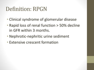
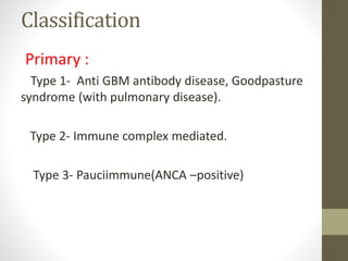
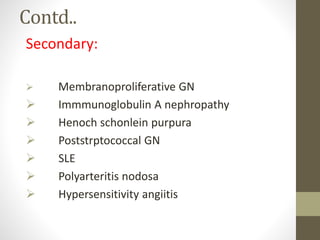
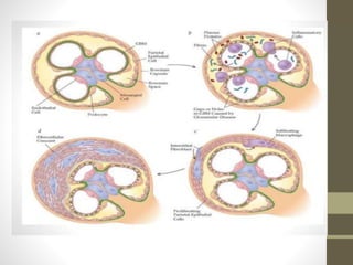
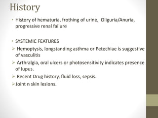
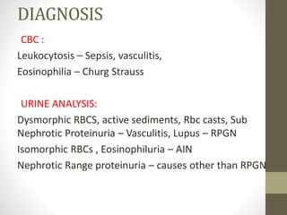
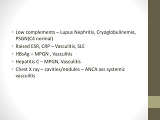
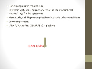
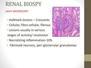
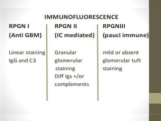
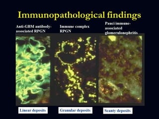
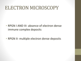
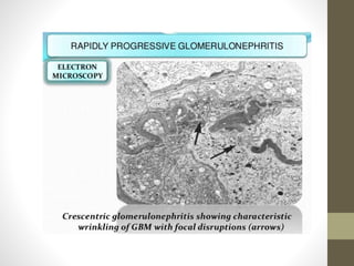
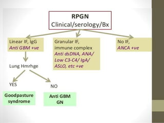
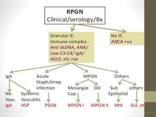
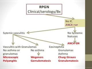
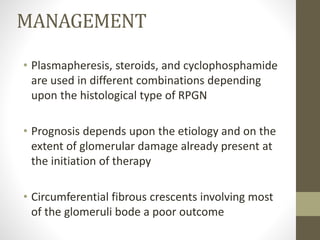
RAPIDLY
PROGRESSIVE(CRESCENTIC)
GLOMERULONEPHRITIS
DR. DR.RASIKAPRIYA
MD PAEDIATRICS
FIRST YEAR
OBJECTIVES
•Case scenario
•Introduction
•Classification
•Pathogenesis
•Clinical manifestations
•Diagnosis
•Treatment and prognosis
CASE SCENARIO
INTRODUCTION
• RPGN is characterized clinically by a rapid
decrease in the glomerular filtration rate
of at least 50% over a short period , from
few days to 3 months.
• Crescent formation.
Definition: RPGN
• Clinical syndrome of glomerular disease
• Rapid loss of renal function > 50% decline
in GFR within 3 months.
• Nephrotic-nephritic urine sediment
• Extensive crescent formation
Classification
Primary :
Type 1- Anti GBM antibody disease, Goodpasture
syndrome (with pulmonary disease).
Type 2- Immune complex mediated.
Type 3- Pauciimmune(ANCA –positive)
Contd..
Secondary:
 Membranoproliferative GN
 Immmunoglobulin A nephropathy
 Henoch schonlein purpura
 Poststrptococcal GN
 SLE
 Polyarteritis nodosa
 Hypersensitivity angiitis
PATHOGENESIS
• Crescents in glomeruli - through proliferation of
parietal epithelial cells in Bowman’s space, may
be the final pathway of any severe inflammatory
glomerular injury.
• Podocytes and renal progenitor cells are
involved in the pathogenesis of CGN.
CONTD..
• The severity of the disease -degree of crescent
formation nonspecific response to severe injury
to the glomerular capillary wall .
• Rents are induced in the glomerular capillary
wall- movement of plasma products, including
fibrinogen, into Bowman's space with
subsequent
• Fibrin formation, the influx of macrophages and
T cells, release of proinflammatory cytokines-IL-1
and TNF a
CLINICAL MANIFESTATION
• Acute nephritis- hematuria, hypertension and renal
insufficiency
• Proteinuria
• Later- oliguric renal failure
• Extra renal manifestations.
History
• History of hematuria, frothing of urine, Oliguria/Anuria,
progressive renal failure
• SYSTEMIC FEATURES
 Hemoptysis, longstanding asthma or Petechiae is suggestive
of vasculitis
 Arthralgia, oral ulcers or photosensitivity indicates presence
of lupus.
 Recent Drug history, fluid loss, sepsis.
Joint n skin lesions.
Examination
• General examination
• Skin lesions
• Respiratory system
DIAGNOSIS
CBC :
Leukocytosis – Sepsis, vasculitis,
Eosinophilia – Churg Strauss
URINE ANALYSIS:
Dysmorphic RBCS, active sediments, Rbc casts, Sub
Nephrotic Proteinuria – Vasculitis, Lupus – RPGN
Isomorphic RBCs , Eosinophiluria – AIN
Nephrotic Range proteinuria – causes other than RPGN
• Low complements – Lupus Nephritis, Cryoglobulinemia,
PSGN(C4 normal)
• Raised ESR, CRP – Vasculitis, SLE
• HBsAg – MPGN , Vasculitis
• Hepatitis C – MPGN, Vasculitis
• Chest X ray – cavities/nodules – ANCA ass systemic
vasculitis
Laboratory approach
ANCA-veANCA+ve
Low C3Pauci immune GN
Immune complex
GN
Normal C3
Normal C3
Anti-GBM disease
SEROLOGIAL TESTS
• ANA, APLA – Lupus Nephritis,
• Anti GBM – Good pasture's Syndrome/Anti GBM dis
• ASLO, Anti DNAse- PSGN
Serum C3
normal
Low
IgA
Hereditary disease
Wegener granulomatosis
Good pasture’s syndrome
Henoch-Schonlein purpura
MPGN
Shunt
nephritis
IE
Hepatitis
B,C
Low
normal
Serum C4
SLEAPSGN
Normal Serum C3 83-177 mg/dL
• Rapid progressive renal failure
• Systemic features – Pulmonary renal/ rashes/ peripheral
neuropathy/ flu like syndrome
• Hematuria, sub Nephrotic proteinuria, active urinary sediment
• Low complement
• ANCA/ ANA/ Anti GBM/ ASLO – positive
RENAL BIOPSY
RENAL BIOSPY
LIGHT MICROSCOPY:
• Hallmark lesions – Crescents
• Cellular, fibro cellular, fibrous
• Lesions usually in various
stages of activity/ resolution
• Necrotizing inflammation-10%
• Fibrinoid necrosis, peri glomerular granulomas
ELECTRON MICROSCOPY
• RPGN I AND III- absence of electron dense
immune complex deposits
• RPGN II- multiple electron dense deposits
MANAGEMENT
• Plasmapheresis, steroids, and cyclophosphamide
are used in different combinations depending
upon the histological type of RPGN
• Prognosis depends upon the etiology and on the
extent of glomerular damage already present at
the initiation of therapy
• Circumferential fibrous crescents involving most
of the glomeruli bode a poor outcome
Take home message
THANK U

5 1093438501069783057

  • 1.
  • 2.
  • 3.
  • 4.
    INTRODUCTION • RPGN ischaracterized clinically by a rapid decrease in the glomerular filtration rate of at least 50% over a short period , from few days to 3 months. • Crescent formation.
  • 5.
    Definition: RPGN • Clinicalsyndrome of glomerular disease • Rapid loss of renal function > 50% decline in GFR within 3 months. • Nephrotic-nephritic urine sediment • Extensive crescent formation
  • 6.
    Classification Primary : Type 1-Anti GBM antibody disease, Goodpasture syndrome (with pulmonary disease). Type 2- Immune complex mediated. Type 3- Pauciimmune(ANCA –positive)
  • 7.
    Contd.. Secondary:  Membranoproliferative GN Immmunoglobulin A nephropathy  Henoch schonlein purpura  Poststrptococcal GN  SLE  Polyarteritis nodosa  Hypersensitivity angiitis
  • 8.
    PATHOGENESIS • Crescents inglomeruli - through proliferation of parietal epithelial cells in Bowman’s space, may be the final pathway of any severe inflammatory glomerular injury. • Podocytes and renal progenitor cells are involved in the pathogenesis of CGN.
  • 9.
    CONTD.. • The severityof the disease -degree of crescent formation nonspecific response to severe injury to the glomerular capillary wall . • Rents are induced in the glomerular capillary wall- movement of plasma products, including fibrinogen, into Bowman's space with subsequent • Fibrin formation, the influx of macrophages and T cells, release of proinflammatory cytokines-IL-1 and TNF a
  • 11.
    CLINICAL MANIFESTATION • Acutenephritis- hematuria, hypertension and renal insufficiency • Proteinuria • Later- oliguric renal failure • Extra renal manifestations.
  • 12.
    History • History ofhematuria, frothing of urine, Oliguria/Anuria, progressive renal failure • SYSTEMIC FEATURES  Hemoptysis, longstanding asthma or Petechiae is suggestive of vasculitis  Arthralgia, oral ulcers or photosensitivity indicates presence of lupus.  Recent Drug history, fluid loss, sepsis. Joint n skin lesions.
  • 13.
    Examination • General examination •Skin lesions • Respiratory system
  • 14.
    DIAGNOSIS CBC : Leukocytosis –Sepsis, vasculitis, Eosinophilia – Churg Strauss URINE ANALYSIS: Dysmorphic RBCS, active sediments, Rbc casts, Sub Nephrotic Proteinuria – Vasculitis, Lupus – RPGN Isomorphic RBCs , Eosinophiluria – AIN Nephrotic Range proteinuria – causes other than RPGN
  • 15.
    • Low complements– Lupus Nephritis, Cryoglobulinemia, PSGN(C4 normal) • Raised ESR, CRP – Vasculitis, SLE • HBsAg – MPGN , Vasculitis • Hepatitis C – MPGN, Vasculitis • Chest X ray – cavities/nodules – ANCA ass systemic vasculitis
  • 16.
    Laboratory approach ANCA-veANCA+ve Low C3Pauciimmune GN Immune complex GN Normal C3 Normal C3 Anti-GBM disease
  • 17.
    SEROLOGIAL TESTS • ANA,APLA – Lupus Nephritis, • Anti GBM – Good pasture's Syndrome/Anti GBM dis • ASLO, Anti DNAse- PSGN
  • 18.
    Serum C3 normal Low IgA Hereditary disease Wegenergranulomatosis Good pasture’s syndrome Henoch-Schonlein purpura MPGN Shunt nephritis IE Hepatitis B,C Low normal Serum C4 SLEAPSGN Normal Serum C3 83-177 mg/dL
  • 19.
    • Rapid progressiverenal failure • Systemic features – Pulmonary renal/ rashes/ peripheral neuropathy/ flu like syndrome • Hematuria, sub Nephrotic proteinuria, active urinary sediment • Low complement • ANCA/ ANA/ Anti GBM/ ASLO – positive RENAL BIOPSY
  • 20.
    RENAL BIOSPY LIGHT MICROSCOPY: •Hallmark lesions – Crescents • Cellular, fibro cellular, fibrous • Lesions usually in various stages of activity/ resolution • Necrotizing inflammation-10% • Fibrinoid necrosis, peri glomerular granulomas
  • 23.
    ELECTRON MICROSCOPY • RPGNI AND III- absence of electron dense immune complex deposits • RPGN II- multiple electron dense deposits
  • 28.
    MANAGEMENT • Plasmapheresis, steroids,and cyclophosphamide are used in different combinations depending upon the histological type of RPGN • Prognosis depends upon the etiology and on the extent of glomerular damage already present at the initiation of therapy • Circumferential fibrous crescents involving most of the glomeruli bode a poor outcome
  • 29.
  • 30.