This document provides an overview of rapidly progressive glomerulonephritis (RPGN) for medical students. It defines RPGN as a clinical syndrome characterized by rapid loss of renal function within 3 months. RPGN can be caused by immune complex deposition or associated with anti-neutrophil cytoplasmic antibodies. The document classifies RPGN into 3 types - type I involves anti-GBM antibodies, type II immune complex deposition, and type III is pauci-immune and associated with ANCA-associated vasculitis. The diagnosis of RPGN involves renal biopsy showing crescents, elevated creatinine and urea, hematuria and proteinuria on urine examination.
most of the glomerular diseases , either primary or secondary..touching all the aspects including light microscopy, electron microscopy and immunoflourescence.
Acute kidney injury (AKI) is a sudden episode of kidney failure or kidney damage that happens within a few hours or a few days.It's most common in those who are critically ill and already hospitalized.
- English version of this lecture is available at:
https://youtu.be/XRD-QqGFP18
- Arabic version of this lecture is available at:
https://youtu.be/c9PoavAtNKM
- Visit our website for more lectures: www.NephroTube.com
- Subscribe to our YouTube channel: www.youtube.com/NephroTube
- Join our facebook group: www.facebook.com/groups/NephroTube
- Like our facebook page: www.facebook.com/NephroTube
- Follow us on twitter: www.twitter.com/NephroTube
Etiology- genetic mutations, infection, toxin exposure, autoimmunity, atherosclerosis, hypertension, emboli, thrombosis, or diabetes mellitus.
Even after careful study, however, the cause often remains unknown, and the lesion is called idiopathic.
Inflammation of the glomerular capillaries is called glomerulonephritis.
Persistent glomerulonephritis that worsens renal function is always accompanied by interstitial nephritis, renal fibrosis, and tubular atrophy.
Rapidly progressive glomerulonephritis in childrenNishatTasnim46
Rapidly progressive or crescentic glomerulonephritis is a medical emergency and diagnostic challenge in paediatric population. There is a significant risk of development of complications such as CKD in the long term. This seminar was prepared to increase knowledge about early diagnosis and management of this condition in a tertiary level hospital.
High levels of toxic bilirubin in your blood cause Crigler Najjar syndrome, a rare genetic condition. Bilirubin is a substance that forms when red blood cells reach the end of their lifespan. Your liver breaks down bilirubin from a toxic substance to a nontoxic substance that you get rid of in your stool. If you’re diagnosed with Crigler Najjar syndrome, your liver can’t break down bilirubin and it collects in your blood. As a result, Crigler Najjar syndrome can cause life threatening symptoms if left untreated. Dr. Thenmozhi P | Swathi. D "Crigler-Najjar Syndrome: Case Report" Published in International Journal of Trend in Scientific Research and Development (ijtsrd), ISSN: 2456-6470, Volume-7 | Issue-1 , February 2023, URL: https://www.ijtsrd.com/papers/ijtsrd53852.pdf Paper URL: https://www.ijtsrd.com/medicine/nursing/53852/criglernajjar-syndrome-case-report/dr-thenmozhi-p
Recomendações da OMS sobre cuidados maternos e neonatais para uma experiência pós-natal positiva.
Em consonância com os ODS – Objetivos do Desenvolvimento Sustentável e a Estratégia Global para a Saúde das Mulheres, Crianças e Adolescentes, e aplicando uma abordagem baseada nos direitos humanos, os esforços de cuidados pós-natais devem expandir-se para além da cobertura e da simples sobrevivência, de modo a incluir cuidados de qualidade.
Estas diretrizes visam melhorar a qualidade dos cuidados pós-natais essenciais e de rotina prestados às mulheres e aos recém-nascidos, com o objetivo final de melhorar a saúde e o bem-estar materno e neonatal.
Uma “experiência pós-natal positiva” é um resultado importante para todas as mulheres que dão à luz e para os seus recém-nascidos, estabelecendo as bases para a melhoria da saúde e do bem-estar a curto e longo prazo. Uma experiência pós-natal positiva é definida como aquela em que as mulheres, pessoas que gestam, os recém-nascidos, os casais, os pais, os cuidadores e as famílias recebem informação consistente, garantia e apoio de profissionais de saúde motivados; e onde um sistema de saúde flexível e com recursos reconheça as necessidades das mulheres e dos bebês e respeite o seu contexto cultural.
Estas diretrizes consolidadas apresentam algumas recomendações novas e já bem fundamentadas sobre cuidados pós-natais de rotina para mulheres e neonatos que recebem cuidados no pós-parto em unidades de saúde ou na comunidade, independentemente dos recursos disponíveis.
É fornecido um conjunto abrangente de recomendações para cuidados durante o período puerperal, com ênfase nos cuidados essenciais que todas as mulheres e recém-nascidos devem receber, e com a devida atenção à qualidade dos cuidados; isto é, a entrega e a experiência do cuidado recebido. Estas diretrizes atualizam e ampliam as recomendações da OMS de 2014 sobre cuidados pós-natais da mãe e do recém-nascido e complementam as atuais diretrizes da OMS sobre a gestão de complicações pós-natais.
O estabelecimento da amamentação e o manejo das principais intercorrências é contemplada.
Recomendamos muito.
Vamos discutir essas recomendações no nosso curso de pós-graduação em Aleitamento no Instituto Ciclos.
Esta publicação só está disponível em inglês até o momento.
Prof. Marcus Renato de Carvalho
www.agostodourado.com
These simplified slides by Dr. Sidra Arshad present an overview of the non-respiratory functions of the respiratory tract.
Learning objectives:
1. Enlist the non-respiratory functions of the respiratory tract
2. Briefly explain how these functions are carried out
3. Discuss the significance of dead space
4. Differentiate between minute ventilation and alveolar ventilation
5. Describe the cough and sneeze reflexes
Study Resources:
1. Chapter 39, Guyton and Hall Textbook of Medical Physiology, 14th edition
2. Chapter 34, Ganong’s Review of Medical Physiology, 26th edition
3. Chapter 17, Human Physiology by Lauralee Sherwood, 9th edition
4. Non-respiratory functions of the lungs https://academic.oup.com/bjaed/article/13/3/98/278874
micro teaching on communication m.sc nursing.pdfAnurag Sharma
Microteaching is a unique model of practice teaching. It is a viable instrument for the. desired change in the teaching behavior or the behavior potential which, in specified types of real. classroom situations, tends to facilitate the achievement of specified types of objectives.
Local Advanced Lung Cancer: Artificial Intelligence, Synergetics, Complex Sys...Oleg Kshivets
Overall life span (LS) was 1671.7±1721.6 days and cumulative 5YS reached 62.4%, 10 years – 50.4%, 20 years – 44.6%. 94 LCP lived more than 5 years without cancer (LS=2958.6±1723.6 days), 22 – more than 10 years (LS=5571±1841.8 days). 67 LCP died because of LC (LS=471.9±344 days). AT significantly improved 5YS (68% vs. 53.7%) (P=0.028 by log-rank test). Cox modeling displayed that 5YS of LCP significantly depended on: N0-N12, T3-4, blood cell circuit, cell ratio factors (ratio between cancer cells-CC and blood cells subpopulations), LC cell dynamics, recalcification time, heparin tolerance, prothrombin index, protein, AT, procedure type (P=0.000-0.031). Neural networks, genetic algorithm selection and bootstrap simulation revealed relationships between 5YS and N0-12 (rank=1), thrombocytes/CC (rank=2), segmented neutrophils/CC (3), eosinophils/CC (4), erythrocytes/CC (5), healthy cells/CC (6), lymphocytes/CC (7), stick neutrophils/CC (8), leucocytes/CC (9), monocytes/CC (10). Correct prediction of 5YS was 100% by neural networks computing (error=0.000; area under ROC curve=1.0).
Title: Sense of Smell
Presenter: Dr. Faiza, Assistant Professor of Physiology
Qualifications:
MBBS (Best Graduate, AIMC Lahore)
FCPS Physiology
ICMT, CHPE, DHPE (STMU)
MPH (GC University, Faisalabad)
MBA (Virtual University of Pakistan)
Learning Objectives:
Describe the primary categories of smells and the concept of odor blindness.
Explain the structure and location of the olfactory membrane and mucosa, including the types and roles of cells involved in olfaction.
Describe the pathway and mechanisms of olfactory signal transmission from the olfactory receptors to the brain.
Illustrate the biochemical cascade triggered by odorant binding to olfactory receptors, including the role of G-proteins and second messengers in generating an action potential.
Identify different types of olfactory disorders such as anosmia, hyposmia, hyperosmia, and dysosmia, including their potential causes.
Key Topics:
Olfactory Genes:
3% of the human genome accounts for olfactory genes.
400 genes for odorant receptors.
Olfactory Membrane:
Located in the superior part of the nasal cavity.
Medially: Folds downward along the superior septum.
Laterally: Folds over the superior turbinate and upper surface of the middle turbinate.
Total surface area: 5-10 square centimeters.
Olfactory Mucosa:
Olfactory Cells: Bipolar nerve cells derived from the CNS (100 million), with 4-25 olfactory cilia per cell.
Sustentacular Cells: Produce mucus and maintain ionic and molecular environment.
Basal Cells: Replace worn-out olfactory cells with an average lifespan of 1-2 months.
Bowman’s Gland: Secretes mucus.
Stimulation of Olfactory Cells:
Odorant dissolves in mucus and attaches to receptors on olfactory cilia.
Involves a cascade effect through G-proteins and second messengers, leading to depolarization and action potential generation in the olfactory nerve.
Quality of a Good Odorant:
Small (3-20 Carbon atoms), volatile, water-soluble, and lipid-soluble.
Facilitated by odorant-binding proteins in mucus.
Membrane Potential and Action Potential:
Resting membrane potential: -55mV.
Action potential frequency in the olfactory nerve increases with odorant strength.
Adaptation Towards the Sense of Smell:
Rapid adaptation within the first second, with further slow adaptation.
Psychological adaptation greater than receptor adaptation, involving feedback inhibition from the central nervous system.
Primary Sensations of Smell:
Camphoraceous, Musky, Floral, Pepperminty, Ethereal, Pungent, Putrid.
Odor Detection Threshold:
Examples: Hydrogen sulfide (0.0005 ppm), Methyl-mercaptan (0.002 ppm).
Some toxic substances are odorless at lethal concentrations.
Characteristics of Smell:
Odor blindness for single substances due to lack of appropriate receptor protein.
Behavioral and emotional influences of smell.
Transmission of Olfactory Signals:
From olfactory cells to glomeruli in the olfactory bulb, involving lateral inhibition.
Primitive, less old, and new olfactory systems with different path
These lecture slides, by Dr Sidra Arshad, offer a quick overview of the physiological basis of a normal electrocardiogram.
Learning objectives:
1. Define an electrocardiogram (ECG) and electrocardiography
2. Describe how dipoles generated by the heart produce the waveforms of the ECG
3. Describe the components of a normal electrocardiogram of a typical bipolar lead (limb II)
4. Differentiate between intervals and segments
5. Enlist some common indications for obtaining an ECG
6. Describe the flow of current around the heart during the cardiac cycle
7. Discuss the placement and polarity of the leads of electrocardiograph
8. Describe the normal electrocardiograms recorded from the limb leads and explain the physiological basis of the different records that are obtained
9. Define mean electrical vector (axis) of the heart and give the normal range
10. Define the mean QRS vector
11. Describe the axes of leads (hexagonal reference system)
12. Comprehend the vectorial analysis of the normal ECG
13. Determine the mean electrical axis of the ventricular QRS and appreciate the mean axis deviation
14. Explain the concepts of current of injury, J point, and their significance
Study Resources:
1. Chapter 11, Guyton and Hall Textbook of Medical Physiology, 14th edition
2. Chapter 9, Human Physiology - From Cells to Systems, Lauralee Sherwood, 9th edition
3. Chapter 29, Ganong’s Review of Medical Physiology, 26th edition
4. Electrocardiogram, StatPearls - https://www.ncbi.nlm.nih.gov/books/NBK549803/
5. ECG in Medical Practice by ABM Abdullah, 4th edition
6. Chapter 3, Cardiology Explained, https://www.ncbi.nlm.nih.gov/books/NBK2214/
7. ECG Basics, http://www.nataliescasebook.com/tag/e-c-g-basics
Best Ayurvedic medicine for Gas and IndigestionSwastikAyurveda
Here is the updated list of Top Best Ayurvedic medicine for Gas and Indigestion and those are Gas-O-Go Syp for Dyspepsia | Lavizyme Syrup for Acidity | Yumzyme Hepatoprotective Capsules etc
Adv. biopharm. APPLICATION OF PHARMACOKINETICS : TARGETED DRUG DELIVERY SYSTEMSAkankshaAshtankar
MIP 201T & MPH 202T
ADVANCED BIOPHARMACEUTICS & PHARMACOKINETICS : UNIT 5
APPLICATION OF PHARMACOKINETICS : TARGETED DRUG DELIVERY SYSTEMS By - AKANKSHA ASHTANKAR
Ozempic: Preoperative Management of Patients on GLP-1 Receptor Agonists Saeid Safari
Preoperative Management of Patients on GLP-1 Receptor Agonists like Ozempic and Semiglutide
ASA GUIDELINE
NYSORA Guideline
2 Case Reports of Gastric Ultrasound
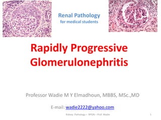
1. Renal Pathology
for medical students
Rapidly Progressive
Glomerulonephritis
Professor Wadie M Y Elmadhoun, MBBS, MSc.,MD
E-mail: wadie2222@yahoo.com
Kidney Pathology – RPGN – Prof. Wadie 1
2. Presentation outlines
1. Intended Learning outcomes (ILOs).
2. Rapidly Progressive Glomerulonephritis
(RPGN): definition, pathogenesis and clinical
course.
3. Types of RPGN: Type I, Type II, Type III.
5. Quiz.
6. Further learning resources.
Kidney Pathology – RPGN – Prof. Wadie
2
3. Intended Learning outcomes (ILOs)
• By the end of this session, the learner should be
able to:
1. Explain what is meant by Rapidly Progress GN,
discuss its nature, clarify its clinical course,
causes and pathogenesis.
2. Differentiate between the various types of
RPGN.
Kidney Pathology –RPGN – Prof. Wadie
3
4. Definition of the Rapidly Progressive
Glomerulonephritis (RPGN)
• A clinical syndrome characterized by:
• Rapid loss of renal function: more than 50%
decline of GFR within 3 months.
• Crescent-shaped scars in most glomeruli,
• If not treated, will progress to acute renal
failure and death within months.
Kidney Pathology – RPGN – Prof. Wadie 4
5. Crescents in 2 glomeruli
Kidney Pathology – RPGN – Prof. Wadie 5
8. Overview of RPGN/Crescentic GN
• RPGN is a clinical definition, NOT a specific
pathologic GN.
• Also known as “CRESCENTIC GN”
• It is caused by either
immunological damage to
glomeruli or associated with anti-
neutrophil cytoplasmic antibody
(ANCA). Kidney Pathology – RPGN – Prof. Wadie 8
9. Rapidly Progressive Glomerulonephritis
(Crescentic GN)
• Type I: anti-GBM antibodies.
• Type II : immune complex
deposition in GBM.
• Type III : No remarkable immune damage,
associated with anti-neutrophil cytoplasmic
antibody (ANCA).
Kidney Pathology – RPGN – Prof. Wadie
10. Signs and symptoms
• Most types of RPGN are characterized by:
• severe and rapid loss of kidney function
• with marked hematuria; red blood cell casts in
the urine; and proteinuria
• Some patients also experience hypertension
and edema.
• Severe disease is characterized by oliguria or
anuria.
Kidney Pathology – RPGN – Prof. Wadie
11. Two urine samples showing gross and microscopic
hematuria: in RPGN
Kidney Pathology – RPGN – Prof. Wadie
11
12. CLASSIFICATION
• Crescentic glomerulonephritis (CrGN)
• RPGN can be classified into three types, based
upon the pathogenesis and
immunofluorescence patterns:
Kidney Pathology – RPGN – Prof. Wadie 12
13. 1. Type I CrGN
• also called anti-GBM antibody Crescentic GN: is
characterized by the presence of autoantibodies
directed against type IV collagen in the (GBM).
• Some cases are associated with antibodies
directed against the basement membrane of lung
alveoli, producing Goodpasture syndrome.
• Type I accounts for less than 20% of RPGN.
• Plasma exchange or plasma-pheresis benefit
patients with type I CrGN, but not types II or III.
Kidney Pathology – RPGN – Prof. Wadie 13
14. Anti-GBM Antibody, type I RPGN
(Linear immunoflourescence in Type I CrGN)
Kidney Pathology – RPGN – Prof. Wadie 14
15. 2. Type II CrGN
• Characterized by deposition of immune
complexes in glomerular tissues. (Granular IF)
• Any immune complex disease—including SLE,
acute proliferative glomerulonephritis,
Henoch–Schönlein purpura, and IgA
nephropathy may progress to RPGN.
• Type II RPGN accounts for about 40% of cases
Kidney Pathology – RPGN – Prof. Wadie 15
18. RPGN type II: secondary to IgA nephropathy
Kidney Pathology – RPGN – Prof. Wadie 18
19. 3. Type III CrGN
• Also known as pauci-immune RPGN, and features
neither immune complex deposition nor anti-GBM
antibodies.
• Instead, the glomeruli are damaged perhaps
through the activation of neutrophils in response to
ANCA.
• Type III RPGN may be (primary, or idiopathic) or
associated with a systemic disease (secondary) to
ANCA-associated vasculitis such as granulomatosis
with polyangiitis.
• Type III RPGN accounts for more than 40% of RPGN
Kidney Pathology – RPGN – Prof. Wadie 19
20. Diagnosis of RPGN
• Impaired kidney function in an individual who has
had the condition for fewer than three months is
characteristic of RPGN.
1. Renal biopsy: is the most important
investigation. Crescents are identified on
microscopy + linear or granular pattern in
immunofluorescence.
2. Raised serum creatinine and urea.
3. Urine examination: oliguria or anuria, hematuria
and proteinuria.
Kidney Pathology – RPGN – Prof. Wadie 20
22. Diagnosis
• Serum analysis often aids in the diagnosis of a
specific underlying disease.
1. The presence of anti-glomerular basement
membrane (GBM) antibodies suggests type I
RPGN.
2. Antinuclear antibodies (ANA) may support a
diagnosis of systemic lupus erythematosus and
type II RPGN;
3. Type III is associated with anti-neutrophil
cytoplasmic antibodies (ANCA)-positive serum.
Kidney Pathology – RPGN – Prof. Wadie 22
25. ASSIGNMENT
•You are asked to revise
the next slide carefully,
then write a two-page
essay about its topic.
Kidney Pathology – RPGN – Prof. Wadie
25