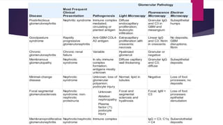
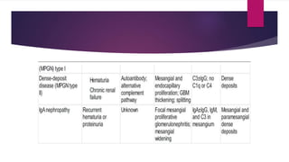
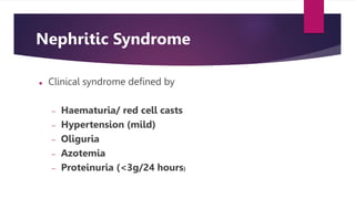
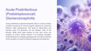
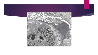
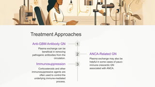
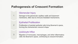
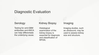
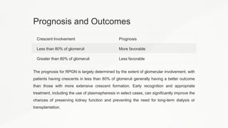
GLOMERULAR DISEASES
AND SYNDROMES
DR. SARA REZA
Specific learning objectives
By the end of this class, the students should be able to understand
the pathogenesis and clinical presentation of various glomerular
diseases.
 Which of the following characteristics is used to define a
nephritic syndrome?
A. Red Blood Cell casts in urine with a decreased BUN and
creatinine
B. Fatty casts in urine
C. Polyuria and hypotension
D. Protein in the urine (under 3.5 grams per day)
Nephritic Syndrome
 Clinical syndrome defined by
 Haematuria/ red cell casts
 Hypertension (mild)
 Oliguria
 Azotemia
 Proteinuria (<3g/24 hours)
CAUSES OF NEPHRITIC SYNDROME
Acute Postinfectious
(Poststreptococcal)
Glomerulonephritis
Acute postinfectious glomerulonephritis (GN) is a serious kidney
condition caused by an immune system response to a previous
infection, often streptococcal. This leads to inflammation and
damage within the glomeruli, the tiny filtration units of the
kidneys. While most cases resolve on their own, some can
progress to chronic kidney disease if not properly managed.
Understanding the underlying pathogenesis and clinical features
of this condition is crucial for effective diagnosis and treatment.
Pathogenesis of Postinfectious GN
1 Infection
Certain "nephritogenic" strains of streptococcal bacteria, as well as other
pathogens like pneumococcus and hepatitis viruses, can trigger an immune
response that leads to postinfectious GN.
2 Immune Complex Formation
Antibodies produced against bacterial antigens form immune complexes
that deposit in the glomeruli, activating the complement system and causing
inflammation.
3 Glomerular Injury
The immune complexes and complement activation lead to proliferation of
glomerular cells, leukocyte infiltration, and damage to the capillary walls,
resulting in the clinical features of postinfectious GN.
Histologic Features
Light Microscopy
Postinfectious GN is
characterized by diffuse
proliferation of glomerular
cells, including
endothelial, mesangial,
and infiltrating
inflammatory cells like
neutrophils. There may
also be necrosis of
capillary walls and
crescent formation in
severe cases.
Electron Microscopy
Electron microscopy
reveals immune complex
deposits, often in a
subepithelial "hump"
pattern, along the
glomerular basement
membrane. Mesangial
deposits may also be
present.
Immunofluorescence
Granular deposits of IgG
and complement
components are seen
along the capillary walls
and in the mesangium,
corresponding to the
immune complexes
visualized by electron
microscopy.
immunofluorescence microscopy
IgG, IgM, and C3 in in the mesangium and along the GBM
Clinical Presentation
1 Acute Nephritic Syndrome
The most common clinical presentation,
characterized by edema, hypertension,
and hematuria.
2 Proteinuria
Varying degrees of proteinuria,
occasionally severe enough to cause
nephrotic syndrome.
3 Complement Consumption
Decreased serum complement levels
during the active phase of the disease.
4 Serologic Markers
Elevated anti-streptolysin O (ASO)
antibody titers in poststreptococcal cases.
Prognosis and Outcomes
1 Acute Resolution
In most children with poststreptococcal GN, the condition resolves with
supportive care and the inflammation subsides over 2-3 months.
2 Rapid Progression
Some cases can develop rapidly progressive GN due to severe glomerular
injury and crescent formation, leading to rapid renal function decline.
3 Chronic Kidney Disease
In adults, 15-50% of cases progress to end-stage renal disease over years,
depending on the initial severity. Children have a lower risk of chronicity.
Treatment and Management
Supportive Care
The mainstay of treatment is supportive
care, including management of
hypertension, edema, and electrolyte
imbalances. Antibiotics may be used to
treat the underlying infection.
Immunosuppression
In severe or rapidly progressive cases,
immunosuppressive medications like
corticosteroids may be used to
suppress the aberrant immune
response.
Dialysis and Transplant
Patients who develop end-stage renal disease may require dialysis or kidney
transplantation as definitive treatment.
Prevention and Risk Factors
Streptococcal
Infections
Infection with certain
"nephritogenic" strains of
group A Streptococcus is
the most common trigger
for postinfectious GN.
Vaccination
Vaccination against
streptococcal and other
bacterial infections may
help prevent some cases of
postinfectious GN.
Good Hygiene
Proper hand hygiene and
prompt treatment of
streptococcal throat or skin
infections can reduce the
risk of postinfectious GN.
Future Directions and Research
Pathogenesis
Ongoing research aims to further elucidate the specific antigens and immune
mechanisms involved in the development of postinfectious GN.
Novel Therapies
Exploration of targeted immunomodulatory treatments to interrupt the
aberrant immune response and prevent progression to chronic kidney
disease.
Risk Stratification
Developing better predictive models to identify patients at high risk of
complications, informing individualized treatment approaches.
Rapidly Progressive
Glomerulonephritis
(RPGN)
Rapidly Progressive Glomerulonephritis (RPGN) is a clinical
syndrome characterized by the rapid loss of renal function, often
leading to kidney failure within weeks or months if left untreated.
It is typically associated with the presence of crescents, or
proliferative lesions outside the glomerular capillary loops, which
are a hallmark of this condition.
Causes of RPGN
Anti-GBM
Antibody-
Mediated
Also known as
Goodpasture's
disease, this form is
characterized by
linear deposits of IgG
and C3 along the
glomerular basement
membrane (GBM).
Patients may also
experience
pulmonary
hemorrhage.
Immune
Complex-
Mediated
Immune complexes
can accumulate in the
glomeruli, leading to
cellular proliferation
and crescent
formation. This can
occur in conditions
like post-
streptococcal
glomerulonephritis,
lupus, and IgA
nephropathy.
Pauci-Immune
In this form, there is a
lack of significant
immune complex
deposition or anti-
GBM antibodies. Anti-
neutrophil
cytoplasmic
antibodies (ANCA)
are often present,
and it may be a
component of
systemic vasculitis.
Pathological Features
1 Cellular Proliferation
Proliferation of epithelial cells
and migration of
monocytes/macrophages into
Bowman's space, forming the
characteristic crescent shape.
2 Glomerular Injury
Segmental capillary necrosis,
breaks in the GBM, and
deposition of fibrin in Bowman's
space, indicating severe
glomerular damage.
3 Immunofluorescence Patterns
Linear staining of IgG and C3 along the GBM in anti-GBM disease,
granular staining in immune complex-mediated GN, and negative staining
in pauci-immune GN.
Clinical Presentation
Rapid Onset
The onset of RPGN is similar to the
nephritic syndrome, but with more
pronounced oliguria and azotemia.
Proteinuria
Proteinuria may approach the nephrotic
range in some cases.
Dialysis or Transplant
Some patients become anuric and
require long-term dialysis or kidney
transplantation.
Prognosis
The prognosis is related to the
percentage of glomeruli involved, with a
better outlook for those with less than
80% crescent formation.
Treatment Approaches
1
Anti-GBM Antibody GN
Plasma exchange can be
beneficial in removing
pathogenic antibodies from the
circulation.
2 ANCA-Related GN
Plasma exchange may also be
helpful in some cases of pauci-
immune crescentic GN
associated with ANCA.
3
Immunosuppression
Corticosteroids and other
immunosuppressive agents are
often used to control the
underlying immune-mediated
process.
Pathogenesis of Crescent Formation
Glomerular Injury
Damage to the glomerular capillary walls and basement
membrane, often due to immune-mediated mechanisms.
Epithelial Proliferation
Proliferation of parietal epithelial cells lining Bowman's space,
forming the characteristic crescent shape.
Leukocyte Influx
Migration of monocytes, macrophages, and other inflammatory
cells into Bowman's space, contributing to crescent formation.
Diagnostic Evaluation
Serology
Testing for anti-GBM
antibodies and ANCA
can help differentiate
the underlying cause.
Kidney Biopsy
Histological
examination of the
kidney biopsy is
essential for diagnosis
and classification of
RPGN.
Imaging
Imaging studies, such
as ultrasound, may be
used to assess kidney
size and structure.
Prognosis and Outcomes
Crescent Involvement Prognosis
Less than 80% of glomeruli More favorable
Greater than 80% of glomeruli Less favorable
The prognosis for RPGN is largely determined by the extent of glomerular involvement, with
patients having crescents in less than 80% of glomeruli generally having a better outcome
than those with more extensive crescent formation. Early recognition and appropriate
treatment, including the use of plasmapheresis in select cases, can significantly improve the
chances of preserving kidney function and preventing the need for long-term dialysis or
transplantation.

Acute-Postinfectious-Poststreptococcal-Glomerulonephritis.pptx

  • 1.
  • 2.
    Specific learning objectives Bythe end of this class, the students should be able to understand the pathogenesis and clinical presentation of various glomerular diseases.
  • 5.
     Which ofthe following characteristics is used to define a nephritic syndrome? A. Red Blood Cell casts in urine with a decreased BUN and creatinine B. Fatty casts in urine C. Polyuria and hypotension D. Protein in the urine (under 3.5 grams per day)
  • 6.
    Nephritic Syndrome  Clinicalsyndrome defined by  Haematuria/ red cell casts  Hypertension (mild)  Oliguria  Azotemia  Proteinuria (<3g/24 hours)
  • 7.
  • 8.
    Acute Postinfectious (Poststreptococcal) Glomerulonephritis Acute postinfectiousglomerulonephritis (GN) is a serious kidney condition caused by an immune system response to a previous infection, often streptococcal. This leads to inflammation and damage within the glomeruli, the tiny filtration units of the kidneys. While most cases resolve on their own, some can progress to chronic kidney disease if not properly managed. Understanding the underlying pathogenesis and clinical features of this condition is crucial for effective diagnosis and treatment.
  • 9.
    Pathogenesis of PostinfectiousGN 1 Infection Certain "nephritogenic" strains of streptococcal bacteria, as well as other pathogens like pneumococcus and hepatitis viruses, can trigger an immune response that leads to postinfectious GN. 2 Immune Complex Formation Antibodies produced against bacterial antigens form immune complexes that deposit in the glomeruli, activating the complement system and causing inflammation. 3 Glomerular Injury The immune complexes and complement activation lead to proliferation of glomerular cells, leukocyte infiltration, and damage to the capillary walls, resulting in the clinical features of postinfectious GN.
  • 10.
    Histologic Features Light Microscopy PostinfectiousGN is characterized by diffuse proliferation of glomerular cells, including endothelial, mesangial, and infiltrating inflammatory cells like neutrophils. There may also be necrosis of capillary walls and crescent formation in severe cases. Electron Microscopy Electron microscopy reveals immune complex deposits, often in a subepithelial "hump" pattern, along the glomerular basement membrane. Mesangial deposits may also be present. Immunofluorescence Granular deposits of IgG and complement components are seen along the capillary walls and in the mesangium, corresponding to the immune complexes visualized by electron microscopy.
  • 12.
    immunofluorescence microscopy IgG, IgM,and C3 in in the mesangium and along the GBM
  • 14.
    Clinical Presentation 1 AcuteNephritic Syndrome The most common clinical presentation, characterized by edema, hypertension, and hematuria. 2 Proteinuria Varying degrees of proteinuria, occasionally severe enough to cause nephrotic syndrome. 3 Complement Consumption Decreased serum complement levels during the active phase of the disease. 4 Serologic Markers Elevated anti-streptolysin O (ASO) antibody titers in poststreptococcal cases.
  • 15.
    Prognosis and Outcomes 1Acute Resolution In most children with poststreptococcal GN, the condition resolves with supportive care and the inflammation subsides over 2-3 months. 2 Rapid Progression Some cases can develop rapidly progressive GN due to severe glomerular injury and crescent formation, leading to rapid renal function decline. 3 Chronic Kidney Disease In adults, 15-50% of cases progress to end-stage renal disease over years, depending on the initial severity. Children have a lower risk of chronicity.
  • 16.
    Treatment and Management SupportiveCare The mainstay of treatment is supportive care, including management of hypertension, edema, and electrolyte imbalances. Antibiotics may be used to treat the underlying infection. Immunosuppression In severe or rapidly progressive cases, immunosuppressive medications like corticosteroids may be used to suppress the aberrant immune response. Dialysis and Transplant Patients who develop end-stage renal disease may require dialysis or kidney transplantation as definitive treatment.
  • 17.
    Prevention and RiskFactors Streptococcal Infections Infection with certain "nephritogenic" strains of group A Streptococcus is the most common trigger for postinfectious GN. Vaccination Vaccination against streptococcal and other bacterial infections may help prevent some cases of postinfectious GN. Good Hygiene Proper hand hygiene and prompt treatment of streptococcal throat or skin infections can reduce the risk of postinfectious GN.
  • 18.
    Future Directions andResearch Pathogenesis Ongoing research aims to further elucidate the specific antigens and immune mechanisms involved in the development of postinfectious GN. Novel Therapies Exploration of targeted immunomodulatory treatments to interrupt the aberrant immune response and prevent progression to chronic kidney disease. Risk Stratification Developing better predictive models to identify patients at high risk of complications, informing individualized treatment approaches.
  • 19.
    Rapidly Progressive Glomerulonephritis (RPGN) Rapidly ProgressiveGlomerulonephritis (RPGN) is a clinical syndrome characterized by the rapid loss of renal function, often leading to kidney failure within weeks or months if left untreated. It is typically associated with the presence of crescents, or proliferative lesions outside the glomerular capillary loops, which are a hallmark of this condition.
  • 21.
    Causes of RPGN Anti-GBM Antibody- Mediated Alsoknown as Goodpasture's disease, this form is characterized by linear deposits of IgG and C3 along the glomerular basement membrane (GBM). Patients may also experience pulmonary hemorrhage. Immune Complex- Mediated Immune complexes can accumulate in the glomeruli, leading to cellular proliferation and crescent formation. This can occur in conditions like post- streptococcal glomerulonephritis, lupus, and IgA nephropathy. Pauci-Immune In this form, there is a lack of significant immune complex deposition or anti- GBM antibodies. Anti- neutrophil cytoplasmic antibodies (ANCA) are often present, and it may be a component of systemic vasculitis.
  • 22.
    Pathological Features 1 CellularProliferation Proliferation of epithelial cells and migration of monocytes/macrophages into Bowman's space, forming the characteristic crescent shape. 2 Glomerular Injury Segmental capillary necrosis, breaks in the GBM, and deposition of fibrin in Bowman's space, indicating severe glomerular damage. 3 Immunofluorescence Patterns Linear staining of IgG and C3 along the GBM in anti-GBM disease, granular staining in immune complex-mediated GN, and negative staining in pauci-immune GN.
  • 24.
    Clinical Presentation Rapid Onset Theonset of RPGN is similar to the nephritic syndrome, but with more pronounced oliguria and azotemia. Proteinuria Proteinuria may approach the nephrotic range in some cases. Dialysis or Transplant Some patients become anuric and require long-term dialysis or kidney transplantation. Prognosis The prognosis is related to the percentage of glomeruli involved, with a better outlook for those with less than 80% crescent formation.
  • 25.
    Treatment Approaches 1 Anti-GBM AntibodyGN Plasma exchange can be beneficial in removing pathogenic antibodies from the circulation. 2 ANCA-Related GN Plasma exchange may also be helpful in some cases of pauci- immune crescentic GN associated with ANCA. 3 Immunosuppression Corticosteroids and other immunosuppressive agents are often used to control the underlying immune-mediated process.
  • 26.
    Pathogenesis of CrescentFormation Glomerular Injury Damage to the glomerular capillary walls and basement membrane, often due to immune-mediated mechanisms. Epithelial Proliferation Proliferation of parietal epithelial cells lining Bowman's space, forming the characteristic crescent shape. Leukocyte Influx Migration of monocytes, macrophages, and other inflammatory cells into Bowman's space, contributing to crescent formation.
  • 27.
    Diagnostic Evaluation Serology Testing foranti-GBM antibodies and ANCA can help differentiate the underlying cause. Kidney Biopsy Histological examination of the kidney biopsy is essential for diagnosis and classification of RPGN. Imaging Imaging studies, such as ultrasound, may be used to assess kidney size and structure.
  • 28.
    Prognosis and Outcomes CrescentInvolvement Prognosis Less than 80% of glomeruli More favorable Greater than 80% of glomeruli Less favorable The prognosis for RPGN is largely determined by the extent of glomerular involvement, with patients having crescents in less than 80% of glomeruli generally having a better outcome than those with more extensive crescent formation. Early recognition and appropriate treatment, including the use of plasmapheresis in select cases, can significantly improve the chances of preserving kidney function and preventing the need for long-term dialysis or transplantation.

Editor's Notes

  • #4 Summary of Major Primary Glomerulonephritides
  • #6 D One of the defining features used to determine the difference between nephritic and nephrotic syndromes is the amount of protein in the urine. Nephritic syndromes have under 3.5 g/day. Nephrotic syndromes have over 3.5 g/day. Fatty casts are a feature of nephrotic syndromes.
  • #7 Glomerular diseases presenting with nephritic syndrome are often characterized by inflammation in the glomeruli. There is hypercellularity. The proliferation of cells compress the blood vessels resulting in hypertension and oliguria and injury to endothelial cells causes hematuria. Proteinuria and edema are common, but these are not as severe as those encountered in the nephrotic syndrome
  • #12 A, Normal glomerulus. B, Glomerular hypercellularity is due to intracapillary leukocytes and proliferation of intrinsic glomerular cells.
  • #14 Typical electron-dense subepithelial “hump”
  • #24 Crescentic glomerulonephritis (PAS stain). Note the collapsed glomerular tufts and the crescent-shaped mass of proliferating parietal epithelial cells and leukocytes internal to Bowman capsule.