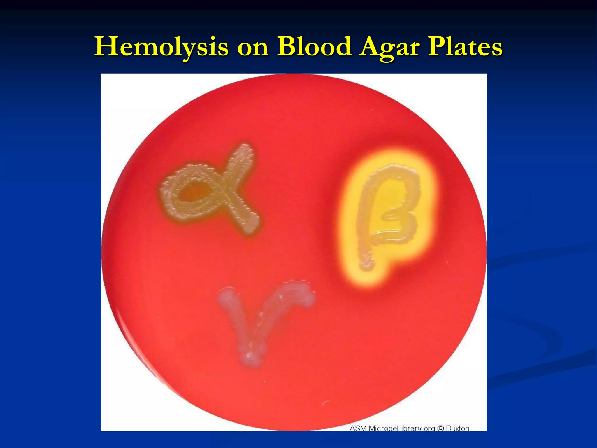
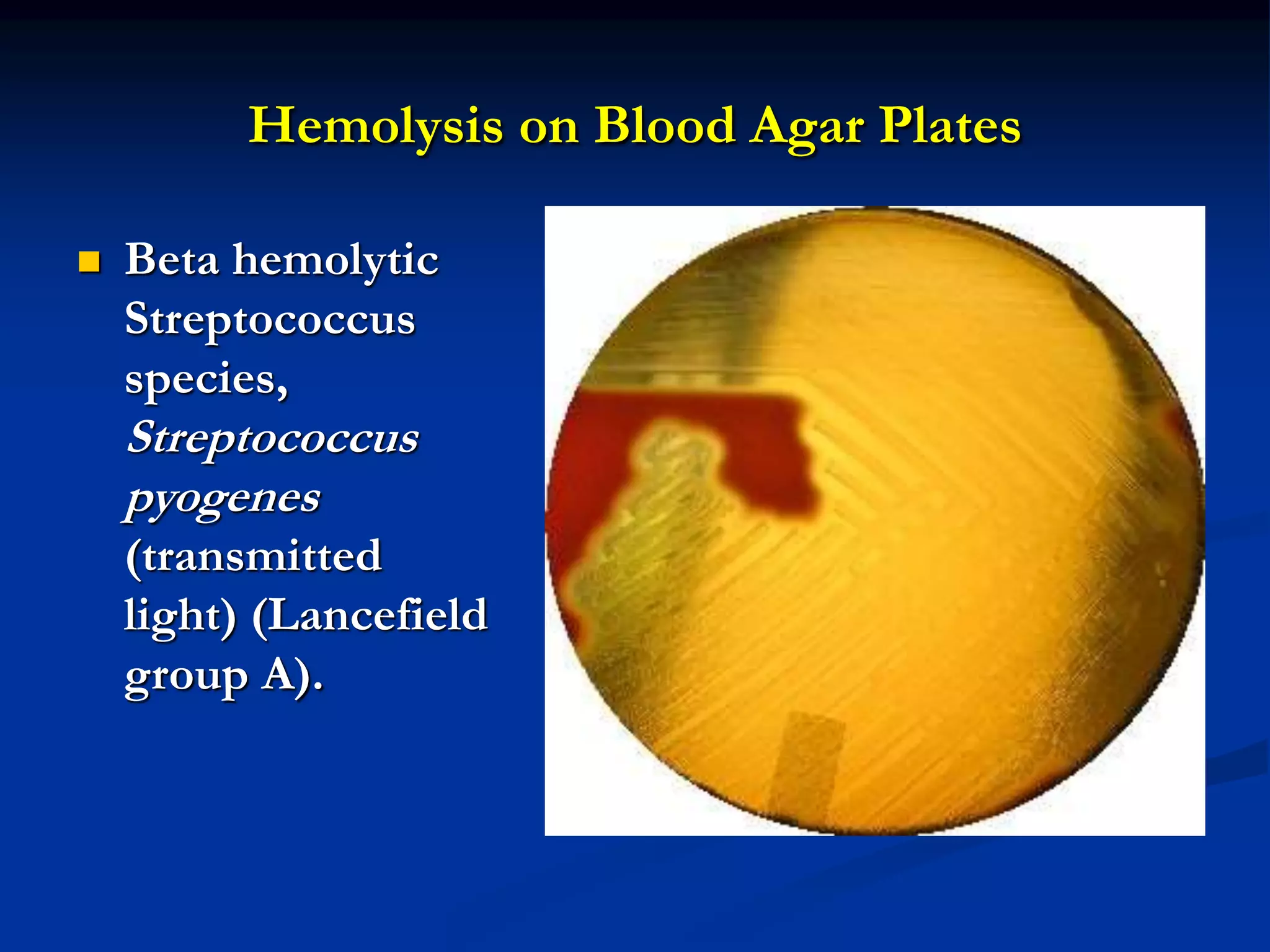
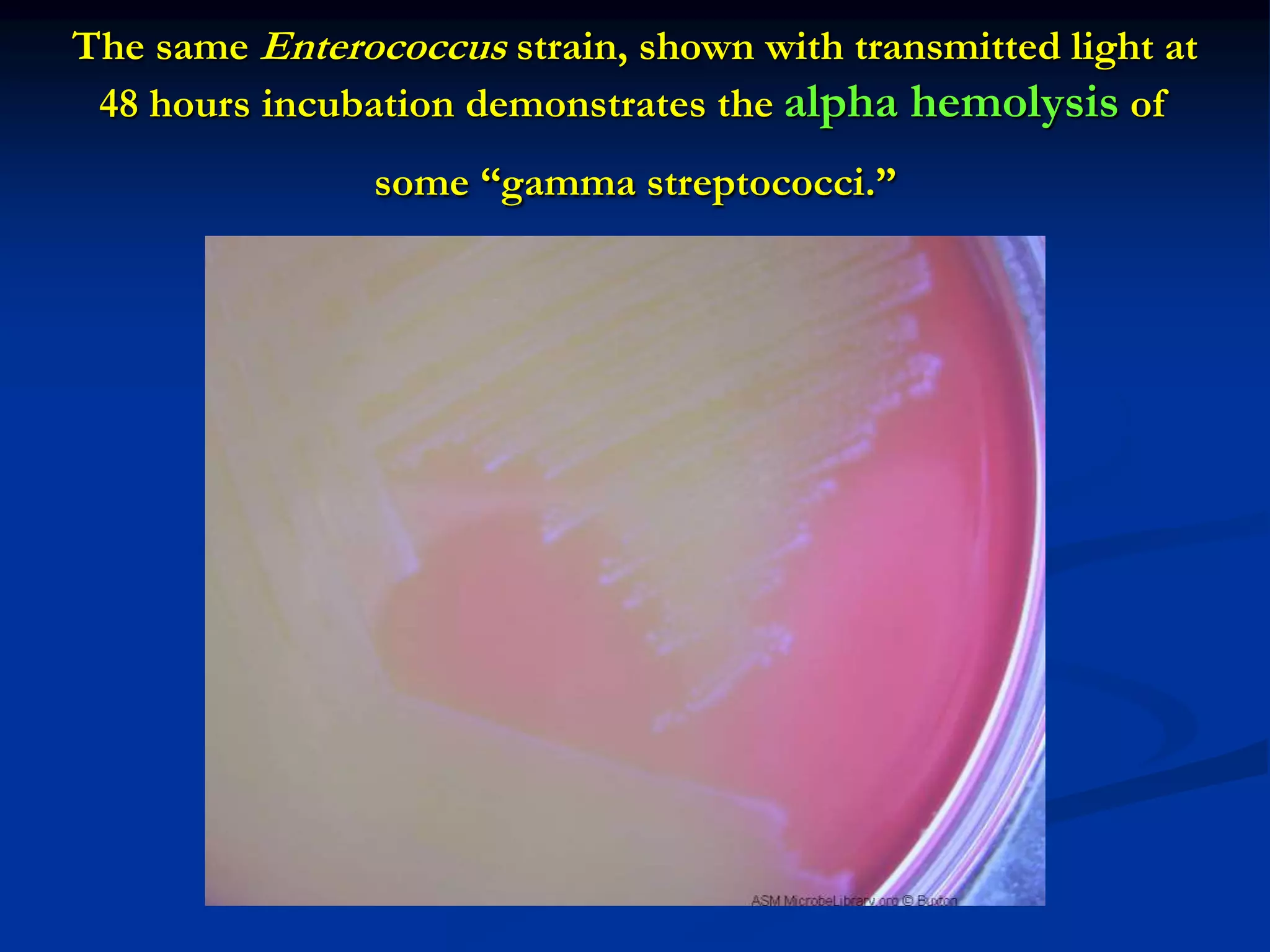
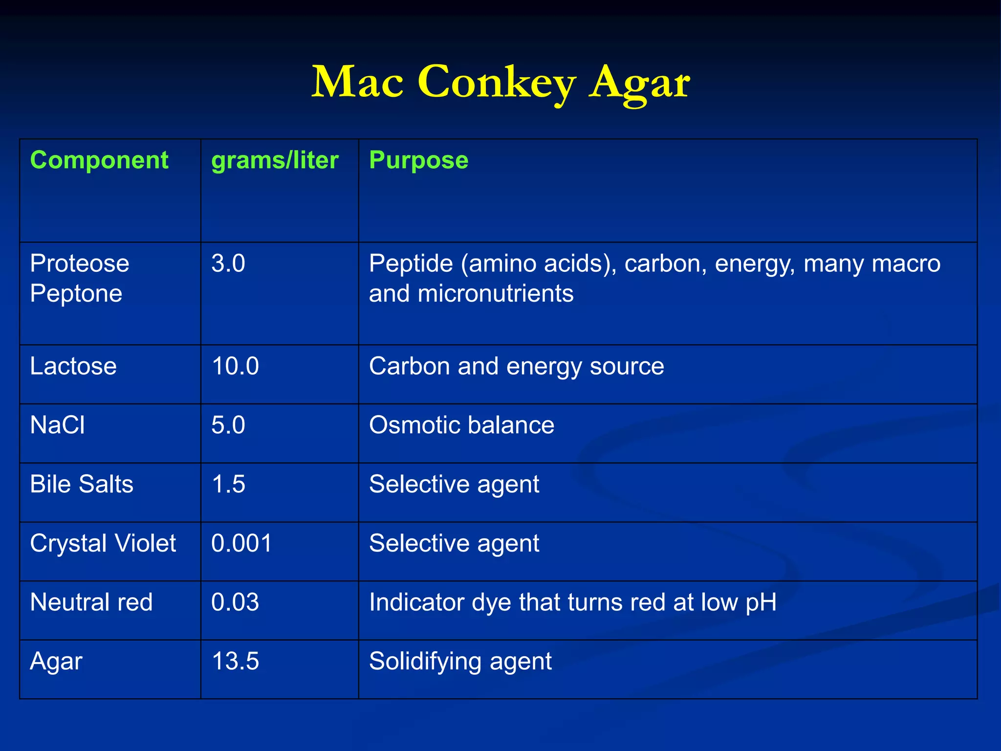
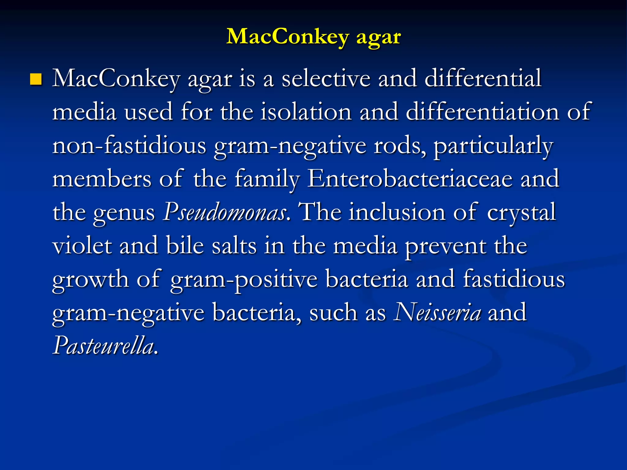
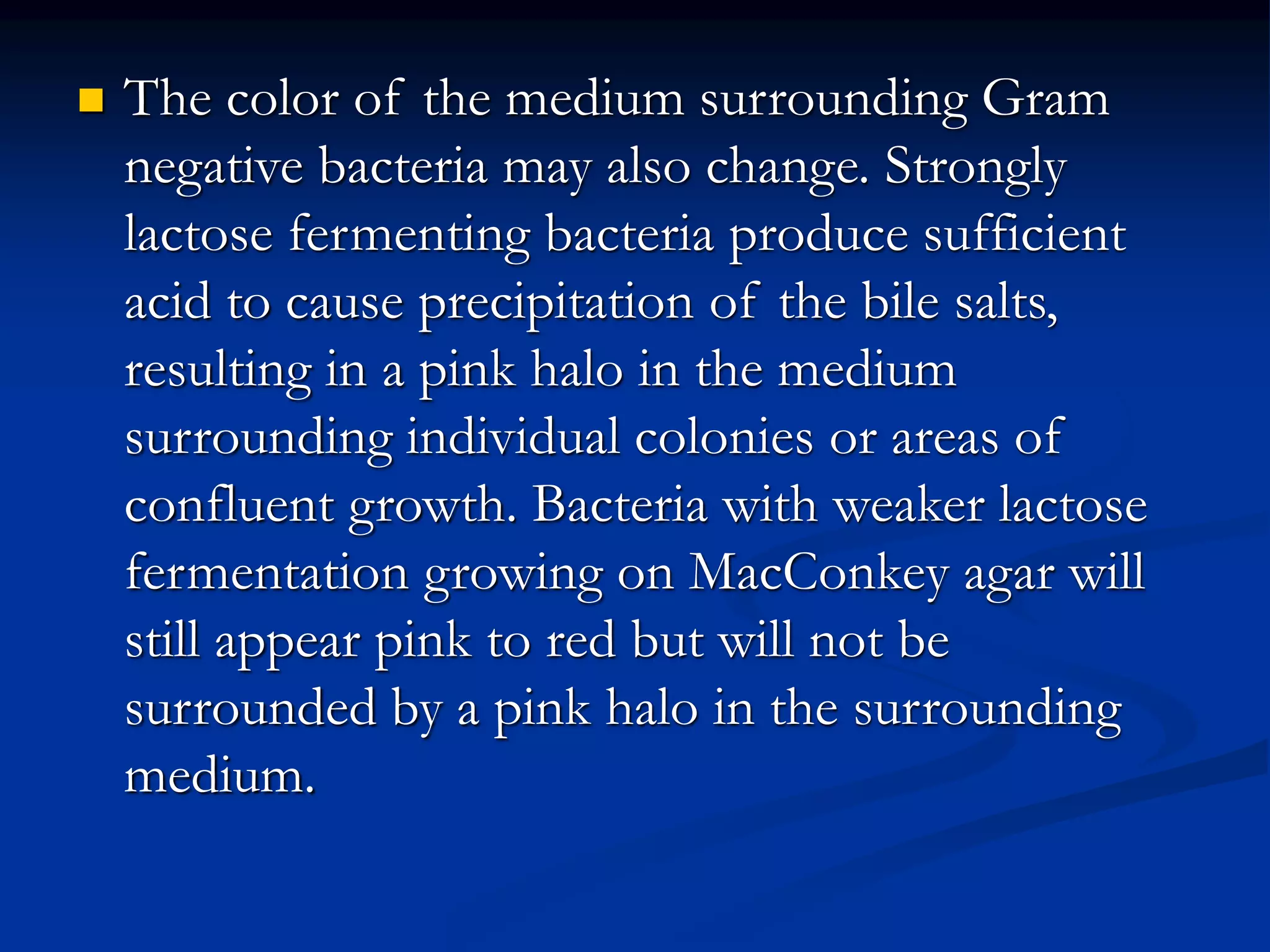
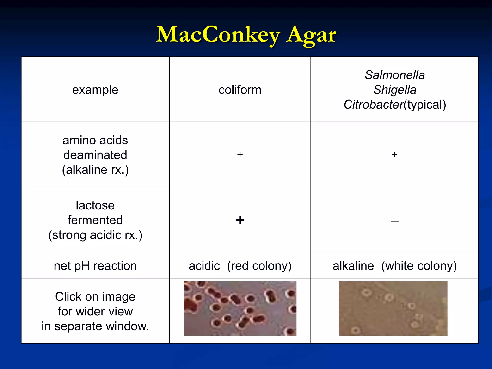
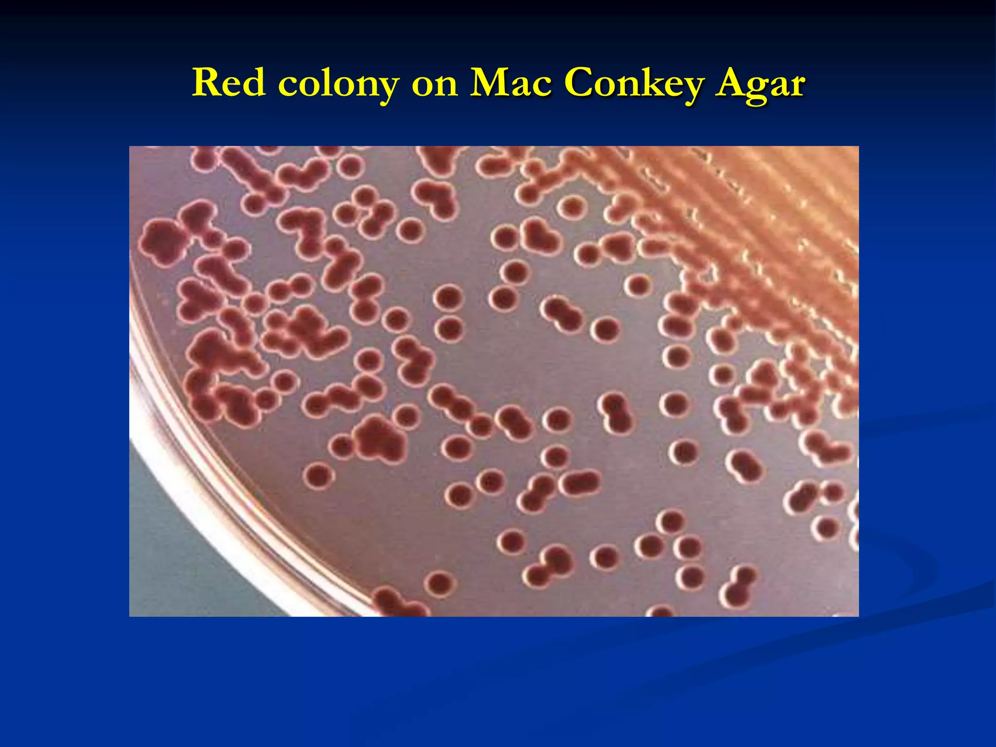
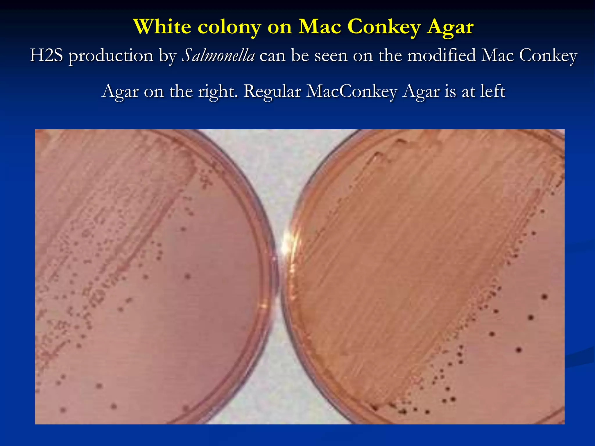
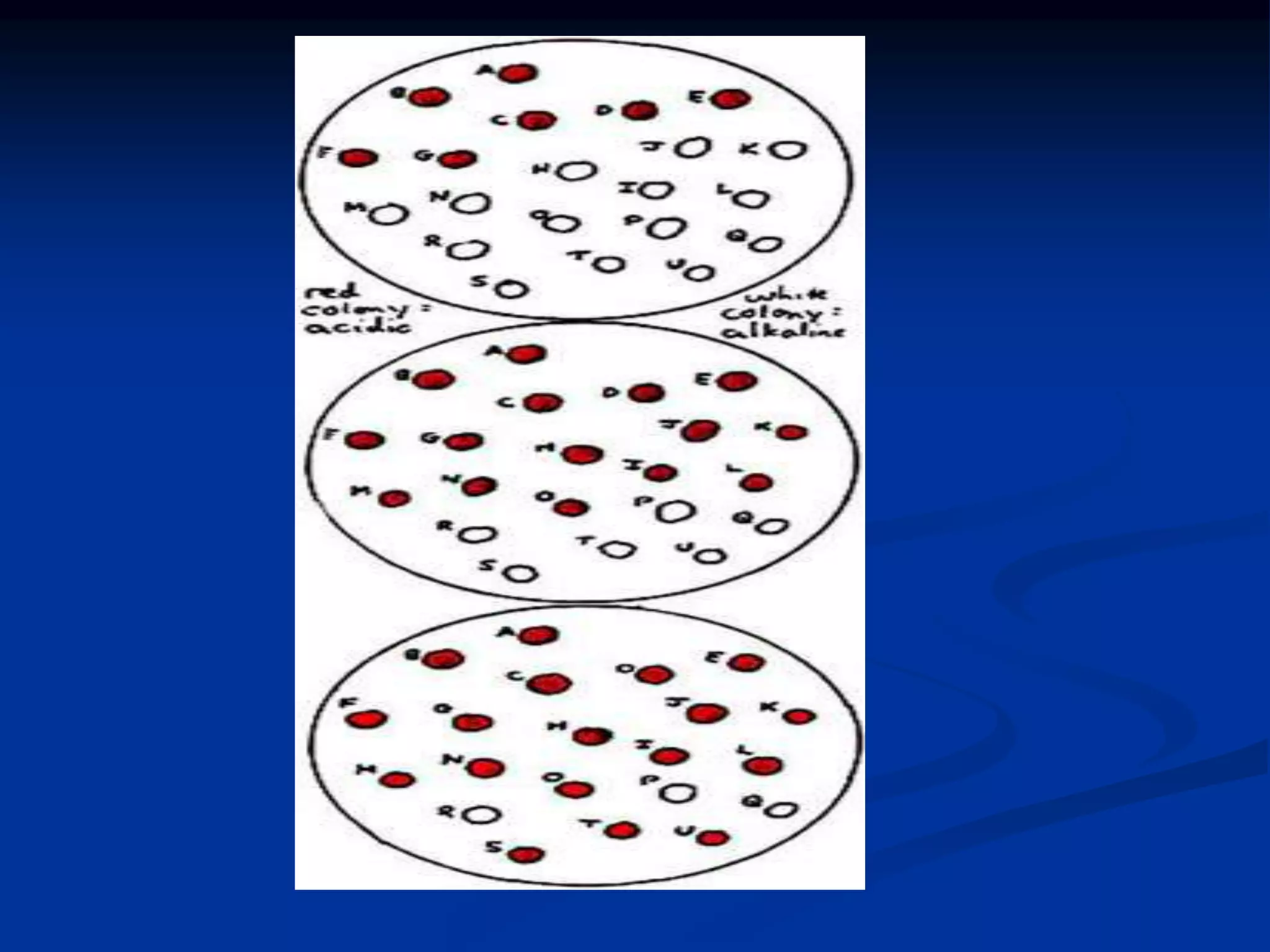
Blood Agar
 To sterile Blood Agar Base which has been
melted and cooled to 45 to 50°C, add 5%
(vol/vol) sterile defibrinated blood that has been
warmed to room temperature.
 Swirl the flask to mix thoroughly, avoiding the
formation of bubbles, and dispense into sterile
plates, continuing to avoid bubbles and froth on
the surface.
 NOTE: Cooling the agar and warming the
blood are essential steps in this procedure.
 Hot agar can damage red blood cells, and cold
blood can cause the agar to gel before pouring.
Tryptic Soy Agar with and without sheep blood
Hemolysis on Blood Agar Plates
 Beta hemolytic
Streptococcus
species,
Streptococcus
pyogenes
(transmitted
light) (Lancefield
group A).
Beta hemolysis
 is defined as complete or true lysis of red blood
cells. A clear zone, approaching the color and
transparency of the base medium, surrounds the
colony. Many species of bacteria produce toxic
by-products that are capable of destroying red
blood cells
Beta-hemolytic
 Normal Upper
respiratory flora
mixed with beta-
hemolytic Streptococcus
species. (The
presence of beta-
hemolytic colonies
indicates the
possibility of
Streptococcus
pyogenes infection.)
Beta hemolysis
 Same blood agar
plate as Figure 2
demonstrating that
the beta hemolysis
of Streptococcus
pyogenes is so
complete that print
my be read through
the resulting
transparent medium
Alpha hemolysis
 is the reduction of the red blood cell
hemoglobin to methemoglobin in the medium
surrounding the colony. This causes a green or
brown discoloration in the medium. The color
can be equated with "bruising" the cells.
Microscopic inspection of alpha-hemolyzed red
blood cells shows that the cell membrane is
intact, so it is not, in fact, true lysis. Some text
book authors refer to alpha as “partial
hemolysis,” which may be confusing to the
student.
Alpha hemolysis
 It is most important to not confuse this “partial”
or “incomplete” hemolysis with the “weak” or
“subtle” lysis of Streptococcus agalactiae or Listeria
monocytogenes, as seen above. Beta hemolysis will
never include the brown or green discoloration
of the cells in the surrounding medium. On
prolonged incubation, many alpha hemolytic
organisms will begin to appear more clear, but if
the surrounding medium contains any shades of
brown or green the “hemolysis” is still
considered “alpha.”
Alpha-hemolytic
 Alpha-hemolytic
Streptococcus species
“Viridans group”
streptococci, including
species such as the
Streptococcus mutans, mitis,
and salivarius groups
display alpha hemolysis
Alpha hemolysis
 Alpha hemolysis
of Streptococcus
pneumoniae
(Encapsulated
strain).
Gamma hemolysis
 is somewhat self-contradictory. Gamma
indicates the lack of hemolysis. There should be
no reaction in the surrounding medium.
Gamma hemolysis
 Gamma Streptococcus" or
Enterococcus faecalis (24
hours, non-hemolytic).
"Gamma streptococcus" are
usually non-hemolytic after
24 hours of incubation,
but many eventually
display weak alpha
hemolysis. (The genus
Enterococcus was once a part
of the Streptococcus genus,
and was considered a
"gamma Streptococcus
species".
The same Enterococcus strain, shown with transmitted light at
48 hours incubation demonstrates the alpha hemolysis of
some “gamma streptococci.”
Mac Conkey Agar
Component grams/liter Purpose
Proteose
Peptone
3.0 Peptide (amino acids), carbon, energy, many macro
and micronutrients
Lactose 10.0 Carbon and energy source
NaCl 5.0 Osmotic balance
Bile Salts 1.5 Selective agent
Crystal Violet 0.001 Selective agent
Neutral red 0.03 Indicator dye that turns red at low pH
Agar 13.5 Solidifying agent
MacConkey agar
 MacConkey agar was the first solid differential
media to be formulated. It was developed at the
turn of the 20th century by Alfred Theodore
MacConkey, M.D, then Assistant Bacteriologist
to the Royal Commission on Sewage Disposal,
in the Thompson-Yates Laboratories of
Liverpool University, England. The goal was to
formulate a medium that would select for the
growth of gram-negative microorganisms and
inhibit the growth of gram-positive
microorganisms.
MacConkey agar
 Dr. MacConkey first developed a bile salt medium
containing glycocholate, lactose and litmus, to be
incubated at 22°C (MacConkey, 1900). This formula
was soon altered by the replacement of glycocholate
with taurocholate and the incubation temperature was
raised to 42°C.
 MacConkey later changed the recipe again by
substituting neutral red for litmus (following the
suggestion that neutral red be used as an indicator in
bile salt lactose medium (Grunbaum and Hume, 1902).
The final media formulation was designed to support
growth of Shigella and is the one that is most commonly
used today.
Purpose
 MacConkey agar is used for the isolation of
gram-negative enteric bacteria and the
differentiation of lactose fermenting from
lactose non-fermenting gram-negative bacteria.
 It has also become common to use the media to
differentiate bacteria by their abilities to ferment
sugars other than lactose. In these cases lactose
is replaced in the medium by another sugar.
These modified media are used to differentiate
gram-negative bacteria.
MacConkey agar
 MacConkey agar is a selective and differential
media used for the isolation and differentiation of
non-fastidious gram-negative rods, particularly
members of the family Enterobacteriaceae and
the genus Pseudomonas. The inclusion of crystal
violet and bile salts in the media prevent the
growth of gram-positive bacteria and fastidious
gram-negative bacteria, such as Neisseria and
Pasteurella.
 The tolerance of gram-negative enteric bacteria to
bile is partly a result of the relatively bile-resistant
outer membrane, which hides the bile-sensitive
cytoplasmic membrane. Other species specific
bile-resistance mechanisms have also been
identified.
 Gram-negative bacteria growing on the media are
differentiated by their ability to ferment the sugar
lactose. Bacteria that ferment lactose cause the pH
of the media to drop and the resultant change in
pH is detected by neutral red, which is red in color
at pH's below 6.8. As the pH drops, neutral red is
absorbed by the bacteria, which appear as bright
pink to red colonies on the agar.
 The color of the medium surrounding Gram
negative bacteria may also change. Strongly
lactose fermenting bacteria produce sufficient
acid to cause precipitation of the bile salts,
resulting in a pink halo in the medium
surrounding individual colonies or areas of
confluent growth. Bacteria with weaker lactose
fermentation growing on MacConkey agar will
still appear pink to red but will not be
surrounded by a pink halo in the surrounding
medium.
 Gram-negative bacteria that grow on MacConkey
agar but do not ferment lactose appear colorless
on the medium and the agar surrounding the
bacteria remains relatively transparent.
 Lactose can be replaced in the medium by other
sugars and the abilities of gram-negative bacteria
to ferment these replacement sugars is detectable
in the same way as is lactose fermentation (for
example Farmer and Davis, 1985).
MacConkey Agar
 MacConkey Agar differentiates between lactose
fermenters (e.g., coliforms) and non-lactose-
fermenters (e.g., most strains of Citrobacter and
typical enteric pathogens such as Salmonella and
Shigella). Shigella).
MacConkey Agar
example coliform
Salmonella
Shigella
Citrobacter(typical)
amino acids
deaminated
(alkaline rx.)
+ +
lactose
fermented
(strong acidic rx.)
+ –
net pH reaction acidic (red colony) alkaline (white colony)
Click on image
for wider view
in separate window.
Red colony on Mac Conkey Agar
<
Red colony on Mac Conkey Agar
White colony on Mac Conkey Agar
H2S production by Salmonella can be seen on the modified Mac Conkey
Agar on the right. Regular MacConkey Agar is at left
Colony isolation
 The basis of pure culture technique is the
isolation, in colonies, of individual cells, and
their descendants, from other colonies of
individuals.
 This is usually done by culturing methods
employing petri dishes such as:
 streaking
 pouring
 spreading
Spreading a plate
 Quantification technique:
 Spreading a plate is an additional method of
quantifying microorganisms on solid medium.
 Instead of embedding microorganisms into agar,
as is done with the pour plate method, liquid
cultures are spread on the agar surface using a
devise that looks more or less like a hockey stick.
 An advantage of spreading a plate over the pour
plate method is that cultures are never exposed
to 45°C+ melted agar temperatures.
Colony morphology
 Differentiating colonies:
 Colony morphology gives important clues as to
the identity of their constituent microorganisms.
 Important classes of characteristics include:
 size
 type of margin
 colony elevation
 colony texture
 light transmission
 colony pigmentation
Colony elevation
 Colonies can vary in their elevations both
between microorganisms and growth conditions,
and within individual colonies themselves.
Colony size
 Colony size is dependent not just on the type of
organism but also on the growth medium and the
number of colonies present on a plate (that is,
colonies tend to be smaller when greater than
ascertain amount are present) and on culture medium
characteristics.
 Usually stabilizes after few days:
 Colony size usually stabilizes after a day or two of
incubation.
 Exceptions include:
 slow growing microorganisms
 during growth under conditions that promote slow growth
 With slow growth colonies may continue to
experience growth past this time, especially if an
effort is made to prevent solid medium from drying
out
Illustration, variation in colony margins

4 blood agar and hemolysis and mac-conkey.ppt

  • 1.
    Blood Agar  Tosterile Blood Agar Base which has been melted and cooled to 45 to 50°C, add 5% (vol/vol) sterile defibrinated blood that has been warmed to room temperature.  Swirl the flask to mix thoroughly, avoiding the formation of bubbles, and dispense into sterile plates, continuing to avoid bubbles and froth on the surface.  NOTE: Cooling the agar and warming the blood are essential steps in this procedure.  Hot agar can damage red blood cells, and cold blood can cause the agar to gel before pouring.
  • 2.
    Tryptic Soy Agarwith and without sheep blood
  • 3.
    Hemolysis on BloodAgar Plates
  • 4.
    Hemolysis on BloodAgar Plates  Beta hemolytic Streptococcus species, Streptococcus pyogenes (transmitted light) (Lancefield group A).
  • 5.
    Beta hemolysis  isdefined as complete or true lysis of red blood cells. A clear zone, approaching the color and transparency of the base medium, surrounds the colony. Many species of bacteria produce toxic by-products that are capable of destroying red blood cells
  • 6.
    Beta-hemolytic  Normal Upper respiratoryflora mixed with beta- hemolytic Streptococcus species. (The presence of beta- hemolytic colonies indicates the possibility of Streptococcus pyogenes infection.)
  • 7.
    Beta hemolysis  Sameblood agar plate as Figure 2 demonstrating that the beta hemolysis of Streptococcus pyogenes is so complete that print my be read through the resulting transparent medium
  • 8.
    Alpha hemolysis  isthe reduction of the red blood cell hemoglobin to methemoglobin in the medium surrounding the colony. This causes a green or brown discoloration in the medium. The color can be equated with "bruising" the cells. Microscopic inspection of alpha-hemolyzed red blood cells shows that the cell membrane is intact, so it is not, in fact, true lysis. Some text book authors refer to alpha as “partial hemolysis,” which may be confusing to the student.
  • 9.
    Alpha hemolysis  Itis most important to not confuse this “partial” or “incomplete” hemolysis with the “weak” or “subtle” lysis of Streptococcus agalactiae or Listeria monocytogenes, as seen above. Beta hemolysis will never include the brown or green discoloration of the cells in the surrounding medium. On prolonged incubation, many alpha hemolytic organisms will begin to appear more clear, but if the surrounding medium contains any shades of brown or green the “hemolysis” is still considered “alpha.”
  • 10.
    Alpha-hemolytic  Alpha-hemolytic Streptococcus species “Viridansgroup” streptococci, including species such as the Streptococcus mutans, mitis, and salivarius groups display alpha hemolysis
  • 11.
    Alpha hemolysis  Alphahemolysis of Streptococcus pneumoniae (Encapsulated strain).
  • 12.
    Gamma hemolysis  issomewhat self-contradictory. Gamma indicates the lack of hemolysis. There should be no reaction in the surrounding medium.
  • 13.
    Gamma hemolysis  GammaStreptococcus" or Enterococcus faecalis (24 hours, non-hemolytic). "Gamma streptococcus" are usually non-hemolytic after 24 hours of incubation, but many eventually display weak alpha hemolysis. (The genus Enterococcus was once a part of the Streptococcus genus, and was considered a "gamma Streptococcus species".
  • 14.
    The same Enterococcusstrain, shown with transmitted light at 48 hours incubation demonstrates the alpha hemolysis of some “gamma streptococci.”
  • 15.
    Mac Conkey Agar Componentgrams/liter Purpose Proteose Peptone 3.0 Peptide (amino acids), carbon, energy, many macro and micronutrients Lactose 10.0 Carbon and energy source NaCl 5.0 Osmotic balance Bile Salts 1.5 Selective agent Crystal Violet 0.001 Selective agent Neutral red 0.03 Indicator dye that turns red at low pH Agar 13.5 Solidifying agent
  • 16.
    MacConkey agar  MacConkeyagar was the first solid differential media to be formulated. It was developed at the turn of the 20th century by Alfred Theodore MacConkey, M.D, then Assistant Bacteriologist to the Royal Commission on Sewage Disposal, in the Thompson-Yates Laboratories of Liverpool University, England. The goal was to formulate a medium that would select for the growth of gram-negative microorganisms and inhibit the growth of gram-positive microorganisms.
  • 17.
    MacConkey agar  Dr.MacConkey first developed a bile salt medium containing glycocholate, lactose and litmus, to be incubated at 22°C (MacConkey, 1900). This formula was soon altered by the replacement of glycocholate with taurocholate and the incubation temperature was raised to 42°C.  MacConkey later changed the recipe again by substituting neutral red for litmus (following the suggestion that neutral red be used as an indicator in bile salt lactose medium (Grunbaum and Hume, 1902). The final media formulation was designed to support growth of Shigella and is the one that is most commonly used today.
  • 18.
    Purpose  MacConkey agaris used for the isolation of gram-negative enteric bacteria and the differentiation of lactose fermenting from lactose non-fermenting gram-negative bacteria.  It has also become common to use the media to differentiate bacteria by their abilities to ferment sugars other than lactose. In these cases lactose is replaced in the medium by another sugar. These modified media are used to differentiate gram-negative bacteria.
  • 19.
    MacConkey agar  MacConkeyagar is a selective and differential media used for the isolation and differentiation of non-fastidious gram-negative rods, particularly members of the family Enterobacteriaceae and the genus Pseudomonas. The inclusion of crystal violet and bile salts in the media prevent the growth of gram-positive bacteria and fastidious gram-negative bacteria, such as Neisseria and Pasteurella.
  • 20.
     The toleranceof gram-negative enteric bacteria to bile is partly a result of the relatively bile-resistant outer membrane, which hides the bile-sensitive cytoplasmic membrane. Other species specific bile-resistance mechanisms have also been identified.  Gram-negative bacteria growing on the media are differentiated by their ability to ferment the sugar lactose. Bacteria that ferment lactose cause the pH of the media to drop and the resultant change in pH is detected by neutral red, which is red in color at pH's below 6.8. As the pH drops, neutral red is absorbed by the bacteria, which appear as bright pink to red colonies on the agar.
  • 21.
     The colorof the medium surrounding Gram negative bacteria may also change. Strongly lactose fermenting bacteria produce sufficient acid to cause precipitation of the bile salts, resulting in a pink halo in the medium surrounding individual colonies or areas of confluent growth. Bacteria with weaker lactose fermentation growing on MacConkey agar will still appear pink to red but will not be surrounded by a pink halo in the surrounding medium.
  • 22.
     Gram-negative bacteriathat grow on MacConkey agar but do not ferment lactose appear colorless on the medium and the agar surrounding the bacteria remains relatively transparent.  Lactose can be replaced in the medium by other sugars and the abilities of gram-negative bacteria to ferment these replacement sugars is detectable in the same way as is lactose fermentation (for example Farmer and Davis, 1985).
  • 23.
    MacConkey Agar  MacConkeyAgar differentiates between lactose fermenters (e.g., coliforms) and non-lactose- fermenters (e.g., most strains of Citrobacter and typical enteric pathogens such as Salmonella and Shigella). Shigella).
  • 24.
    MacConkey Agar example coliform Salmonella Shigella Citrobacter(typical) aminoacids deaminated (alkaline rx.) + + lactose fermented (strong acidic rx.) + – net pH reaction acidic (red colony) alkaline (white colony) Click on image for wider view in separate window.
  • 25.
    Red colony onMac Conkey Agar <
  • 26.
    Red colony onMac Conkey Agar
  • 27.
    White colony onMac Conkey Agar H2S production by Salmonella can be seen on the modified Mac Conkey Agar on the right. Regular MacConkey Agar is at left
  • 29.
    Colony isolation  Thebasis of pure culture technique is the isolation, in colonies, of individual cells, and their descendants, from other colonies of individuals.  This is usually done by culturing methods employing petri dishes such as:  streaking  pouring  spreading
  • 30.
    Spreading a plate Quantification technique:  Spreading a plate is an additional method of quantifying microorganisms on solid medium.  Instead of embedding microorganisms into agar, as is done with the pour plate method, liquid cultures are spread on the agar surface using a devise that looks more or less like a hockey stick.  An advantage of spreading a plate over the pour plate method is that cultures are never exposed to 45°C+ melted agar temperatures.
  • 31.
    Colony morphology  Differentiatingcolonies:  Colony morphology gives important clues as to the identity of their constituent microorganisms.  Important classes of characteristics include:  size  type of margin  colony elevation  colony texture  light transmission  colony pigmentation
  • 32.
    Colony elevation  Coloniescan vary in their elevations both between microorganisms and growth conditions, and within individual colonies themselves.
  • 33.
    Colony size  Colonysize is dependent not just on the type of organism but also on the growth medium and the number of colonies present on a plate (that is, colonies tend to be smaller when greater than ascertain amount are present) and on culture medium characteristics.  Usually stabilizes after few days:  Colony size usually stabilizes after a day or two of incubation.  Exceptions include:  slow growing microorganisms  during growth under conditions that promote slow growth  With slow growth colonies may continue to experience growth past this time, especially if an effort is made to prevent solid medium from drying out
  • 34.