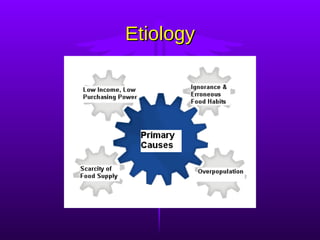
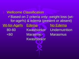
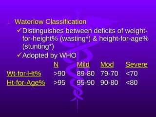
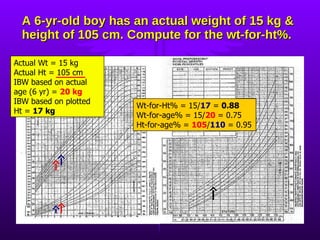
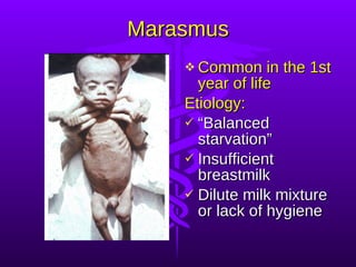
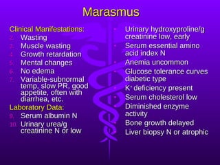
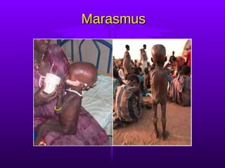
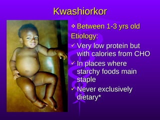
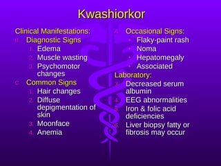
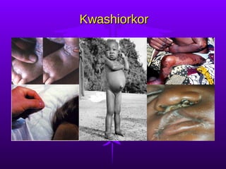
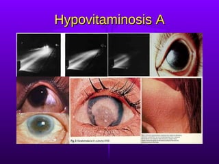
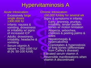
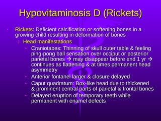
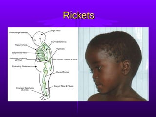
NUTRITIONAL DISORDERS Myrna DC San Pedro, MD, FPPS
MALNUTRITION A pathological state resulting from Relative or absolute deficiency  Or excess of one or more nutrients and Clinically detected only by biochemical,  anthropometric or physiological tests
Forms of Malnutrition Undernutrition:  Marasmus Overnutrition:  Obesity, Hypervitaminoses Specific Deficiency:  Kwashiorkor, Hypovitaminoses, Mineral Deficiencies Imbalance:  Electrolyte Imbalance
Etiology
 
 
Protein Energy Malnutrition (PEM)
Classification of Undernutrition Gomez Classification Uses weight-for-age measurements Provides prognosis Weight-for-Age% Status 91-100 Normal 76-90 1 st  degree 61-75 2 nd  degree <60 3 rd  degree
Wellcome Classification Based on 2 criteria only: weight loss (wt-for-age%) & edema (present or absent) Wt-for-Age% Edema No Edema 80-60 Kwashiorkor Undernutrition <60 Marasmic- Marasmus Kwashiorkor
Waterlow Classification Distinguishes between deficits of weight-for-height% (wasting*) & height-for-age% (stunting*) Adopted by WHO N   Mild Mod   Severe Wt-for-Ht% >90   89-80  79-70  <70 Ht-for-Age% >95   95-90 90-80  <80
A 6-yr-old boy has an actual weight of 15 kg & height of 105 cm. Compute for the wt-for-ht%. Actual Wt = 15 kg Actual Ht = 105 cm IBW based on actual age (6 yr) =  20 kg IBW based on plotted Ht =  17 kg Wt-for-Ht% = 15/ 17  =  0.88 Wt-for-age% = 15/ 20   = 0.75 Ht-for-age% =  105 / 110   = 0.95
Marasmus Common in the 1st year of life Etiology: “ Balanced starvation” Insufficient breastmilk Dilute milk mixture or lack of hygiene
Marasmus Clinical Manifestations: Wasting Muscle wasting Growth retardation Mental changes No edema Variable-subnormal temp, slow PR, good appetite, often with diarrhea, etc. Laboratory Data: Serum albumin N Urinary urea/g creatinine N or low Urinary hydroxyproline/g creatinine low, early Serum essential amino acid index N Anemia uncommon Glucose tolerance curves diabetic type K +  deficiency present Serum cholesterol low Diminished enzyme activity Bone growth delayed Liver biopsy N or atrophic
Marasmus
Kwashiorkor Between 1-3 yrs old Etiology: Very low protein but with calories from CHO In places where starchy foods main staple Never exclusively dietary*
Kwashiorkor Clinical Manifestations: Diagnostic Signs Edema Muscle wasting Psychomotor changes Common Signs Hair changes Diffuse depigmentation of skin Moonface Anemia Occasional Signs: Flaky-paint rash Noma Hepatomegaly Associated Laboratory: Decreased serum albumin EEG abnormalities Iron & folic acid deficiencies Liver biopsy fatty or fibrosis may occur
Kwashiorkor
Treatment of PEM Severe PEM is an emergency, 1-3 months hospitalization desirable On admission, treat dehydration, infections & vitamin deficiencies During 1 st  wk, the acute phase, stabilize & start feeding as soon as rehydrated When edema subsides, give full-strength feeds of maintenance calories & protein, recovery after 2-3 wk Once full-strength feeds tolerated, start rehabilitation with high energy feeds (150-200 kcal/kg/day), recovery expected within 4-6 wk
Prognosis of PEM If severe & early, <6 months old, permanent impairment of physical & mental growth First 48 hours critical, with poor treatment mortality may be >50% Even with thorough treatment, 10% mortality may still occur Some mortality causes: endocrine, cardiac or liver failure, electrolyte imbalance, hypoglycemia & hypothermia
Obesity
Obesity Definition:  Generalized, excessive accumulation of fat in subcutaneous & other tissues Classification according to “desirable” weight standard: Overweight =  > 10% IBW or “desirable” weight   Obese =  > 20% IBW or “desirable” weight   The Centers for Disease Control (CDC)  avoids using &quot;obesity&quot; instead suggest  two levels of overweight: “ At risk&quot; level = BMI 85 th % More severe level = BMI 95 th % The American Obesity Association: Overweight = BMI 85 th % because ~ BMI of 25, overweight for adults Obesity = BMI 95 th % because ~ BMI of 30, obesity in adults
Obesity Appears most frequently in the 1 st  yr, 5-6 yr & adolescence Etiology: Excessive food intake compared with use Genetic constitution Psychic disturbance  Endocrine & metabolic disturbances rare Insufficient exercise or lack of activity
Obesity Clinical Manifestations: Fine facial features on a heavy-looking taller child Larger upper arms & thighs Genu valgum common Relatively small hands & fingers tapering Adiposity in mammary regions Pendulous abdomen with striae In boys, external genitalia appears small but actually average in size In girls, external genitalia normal & menarche not delayed Psychologic disturbances common Bone age advanced
Obesity
Treatment of Obesity 1 st  principle: Decrease energy intake Initial med exam to R/O pathological causes 3-day food recall to itemize child’s diet Plan right diet Avoid all sweets, fried foods & fats Limit milk intake to not >2 glasses/day For 10-14 yrs, limit to 1,100-1300 kcal diet for several months Child must be properly motivated & family involvement essential 2 nd  principle: Increase energy output Obtain an activity history Increase physical activity Involve in hobbies to prevent boredom
Complication of Obesity Pickwickian Syndrome Rare complication of extreme exogenous obesity Severe cardiorespiratory distress & alveolar hypoventilation Includes polycythemia, hypoxemia, cyanosis, CHF & somnolence High O 2  dangerous in cyanosis Weight reduction ASAP & quick
The Fat-Soluble  Vitamins
Vitamin A (Retinol) Active forms: retinol, retinaldehyde & retinoic acid Plants synthesize  carotenoids     cleaved to  retinol  by most animals    stored in liver as  retinyl palmitate Retinol,  alcohol form of vitamin A: N plasma values = 20–50 μg/dl in infants & 30–225 μg/dL in older children & adults Retinal , the aldehyde form, functions in vision Retinoic acid , the physiologically most important vitamin A metabolite, regulates many genes involved in fundamental biologic activities of cells*
Vitamin A (Retinol) Deficiency Functions: Retinal is in photosensitive pigment in both rods (rhodopsin) & cones (iodopsin) Needed in lysosomal membrane stability Involved in keratinization, cornification, bone development & cell growth & reproduction Absence in the diet common by 2-3 yrs old Etiology: Poor fetal storage Poor absorption as in low-fat diet, malabsorption syndromes, etc. Low protein intake resulting in deficient carriers Increased excretion as in cancer & UTI
Hypovitaminosis A Eye signs & symptoms Nyctalopia or night blindness, an early symptom   photophobia   pain insensitivity Xerosis conjunctivae, 1 st  clinical sign Bitot’s spots Corneal xerosis or xeropthalmia Corneal ulcers Keratomalacia Blindness
Hypovitaminosis A Skin signs:  Xerosis of the skin & follicular hyperkeratosis or phrynoderma Others:  Apathy, physical & mental growth retardation, faulty epiphyseal bone formation, defective teeth enamel & signs of benign increased ICP Diagnosis: Routine PE Dark adaptation test to detect nyctalopia Absorption test for vitamin A Conjunctival impression cytology to evaluate early xeropthalmia
Hypovitaminosis A RDA:  1800 IU/day (1 IU vitamin A =  0.3 mcg retinol) Prevention: Pregnant in last trimester be given 5000 IU p. o. Every 6 months, infants <1 yr retinol palmitate 55mg or 33mg retinol acetate (100,000 IU) p. o. Every 4-6 months, older children 110mg retinol palmitate or 66 mg retinol acetate (200,000 IU) p. o. In prevalent areas, 100,000 IU p. o. q 3 mo For malnourished children 1-6 yrs, 250,000 IU p. o. q 6 mo Treatment: > 1 yr: 110mg retinol palmitate or 66mg retinol acetate (200,000 IU) orally or preferably 33mg (100,000 IU) of retinyl palmitate by IM The oral dose should be repeated on 2nd day and on discharge from hospital 7-30 days after 1st dose Above doses halved for infants For corneal involvement, apply antibiotic ointment like topical  bacitracin  to both eyes 6x/day and give also systemic antibiotics
Hypovitaminosis A
Hypervitaminosis A Acute Intoxication: Excessively  large single doses  > 300,000 IU Infants: nausea & vomiting, drowsiness or irritability w/ signs of increased ICP Adults: drowsiness, irritability, headache & vomiting Serum vitamin A values = 200-1000 IU/dl (N: 50-100 IU/dl) Chronic Intoxication: > 50,000 IU/day for several wk Signs & symptoms in infants: Early: anorexia, pruritus, irritability, tender swollen bones w/ motion limitation Alopecia, seborrhea, cheilosis & peeling palms & soles Hepatomegaly & hypercalcemia Craniotabes & hyperostosis of long bones (differentiate from Caffey’s disease) Elevated serum vitamin A Reversible manifestations when vitamin A discontinued
Vitamin D (Cholecalciferol)  90%  Vitamin D 3 ,  cholecalciferol , produced in skin by UV irradiation of 7-dehydrocholesterol (mainly an animal sterol)    blood    25 hydroxylation to calcidiol in liver + PTH    di-hydroxylation in kidney to calcitriol 1,25 (OH) 2-cholecalciferol 1,25-dihydroxycholecalciferol  most active form Vitamin D 2 ,  ergocalciferol , taken orally from plants (ergosterol) then irradiated as above Animal derived vitamin D 3  and plant derived vitamin D 2  biologically equal
Vitamin D (Cholecalciferol) Deficiency Functions:  Vitamin D enhances Ca +2  absorption from gut, removal of Ca +2  from bone & PO 4 -3  reabsorption in kidney Rickets appear towards end of 1-2 yr Etiology: Lack in diet or lack of sunlight exposure Rapid growth as in prematures & adolescents Disorders of absorption such as celiac disease, steatorrhea or cystic fibrosis In children with hepatic disease Maternal malnutrition Poverty or ignorance
Hypovitaminosis D (Rickets) Rickets:  Deficient calcification or softening bones in a growing child resulting in deformation of bones Head manifestations Craniotabes: Thinning of skull outer table & feeling ping-pong ball sensation over occiput or posterior parietal bones    may disappear before end 1 yr    continues as flattening & at times permanent head asymmetry Anterior fontanel larger & closure delayed Caput quadratum: Box-like head due to thickened & prominent central parts of parietal & frontal bones Delayed eruption of temporary teeth while permanent with enamel defects
Hypovitaminosis D (Rickets) Thorax signs Palpable enlargement of costochondral junctions called “rachitic rosary” Flattened sides of the thorax with posterior longitudinal grooves Pigeon-breast deformity Harrison groove Spinal column signs Scoliosis common Kyphosis when sitting Lordosis in the erect position Pelvis narrowed  due to changes in promontory, sacrum & coccyx which if permanent, add to hazards of childbirth in female
Hypovitaminosis D (Rickets) Extremities in children >2 years Thickened & enlarged wrists & ankles Bowlegs or knock-knees as a result of the bending of the softened shafts of the femur, tibia & fibula Coxa vara or pronated feet Greenstick fractures Muscles  poorly developed & lack tone Delay in sitting, standing & walking Potbelly due to weakness of abdominal muscles Other manifestations Underweight Mental retardation
Rickets
Rickets
Rickets
Rickets A teenage male w/ rickets. Note bow legs & compromised height.  Distal femur, proximal tibia and fibula in rickets. Note widening epiphysis, resorption of provisional zone of calcification, flaring metaphysis & bone deformity.
Hypovitaminosis D (Osteomalacia) Osteomalacia:  Accumulation of uncalcified osteoid tissue in rib joints of an adult resulting in Pain in pelvis, lower back and legs Tenderness in shins and in other bones Waddling gait Deformities of pelvis Tetany may occur manifested by involuntary twitching of facial muscles or by carpopedal spasm Spontaneous fractures may occur Osteomalacia should not be confused with osteoporosis , a disease of ageing, in which decalcification is also a feature
Osteomalacia A young male w/ osteomalacia. Note a pseudofracture in the medial edge of the upper femoral shaft (arrow). Xray showing a pseudofracture (red arrow) from an adult who has x-linked hypophosphatemic rickets. This sign is seen only in osteomalacia, but not in many of the cases. AP pelvis in a patient w/ osteomalacia. The film shows diffuse osteopenia, & a Looser zone (arrow) in the superior ramus of the right obturator ring.
Hypovitaminosis D Diagnosis: History & clinical observation Laboratory findings: Serum Ca +2  may be normal or low Serum phosphorus level <4 mg/dl (N serum phosphorus 4.5-6.5 mg/dl but in rachitic infants reduced to  < 1.5-3.5 mg/dl) Serum alkaline phosphatase elevated (N serum phosphatase 5-15 Bodansky units per 100 ml but elevated to 20-30 in mild rickets & to  > 60 in severe) Serum 25-hydroxycholecalciferol decreased Urinary cyclic AMP elevated
Hypovitaminosis D Roentgenographic changes X-ray of wrist, cupping & fraying of proximal ends of ulna & radius, best for early diagnosis Humeral ossification centers barely visualized Shafts osteoporotic or density decreased Rosary beading of sternal ends of the ribs due to deposited uncalcified osteoid tissue becoming compressed & bulges laterally  Initial healing indicated by appearance of line of preparatory calcification
Hypovitaminosis D Differential Diagnosis: Craniotabes in hydrocephalus & osteogenesis imperfecta “ Rosary” at costochondral junctions in scurvy & chondrodystrophy Epiphyseal lesions in congenital epiphyseal dysplasia, cytomegalic inclusion disease, syphilis, rubella & copper deficiency Congenital pigeon breast deformity Familial bowlegs Metabolic disturbances with osseous lesions Complications: Respiratory infections Chronic gastroenteritis Iron deficiency anemia
Hypovitaminosis D Prognosis: In tropics, usually has tendency to heal spontaneously A possibly deforming disorder Not fatal but complications & intercurrent infections may cause death RDA:  400 IU (1 IU vitamin D = 0.025 mcg cholecalciferol/ ergocalciferol) Prevention: Sunlight prophylaxis effective only in temperate zones during summer months in haze-free areas Daily requirement of vitamin D in 1 quart of fresh whole milk or a can of evaporated milk Natural vitamin D present only in animal foods like egg yolk, liver, cod-liver & other fish-liver oils & fishbody oils
Hypovitaminosis D Prematures or breast-fed infants should receive supplemental vitamin D daily because milk is a poor source unless fortified Vitamin D should also be administered to pregnant & lactating mothers Treatment: Daily administration of 50-150 mcg of vitamin D 3  or 0.5-2 mcg of 1,25-dihydroxycholecalciferol will produce healing seen on X-ray within 2-4 wks Vitamin D 15,000 mcg in a single dose w/o further therapy for several months may be advantageous After healing is complete, the dose of vitamin D should be lowered to 10 mcg/day If no healing occurs, rickets is probably resistant to vitamin D or non-nutritional rickets
Hypervitaminosis D Etiology:  Excessive intakes from  Inadvertently substituting concentrated form for dilute Parents’ increasing prescribed dose Inadequately controlling dosages for children receiving large amounts of vitamin D for chronic hyperphosphatemia Clinical Manifestations:  Symptoms after 1-3 months Anorexia, irritability, hypotonia & constipation Polydipsia, polyuria & pallor Dehydration usually present Aortic valvular stenosis, vomiting & hypertension Retinopathy & clouding of cornea & conjunctiva may occur
Hypervitaminosis D Laboratory Data: Proteinuria Hypercalcemia & hypercalciuria With continued excess, renal damage & metastatic calcifications may occur Roentgenograms of long bones reveal metastatic calcification & generalized osteoporosis Differential Diagnosis: Chronic nephritis Hyperparathyroidism Idiopathic Hypercalcemia Treatment: Discontinue vitamin D & decrease Ca +2  intake For severe: Al(OH)3 p. o., cortisone or sodium versenate may be used
Vitamin K Naturally occurring vitamin K abundant in pork, liver, soybeans & green leafy vegetables Synthesize by intestinal microorganisms Required for normal clotting of blood Vitamin K-dependent clotting factors: Prothrombin (Factor II) Proconvertin (Factor VII) Plasma thromboplastin component or PTC (Factor IX) Stuart-Prower factor (Factor X)
Vitamin K Deficiency (Hypoprothrombinemia)   Etiology: The fetus depends on mother for supply & at birth, bacterial flora of GIT not yet produce Exclusively breast-fed infants lower vitamin K compared to formula-fed Faulty intestinal absorption as in diarrhea, celiac disease, gastrointestinal malformation & steatorrhea Obstructive jaundice, biliary fistula, insufficient production of bile acids or pancreatic insufficiency lead to inadequate intestinal absorption Administration of antibiotics inhibit intestinal bacteria In sepsis, deficiency from disease affecting hepatobiliary functions & therapy Drugs like coumarin, salicylates & anticonvulsants
Hypoprothrombinemia Clinical Manifestations: Hemorrhagic manifestations are the hallmark Bleeding in the newborn from the cord or circumcision site GIT bleeding, hematuria & intracranial hemorrhage Anemia & shock may ensue from severe blood loss Laboratory Test:  Most useful test is  1-stage prothrombin time test (Quick) , prolongation presumptive evidence deficiency
Hypoprothrombinemia Prevention and Treatment: 4 requirements to prevent & control a potentially fatal hemorrhagic state: Bile of normal composition in the GIT Adequate diet Normal absorptive surface in the small intestines Functioning liver capable of synthesizing In the newborn, vitamin K 1  is being used because: Greater margin of safety  Acts rapidly with therapeutic levels within 2-4 hours
Hypoprothrombinemia Prevention and Treatment: The AAP Committee on Nutrition recommends: Prophylactic dose: Vitamin K 0.5-1 mg as single parenteral dose or 1-2 mg single p. o. dose Mild prothrombin deficiency: Vitamin K 1-2 mg p. o. OD In severe cases with hemorrhages: Vitamin K 1  5 mg daily parenterally Whole blood if due to liver damage Avoid excessive doses in prematures & G-6-PD deficient newborns due to hemolytic action & hyperbilirubinemia Vitamin K prophylaxis to woman in labor may be followed by hemolytic anemia, hyperbilirubinemia, kernicterus & death in infant
Hypoprothrombinemia
Vitamin E (Tocopherol) Denotes a group of 8 compounds with similar structures and antioxidant activity The most potent member is   tocopherol , the main form in humans Functions as antioxidant mainly located within cell membranes where it prevents lipid peroxidation and formation of free radicals Best dietary sources: vegetable oils, seeds, nuts, green leafy vegetables and margarine
Hypovitaminosis E Etiology: Poor absorption in premature infants* In children with fat malabsorption, most common in cholestatic liver disease & a complication of autosomal recessive abetalipoproteinemia  Other malabsorptive states such as cystic fibrosis, acanthocytosis, celiac disease, short-bowel syndrome or Crohn disease In a rare autosomal recessive disorder, Ataxia with isolated Vitamin E Deficiency (AVED),  that affects vitamin E transport Clinical Manifestations: Appear after 1 yr of age in prolonged deficiency: degenerative neurologic syndrome of cerebellar disease, posterior column dysfunction and retinal disease** In premature infants, hemolysis typically develops during 2 nd  mo with edema and thrombocytosis potentially causing anemia Diagnosis: Best by measuring ratio of vitamin E to serum lipids: abnormal ratio <0.8 mg/g
Hypovitaminosis E Electroretinography abnormalities may precede PE findings in those with retinal involvement Premature infants with unexplained hemolytic anemia after 1 st  mo, especially if thrombocytosis present Prognosis: Hemolytic anemia in infants resolves with correction of deficiency Treatment prevents progression of neurologic manifestations Prevention & Treatment:   Prevention in premature infants: sufficient vitamin E, 7mg/g of unsaturated fat in the diet, and formula without high content PUFAs Treatment dose in neonates: 25–50 units/day for 1 wk, follow with adequate dietary intake Severe malabsorption:   -Tocopheryl polyethylene glycol succinate (TPGS), a water-soluble preparation absorbed in absence of bile salts 20–25 units/kg/day Children with AVED: high doses of vitamin E
The  Energy-Releasing  Vitamins
Thiamine (Vitamin B1) Deficiency Beriberi Pathology:  Biochemical accumulation of pyruvic and lactic acid in body fluids causing: Cardiac dysfunction such as cardiac enlargement esp. right side, edema of interstitial tissue & fatty degeneration of myocardium Degeneration of myelin & axon cylinders resulting in peripheral neuropathy  In chronic deficiency states, vascular dilatation & brain hemorrhages of Wernicke’s Disease resulting in weakness of eye movement, ataxia of gait and mental disturbance
Beriberi Three forms: Wet beriberi:  generalized edema, acute cardiac symptoms and prompt response to thiamine administration Dry beriberi:  edema not present, condition similar to peripheral neuritis w/ neurological disorders present Infantile beriberi  divided into: Acute cardiac:  Ages 2-4 months; sudden onset of cardiac s/sx such as cyanosis, dyspnea, systolic murmur & pulmonary edema w/ rales Aphonic:  Ages 5-7 months; insidious onset of hoarseness, dysphonia or aphonia Pseudomeningeal:  Ages 8-10 months; signs of meningeal irritation w/ apathy, drowsiness & even unconsciousness; occurs more often
Beriberi Diagnosis: Clinical manifestations not conclusive Therapeutic test, parenteral thiamine = dramatic improvement Blood lactic & pyruvic acid levels elevated after oral glucose load Decreased red cell hemolysate transketolase RDA:   Infants 0.4mg  Older children 0.6-1.2mg Nursing mothers 1.5mg Adults 1-1.3mg Prevention: Richest sources: pork, whole grain, enriched cereal grains and legumes Improved milling of rice conserve thiamine Excessive cooking of vegetables or polishing of cereals destroy it In breast-fed infants, prevention achieved by maternal diet w/ sufficient amounts Treatment: Children: 10mg p. o. daily for several weeks Adults: 50mg
Beriberi
Beriberi
Riboflavin (Vitamin B2) Deficiency Functions: Coenzyme of flavoprotein important in a. a., f. a. & CHO metabolism & cellular respiration Needed also by retinal eye pigments for light adaptation Clinical Manifestations: Lesions of the lips characteristic, most common are angular stomatitis and cheilosis  Localized seborrheic dermatitis of the face such as nasolabial seborrhea or dyssebacia & angular palpebritis Scrotal or vulvar dermatosis may also occur Ocular s/sx are photophobia, blurred vision, itching of eyes, lacrimation & corneal vascularization
Riboflavin Deficiency Diagnosis: Urinary riboflavin determination RBC riboflavin load test RDA: Infants & children <10yrs 0.6-1.4mg Children >10yrs 1.4-2mg Adults 0.025mg/gm dietary protein Prevention: Best sources: eggs, liver, meat, fish, milk, whole or enriched ground cereals, legumes, green leafy vegetables Also present in beer Impaired absorption in achlorhydria, diarrhea & vomiting Treatment: Riboflavin 2-5mg p. o. daily w/ increased B complex Parenteral administration  if  relief not obtained
Riboflavin Deficiency
Niacin (Vitamin B3) Deficiency Pellagra Etiology: Diets low in niacin &/or tryptophan Amino acid imbalance or as result of malabsorption Excessive corn consumption Clinical Manifestations: Starts w/ anorexia, weakness, irritability, numbness & dizziness Classical triad of dermatitis, diarrhea & dementia Dermatitis may develop insidiously to sunlight or heat First as symmetrical erythema Followed by drying, scaling & pigmentation w/ vesicles or bullae Predilection for back of hands, wrists, forearms (pellagrous glove), neck (Casal’s necklace) & lower legs (pellagrous boot) GIT s/sx are stomatitis, glossitis or diarrhea w/ feces pale, foul milky, soapy or at times steatorrheic Mental changes include depression, irritability, disorientation, insomnia & delirium
Pellagra Diagnosis: History & manifestations of diet poor in niacin or tryptophan In niacin deficiency, urinary levels of N-methyl-nicotinamide low or absent Differential diagnoses:  Kwashiorkor, Infantile Eczema, Combination deficiencies of amino acids & trace minerals such as zinc RDA: Infants & children <10yrs 6-10mg Older individuals 10-20mg Prevention: Rich sources include meat, peanuts and legumes, whole grain and enriched breads and cereals Avoid too large a proportion of corn Treatment: Niacin 50-300mg daily may be taken for a long time Skin lesions may be covered w/ soothing lotions
Pellagra
Pyridoxine (Vitamin B6) Deficiency Functions:  In the synthesis and catabolism of a. a., synthesis of neurotransmitters, porphyrins and niacin Important role in clinical conditions such as anemia, hyperemesis gravidarum, cardiac decompensation, radiation effects, skin grafting, INH therapy & seborrheic dermatitis Etiology: Losses from refining, processing, cooking & storing Malabsorptive diseases such as celiac disease Direct antagonism between INH & pyridoxal phosphate at the apoenzyme level
Pyridoxine Deficiency Clinical Manifestations: Three different types Neuropathic , due to insufficient neurotransmitter synthesis such as irritability, depression & somnolence Pellagrous , due to low endogenous niacin synthesis such as seborrheic dermatitis, intertrigo, angular stomatitis & glossitis Anemic , due to low porphyrin synthesis such as microcytic anemia & lymphopenia  In genetic diseases involving pyridoxal phosphate enzymes: xanthurenic aciduria, cystathioninuria & homocystinuria
Pyridoxine Deficiency Diagnosis:  As screening test, tryptophan load test -100mg/kg BW tryptophan gives large amount of xanthurenic acid in urine Prevention: Firm requirement not established but usual recommendation: Infant 0.1-0.5mg, Child 0.5-1.5mg & Adult 1.5-2mg Rich sources: yeast, whole wheat, corn, egg yolk, liver and lean meat Toxicity at extremely high doses described; infants whose mothers received large doses during pregnancy should be observed for seizures due to dependency Children receiving INH therapy should be observed for neurologic s/sx in w/c case pyridoxine should be given Treatment: Pyridoxine 100mg IM injection for seizures due to deficiency Children w/ pyridoxine dependency 2-10mg IM injection or 10-100mg oral vitamin B6
The  Hematopoietic  Vitamins
Folic Acid (Vitamin B9) Deficiency Functions: Needed for RBC & DNA formation & cell multiplication esp. GI cells Newly discovered functions: Prevents neural tube defects Prevents heart disease (reduces homocysteine levels) Prevents colon cancer Peak incidence 4-7 months Etiology: Deficient dietary intake:  goat’s milk deficient & powdered milk poor source Deficient absorption  in celiac disease, achlorhydria, w/ anticonvulsant drugs, zinc deficiency & bacterial overgrowth Impaired metabolism  in ascorbic acid deficiency, hypothyroidism, w/ drugs like trimethoprim Increased requirement  during rapid growth & infection Increased excretion/loss  may occur due to vitamin B12 deficiency
Folic Acid Deficiency Clinical Manifestations: Megaloblastic anemia w/ irritability, failure to gain wt & chronic diarrhea Thrombocytopenic hemorrhages advanced cases Scurvy may be present Laboratory Findings: Anemia macrocytic  Serum folic acid <3ng/ml (Normal = 5-20ng/ml) RBC folate levels indicator of chronic deficiency (Normal = 150-600ng/ml) Serum iron & vitamin B12 normal or elevated  Formiminoglutamic acid in urine esp after oral histidine Serum LDH markedly high Bone marrow hypercellular RDA:  20-50mcg/24 hrs Treatment: Parenteral folic acid 2-5mg/24 hr response in 72 hrs, for 3-4 wks Transfusions only if anemia severe Satisfactory responses even w/ low doses of 50mcg/24 hr, no effect on primary vitamin B12 deficiency If pernicious anemia present, prolonged use of folic acid should be avoided
Folic Acid Deficiency
Cobalamine (Vitamin B12) Deficiency Absorption:  Vitamin B12 + glycoprotein (intrinsic factor) from parietal cells of gastric fundus    terminal ileum absorption + intrinsic factor + Ca++    blood Function:  Needed in reactions affecting production of methyl groups Etiology: Congenital Pernicious Anemia : Lack of secretion of intrinsic factor by stomach manifest at  9 mo-10 yr  as uterine stores become exhausted Inadequate intake or dietary deficiency rare Strict vegetarian diet Not commonly seen in kwashiorkor or marasmus Breast-fed infants whose mothers had deficient diets or pernicious anemia Consumption or inhibition of B12-intrinsic factor complex Vitamin B12 malabsorption  from disease of ileal receptor sites or other intestinal causes
Cobalamine Deficiency Clinical Manifestations: Megaloblastic anemia that becomes severe* Neurological includes ataxia, paresthesias, hyporeflexia, Babinski responses, clonus & coma** Tongue smooth, red & painful Laboratory Findings: Anemia macrocytic Serum vitamin B12 <100pg/ml but serum iron & folic acid normal or elevated Serum LDH activity markedly increased Urinary excess of methylmalonic acid, a reliable & sensitive index
Cobalamine Deficiency Schilling test  to assess the absorption of vitamin B12: Normal person ingests small amount of radioactive vitamin B12    none in urine **If flushing dose injected parenterally, 1000mcg of non-radioactive vitamin B12    10-30% of previous radioactive vitamin B12 appears in urine Pernicious anemia    2% or less **If modified: 30 mg intrinsic factor administered along    normal amounts Disease of ileal receptor sites or other intestinal causes    no improvement even w/ intrinsic factor RDA:  Infants 0.5 mcg/day Older children & adults 3mcg/day Treatment: Prompt hematological response w/ parenteral vitamin B12 1-5mcg /24hr If there is neurological involvement 1mg IM daily for at least 2wks Pernicious Anemia: Monthly vitamin B12 1mg IM necessary throughout patient’s life
Cobalamine Deficiency
Ascorbic Acid (Vitamin C) Deficiency Scurvy Functions: Hydroxyproline, found only in collagen, major connective tissue in body, forms from proline requiring ascorbic acid If defective collagen formation, endochondral bone formation stops since oste, intercellular substance, not formed In hydroxylation reactions in synthesis of steroids and epinephrine Aids iron absorption by reducing it to ferrous state in stomach Spares vitamin A, vitamin E and some B vitamins by protecting them from oxidation Enhances utilization of folic acid by aiding conversion of folate to tetrahydrofolate
Ascorbic Acid (Vitamin C) Deficiency Scurvy More common 6-24 months Etiology: May develop in breastfed infant if mother’s diet deficient Improper cooking practices produce significant losses Clinical Manifestations: Early: irritability, digestive disturbances & anorexia Mild deficiency: ecchymoses, corkscrew hairs & petechiae due to increased capillary fragility from weakened collagen Severe deficiency: decreased wound healing, osteoporosis, hemorrhaging, bleeding into skin and friable bleeding gums with loosened teeth
Scurvy Clinical Manifestations: A presenting feature: an infant w/ painful, immobile legs (pseudoparalysis), in “frog position” & occasionally w/ mass Depression of sternum w/ a “rosary of scorbutic beads” at costochondral junction due to subluxation of sternal plate Orbital or subdural hemorrhages, melena & hematuria Low grade fever & anemia usually present Impairment of growth & development Diagnosis: History of vitamin C-deficient diet Clinical picture Therapeutic test
Scurvy Diagnosis: X-ray findings in long bones: Most prominent & early change: knee atrophy Shaft trabeculae cannot be distinguished giving “ground glass appearance” Cortex reduced to “pencil-point thinness” Zone of well-calcified cartilage, white line of Fraenkel  irregular & thickened white line Zone of rarefaction, a linear break in bone proximal & parallel to white line under at metaphysis Calcifying subperiosteal hemorrhages cause bone to assume a dumb-bell shape
Scurvy Laboratory tests not helpful: Ascorbic acid concentrate of buffy layer of centrifuged oxalated blood = zero in latent scurvy Diminished urinary excretion of vitamin C after loading Differential Diagnosis: Bleeding, swollen gums: Chronic gingivitis & pyorrhea w/ pus & respond to good dental hygiene Pseudoparalysis: Syphilis negative x-ray; Poliomyelitis absent tenderness of extremities Tenderness of limbs: RF age >2 yrs; Suppurative arthritis & osteomyelitis positive blood cultures Bleeding manifestations: Blood dyscracias positive blood exams “ Rosary of scorbutic beads”: Rickets
Scurvy Prognosis:   Recovery rapid w/ adequate treatment & permanent deformity rare Pain ceases in a few days but swelling may last months Prevention : Minimum daily intake 30mg as recommended by WHO for all ages Every infant should receive supplement starting 2nd wk  Lactating mothers at least 50mg vitamin C daily Guava & papaya richer than citrus fruits, also in most green leafy vegetables, tomatoes & fresh tubers but absent in cereals, most animal products & canned milk Treatment:  Ascorbic acid 200-500mg daily or 100-150ml of fruit juice
Scurvy
Scurvy
Be master of your habits,  Or they will master you. Thank You!

[3] Nutritional Disorders (Combined)

  • 1.
    NUTRITIONAL DISORDERS MyrnaDC San Pedro, MD, FPPS
  • 2.
    MALNUTRITION A pathologicalstate resulting from Relative or absolute deficiency Or excess of one or more nutrients and Clinically detected only by biochemical, anthropometric or physiological tests
  • 3.
    Forms of MalnutritionUndernutrition: Marasmus Overnutrition: Obesity, Hypervitaminoses Specific Deficiency: Kwashiorkor, Hypovitaminoses, Mineral Deficiencies Imbalance: Electrolyte Imbalance
  • 4.
  • 5.
  • 6.
  • 7.
  • 8.
    Classification of UndernutritionGomez Classification Uses weight-for-age measurements Provides prognosis Weight-for-Age% Status 91-100 Normal 76-90 1 st degree 61-75 2 nd degree <60 3 rd degree
  • 9.
    Wellcome Classification Basedon 2 criteria only: weight loss (wt-for-age%) & edema (present or absent) Wt-for-Age% Edema No Edema 80-60 Kwashiorkor Undernutrition <60 Marasmic- Marasmus Kwashiorkor
  • 10.
    Waterlow Classification Distinguishesbetween deficits of weight-for-height% (wasting*) & height-for-age% (stunting*) Adopted by WHO N Mild Mod Severe Wt-for-Ht% >90 89-80 79-70 <70 Ht-for-Age% >95 95-90 90-80 <80
  • 11.
    A 6-yr-old boyhas an actual weight of 15 kg & height of 105 cm. Compute for the wt-for-ht%. Actual Wt = 15 kg Actual Ht = 105 cm IBW based on actual age (6 yr) = 20 kg IBW based on plotted Ht = 17 kg Wt-for-Ht% = 15/ 17 = 0.88 Wt-for-age% = 15/ 20 = 0.75 Ht-for-age% = 105 / 110 = 0.95
  • 12.
    Marasmus Common inthe 1st year of life Etiology: “ Balanced starvation” Insufficient breastmilk Dilute milk mixture or lack of hygiene
  • 13.
    Marasmus Clinical Manifestations:Wasting Muscle wasting Growth retardation Mental changes No edema Variable-subnormal temp, slow PR, good appetite, often with diarrhea, etc. Laboratory Data: Serum albumin N Urinary urea/g creatinine N or low Urinary hydroxyproline/g creatinine low, early Serum essential amino acid index N Anemia uncommon Glucose tolerance curves diabetic type K + deficiency present Serum cholesterol low Diminished enzyme activity Bone growth delayed Liver biopsy N or atrophic
  • 14.
  • 15.
    Kwashiorkor Between 1-3yrs old Etiology: Very low protein but with calories from CHO In places where starchy foods main staple Never exclusively dietary*
  • 16.
    Kwashiorkor Clinical Manifestations:Diagnostic Signs Edema Muscle wasting Psychomotor changes Common Signs Hair changes Diffuse depigmentation of skin Moonface Anemia Occasional Signs: Flaky-paint rash Noma Hepatomegaly Associated Laboratory: Decreased serum albumin EEG abnormalities Iron & folic acid deficiencies Liver biopsy fatty or fibrosis may occur
  • 17.
  • 18.
    Treatment of PEMSevere PEM is an emergency, 1-3 months hospitalization desirable On admission, treat dehydration, infections & vitamin deficiencies During 1 st wk, the acute phase, stabilize & start feeding as soon as rehydrated When edema subsides, give full-strength feeds of maintenance calories & protein, recovery after 2-3 wk Once full-strength feeds tolerated, start rehabilitation with high energy feeds (150-200 kcal/kg/day), recovery expected within 4-6 wk
  • 19.
    Prognosis of PEMIf severe & early, <6 months old, permanent impairment of physical & mental growth First 48 hours critical, with poor treatment mortality may be >50% Even with thorough treatment, 10% mortality may still occur Some mortality causes: endocrine, cardiac or liver failure, electrolyte imbalance, hypoglycemia & hypothermia
  • 20.
  • 21.
    Obesity Definition: Generalized, excessive accumulation of fat in subcutaneous & other tissues Classification according to “desirable” weight standard: Overweight = > 10% IBW or “desirable” weight Obese = > 20% IBW or “desirable” weight The Centers for Disease Control (CDC) avoids using &quot;obesity&quot; instead suggest two levels of overweight: “ At risk&quot; level = BMI 85 th % More severe level = BMI 95 th % The American Obesity Association: Overweight = BMI 85 th % because ~ BMI of 25, overweight for adults Obesity = BMI 95 th % because ~ BMI of 30, obesity in adults
  • 22.
    Obesity Appears mostfrequently in the 1 st yr, 5-6 yr & adolescence Etiology: Excessive food intake compared with use Genetic constitution Psychic disturbance Endocrine & metabolic disturbances rare Insufficient exercise or lack of activity
  • 23.
    Obesity Clinical Manifestations:Fine facial features on a heavy-looking taller child Larger upper arms & thighs Genu valgum common Relatively small hands & fingers tapering Adiposity in mammary regions Pendulous abdomen with striae In boys, external genitalia appears small but actually average in size In girls, external genitalia normal & menarche not delayed Psychologic disturbances common Bone age advanced
  • 24.
  • 25.
    Treatment of Obesity1 st principle: Decrease energy intake Initial med exam to R/O pathological causes 3-day food recall to itemize child’s diet Plan right diet Avoid all sweets, fried foods & fats Limit milk intake to not >2 glasses/day For 10-14 yrs, limit to 1,100-1300 kcal diet for several months Child must be properly motivated & family involvement essential 2 nd principle: Increase energy output Obtain an activity history Increase physical activity Involve in hobbies to prevent boredom
  • 26.
    Complication of ObesityPickwickian Syndrome Rare complication of extreme exogenous obesity Severe cardiorespiratory distress & alveolar hypoventilation Includes polycythemia, hypoxemia, cyanosis, CHF & somnolence High O 2 dangerous in cyanosis Weight reduction ASAP & quick
  • 27.
  • 28.
    Vitamin A (Retinol)Active forms: retinol, retinaldehyde & retinoic acid Plants synthesize carotenoids  cleaved to retinol by most animals  stored in liver as retinyl palmitate Retinol, alcohol form of vitamin A: N plasma values = 20–50 μg/dl in infants & 30–225 μg/dL in older children & adults Retinal , the aldehyde form, functions in vision Retinoic acid , the physiologically most important vitamin A metabolite, regulates many genes involved in fundamental biologic activities of cells*
  • 29.
    Vitamin A (Retinol)Deficiency Functions: Retinal is in photosensitive pigment in both rods (rhodopsin) & cones (iodopsin) Needed in lysosomal membrane stability Involved in keratinization, cornification, bone development & cell growth & reproduction Absence in the diet common by 2-3 yrs old Etiology: Poor fetal storage Poor absorption as in low-fat diet, malabsorption syndromes, etc. Low protein intake resulting in deficient carriers Increased excretion as in cancer & UTI
  • 30.
    Hypovitaminosis A Eyesigns & symptoms Nyctalopia or night blindness, an early symptom  photophobia  pain insensitivity Xerosis conjunctivae, 1 st clinical sign Bitot’s spots Corneal xerosis or xeropthalmia Corneal ulcers Keratomalacia Blindness
  • 31.
    Hypovitaminosis A Skinsigns: Xerosis of the skin & follicular hyperkeratosis or phrynoderma Others: Apathy, physical & mental growth retardation, faulty epiphyseal bone formation, defective teeth enamel & signs of benign increased ICP Diagnosis: Routine PE Dark adaptation test to detect nyctalopia Absorption test for vitamin A Conjunctival impression cytology to evaluate early xeropthalmia
  • 32.
    Hypovitaminosis A RDA: 1800 IU/day (1 IU vitamin A = 0.3 mcg retinol) Prevention: Pregnant in last trimester be given 5000 IU p. o. Every 6 months, infants <1 yr retinol palmitate 55mg or 33mg retinol acetate (100,000 IU) p. o. Every 4-6 months, older children 110mg retinol palmitate or 66 mg retinol acetate (200,000 IU) p. o. In prevalent areas, 100,000 IU p. o. q 3 mo For malnourished children 1-6 yrs, 250,000 IU p. o. q 6 mo Treatment: > 1 yr: 110mg retinol palmitate or 66mg retinol acetate (200,000 IU) orally or preferably 33mg (100,000 IU) of retinyl palmitate by IM The oral dose should be repeated on 2nd day and on discharge from hospital 7-30 days after 1st dose Above doses halved for infants For corneal involvement, apply antibiotic ointment like topical bacitracin to both eyes 6x/day and give also systemic antibiotics
  • 33.
  • 34.
    Hypervitaminosis A AcuteIntoxication: Excessively large single doses > 300,000 IU Infants: nausea & vomiting, drowsiness or irritability w/ signs of increased ICP Adults: drowsiness, irritability, headache & vomiting Serum vitamin A values = 200-1000 IU/dl (N: 50-100 IU/dl) Chronic Intoxication: > 50,000 IU/day for several wk Signs & symptoms in infants: Early: anorexia, pruritus, irritability, tender swollen bones w/ motion limitation Alopecia, seborrhea, cheilosis & peeling palms & soles Hepatomegaly & hypercalcemia Craniotabes & hyperostosis of long bones (differentiate from Caffey’s disease) Elevated serum vitamin A Reversible manifestations when vitamin A discontinued
  • 35.
    Vitamin D (Cholecalciferol) 90% Vitamin D 3 , cholecalciferol , produced in skin by UV irradiation of 7-dehydrocholesterol (mainly an animal sterol)  blood  25 hydroxylation to calcidiol in liver + PTH  di-hydroxylation in kidney to calcitriol 1,25 (OH) 2-cholecalciferol 1,25-dihydroxycholecalciferol most active form Vitamin D 2 , ergocalciferol , taken orally from plants (ergosterol) then irradiated as above Animal derived vitamin D 3 and plant derived vitamin D 2 biologically equal
  • 36.
    Vitamin D (Cholecalciferol)Deficiency Functions: Vitamin D enhances Ca +2 absorption from gut, removal of Ca +2 from bone & PO 4 -3 reabsorption in kidney Rickets appear towards end of 1-2 yr Etiology: Lack in diet or lack of sunlight exposure Rapid growth as in prematures & adolescents Disorders of absorption such as celiac disease, steatorrhea or cystic fibrosis In children with hepatic disease Maternal malnutrition Poverty or ignorance
  • 37.
    Hypovitaminosis D (Rickets)Rickets: Deficient calcification or softening bones in a growing child resulting in deformation of bones Head manifestations Craniotabes: Thinning of skull outer table & feeling ping-pong ball sensation over occiput or posterior parietal bones  may disappear before end 1 yr  continues as flattening & at times permanent head asymmetry Anterior fontanel larger & closure delayed Caput quadratum: Box-like head due to thickened & prominent central parts of parietal & frontal bones Delayed eruption of temporary teeth while permanent with enamel defects
  • 38.
    Hypovitaminosis D (Rickets)Thorax signs Palpable enlargement of costochondral junctions called “rachitic rosary” Flattened sides of the thorax with posterior longitudinal grooves Pigeon-breast deformity Harrison groove Spinal column signs Scoliosis common Kyphosis when sitting Lordosis in the erect position Pelvis narrowed due to changes in promontory, sacrum & coccyx which if permanent, add to hazards of childbirth in female
  • 39.
    Hypovitaminosis D (Rickets)Extremities in children >2 years Thickened & enlarged wrists & ankles Bowlegs or knock-knees as a result of the bending of the softened shafts of the femur, tibia & fibula Coxa vara or pronated feet Greenstick fractures Muscles poorly developed & lack tone Delay in sitting, standing & walking Potbelly due to weakness of abdominal muscles Other manifestations Underweight Mental retardation
  • 40.
  • 41.
  • 42.
  • 43.
    Rickets A teenagemale w/ rickets. Note bow legs & compromised height. Distal femur, proximal tibia and fibula in rickets. Note widening epiphysis, resorption of provisional zone of calcification, flaring metaphysis & bone deformity.
  • 44.
    Hypovitaminosis D (Osteomalacia)Osteomalacia: Accumulation of uncalcified osteoid tissue in rib joints of an adult resulting in Pain in pelvis, lower back and legs Tenderness in shins and in other bones Waddling gait Deformities of pelvis Tetany may occur manifested by involuntary twitching of facial muscles or by carpopedal spasm Spontaneous fractures may occur Osteomalacia should not be confused with osteoporosis , a disease of ageing, in which decalcification is also a feature
  • 45.
    Osteomalacia A youngmale w/ osteomalacia. Note a pseudofracture in the medial edge of the upper femoral shaft (arrow). Xray showing a pseudofracture (red arrow) from an adult who has x-linked hypophosphatemic rickets. This sign is seen only in osteomalacia, but not in many of the cases. AP pelvis in a patient w/ osteomalacia. The film shows diffuse osteopenia, & a Looser zone (arrow) in the superior ramus of the right obturator ring.
  • 46.
    Hypovitaminosis D Diagnosis:History & clinical observation Laboratory findings: Serum Ca +2 may be normal or low Serum phosphorus level <4 mg/dl (N serum phosphorus 4.5-6.5 mg/dl but in rachitic infants reduced to < 1.5-3.5 mg/dl) Serum alkaline phosphatase elevated (N serum phosphatase 5-15 Bodansky units per 100 ml but elevated to 20-30 in mild rickets & to > 60 in severe) Serum 25-hydroxycholecalciferol decreased Urinary cyclic AMP elevated
  • 47.
    Hypovitaminosis D Roentgenographicchanges X-ray of wrist, cupping & fraying of proximal ends of ulna & radius, best for early diagnosis Humeral ossification centers barely visualized Shafts osteoporotic or density decreased Rosary beading of sternal ends of the ribs due to deposited uncalcified osteoid tissue becoming compressed & bulges laterally Initial healing indicated by appearance of line of preparatory calcification
  • 48.
    Hypovitaminosis D DifferentialDiagnosis: Craniotabes in hydrocephalus & osteogenesis imperfecta “ Rosary” at costochondral junctions in scurvy & chondrodystrophy Epiphyseal lesions in congenital epiphyseal dysplasia, cytomegalic inclusion disease, syphilis, rubella & copper deficiency Congenital pigeon breast deformity Familial bowlegs Metabolic disturbances with osseous lesions Complications: Respiratory infections Chronic gastroenteritis Iron deficiency anemia
  • 49.
    Hypovitaminosis D Prognosis:In tropics, usually has tendency to heal spontaneously A possibly deforming disorder Not fatal but complications & intercurrent infections may cause death RDA: 400 IU (1 IU vitamin D = 0.025 mcg cholecalciferol/ ergocalciferol) Prevention: Sunlight prophylaxis effective only in temperate zones during summer months in haze-free areas Daily requirement of vitamin D in 1 quart of fresh whole milk or a can of evaporated milk Natural vitamin D present only in animal foods like egg yolk, liver, cod-liver & other fish-liver oils & fishbody oils
  • 50.
    Hypovitaminosis D Prematuresor breast-fed infants should receive supplemental vitamin D daily because milk is a poor source unless fortified Vitamin D should also be administered to pregnant & lactating mothers Treatment: Daily administration of 50-150 mcg of vitamin D 3 or 0.5-2 mcg of 1,25-dihydroxycholecalciferol will produce healing seen on X-ray within 2-4 wks Vitamin D 15,000 mcg in a single dose w/o further therapy for several months may be advantageous After healing is complete, the dose of vitamin D should be lowered to 10 mcg/day If no healing occurs, rickets is probably resistant to vitamin D or non-nutritional rickets
  • 51.
    Hypervitaminosis D Etiology: Excessive intakes from Inadvertently substituting concentrated form for dilute Parents’ increasing prescribed dose Inadequately controlling dosages for children receiving large amounts of vitamin D for chronic hyperphosphatemia Clinical Manifestations: Symptoms after 1-3 months Anorexia, irritability, hypotonia & constipation Polydipsia, polyuria & pallor Dehydration usually present Aortic valvular stenosis, vomiting & hypertension Retinopathy & clouding of cornea & conjunctiva may occur
  • 52.
    Hypervitaminosis D LaboratoryData: Proteinuria Hypercalcemia & hypercalciuria With continued excess, renal damage & metastatic calcifications may occur Roentgenograms of long bones reveal metastatic calcification & generalized osteoporosis Differential Diagnosis: Chronic nephritis Hyperparathyroidism Idiopathic Hypercalcemia Treatment: Discontinue vitamin D & decrease Ca +2 intake For severe: Al(OH)3 p. o., cortisone or sodium versenate may be used
  • 53.
    Vitamin K Naturallyoccurring vitamin K abundant in pork, liver, soybeans & green leafy vegetables Synthesize by intestinal microorganisms Required for normal clotting of blood Vitamin K-dependent clotting factors: Prothrombin (Factor II) Proconvertin (Factor VII) Plasma thromboplastin component or PTC (Factor IX) Stuart-Prower factor (Factor X)
  • 54.
    Vitamin K Deficiency(Hypoprothrombinemia) Etiology: The fetus depends on mother for supply & at birth, bacterial flora of GIT not yet produce Exclusively breast-fed infants lower vitamin K compared to formula-fed Faulty intestinal absorption as in diarrhea, celiac disease, gastrointestinal malformation & steatorrhea Obstructive jaundice, biliary fistula, insufficient production of bile acids or pancreatic insufficiency lead to inadequate intestinal absorption Administration of antibiotics inhibit intestinal bacteria In sepsis, deficiency from disease affecting hepatobiliary functions & therapy Drugs like coumarin, salicylates & anticonvulsants
  • 55.
    Hypoprothrombinemia Clinical Manifestations:Hemorrhagic manifestations are the hallmark Bleeding in the newborn from the cord or circumcision site GIT bleeding, hematuria & intracranial hemorrhage Anemia & shock may ensue from severe blood loss Laboratory Test: Most useful test is 1-stage prothrombin time test (Quick) , prolongation presumptive evidence deficiency
  • 56.
    Hypoprothrombinemia Prevention andTreatment: 4 requirements to prevent & control a potentially fatal hemorrhagic state: Bile of normal composition in the GIT Adequate diet Normal absorptive surface in the small intestines Functioning liver capable of synthesizing In the newborn, vitamin K 1 is being used because: Greater margin of safety Acts rapidly with therapeutic levels within 2-4 hours
  • 57.
    Hypoprothrombinemia Prevention andTreatment: The AAP Committee on Nutrition recommends: Prophylactic dose: Vitamin K 0.5-1 mg as single parenteral dose or 1-2 mg single p. o. dose Mild prothrombin deficiency: Vitamin K 1-2 mg p. o. OD In severe cases with hemorrhages: Vitamin K 1 5 mg daily parenterally Whole blood if due to liver damage Avoid excessive doses in prematures & G-6-PD deficient newborns due to hemolytic action & hyperbilirubinemia Vitamin K prophylaxis to woman in labor may be followed by hemolytic anemia, hyperbilirubinemia, kernicterus & death in infant
  • 58.
  • 59.
    Vitamin E (Tocopherol)Denotes a group of 8 compounds with similar structures and antioxidant activity The most potent member is  tocopherol , the main form in humans Functions as antioxidant mainly located within cell membranes where it prevents lipid peroxidation and formation of free radicals Best dietary sources: vegetable oils, seeds, nuts, green leafy vegetables and margarine
  • 60.
    Hypovitaminosis E Etiology:Poor absorption in premature infants* In children with fat malabsorption, most common in cholestatic liver disease & a complication of autosomal recessive abetalipoproteinemia Other malabsorptive states such as cystic fibrosis, acanthocytosis, celiac disease, short-bowel syndrome or Crohn disease In a rare autosomal recessive disorder, Ataxia with isolated Vitamin E Deficiency (AVED), that affects vitamin E transport Clinical Manifestations: Appear after 1 yr of age in prolonged deficiency: degenerative neurologic syndrome of cerebellar disease, posterior column dysfunction and retinal disease** In premature infants, hemolysis typically develops during 2 nd mo with edema and thrombocytosis potentially causing anemia Diagnosis: Best by measuring ratio of vitamin E to serum lipids: abnormal ratio <0.8 mg/g
  • 61.
    Hypovitaminosis E Electroretinographyabnormalities may precede PE findings in those with retinal involvement Premature infants with unexplained hemolytic anemia after 1 st mo, especially if thrombocytosis present Prognosis: Hemolytic anemia in infants resolves with correction of deficiency Treatment prevents progression of neurologic manifestations Prevention & Treatment: Prevention in premature infants: sufficient vitamin E, 7mg/g of unsaturated fat in the diet, and formula without high content PUFAs Treatment dose in neonates: 25–50 units/day for 1 wk, follow with adequate dietary intake Severe malabsorption:  -Tocopheryl polyethylene glycol succinate (TPGS), a water-soluble preparation absorbed in absence of bile salts 20–25 units/kg/day Children with AVED: high doses of vitamin E
  • 62.
  • 63.
    Thiamine (Vitamin B1)Deficiency Beriberi Pathology: Biochemical accumulation of pyruvic and lactic acid in body fluids causing: Cardiac dysfunction such as cardiac enlargement esp. right side, edema of interstitial tissue & fatty degeneration of myocardium Degeneration of myelin & axon cylinders resulting in peripheral neuropathy In chronic deficiency states, vascular dilatation & brain hemorrhages of Wernicke’s Disease resulting in weakness of eye movement, ataxia of gait and mental disturbance
  • 64.
    Beriberi Three forms:Wet beriberi: generalized edema, acute cardiac symptoms and prompt response to thiamine administration Dry beriberi: edema not present, condition similar to peripheral neuritis w/ neurological disorders present Infantile beriberi divided into: Acute cardiac: Ages 2-4 months; sudden onset of cardiac s/sx such as cyanosis, dyspnea, systolic murmur & pulmonary edema w/ rales Aphonic: Ages 5-7 months; insidious onset of hoarseness, dysphonia or aphonia Pseudomeningeal: Ages 8-10 months; signs of meningeal irritation w/ apathy, drowsiness & even unconsciousness; occurs more often
  • 65.
    Beriberi Diagnosis: Clinicalmanifestations not conclusive Therapeutic test, parenteral thiamine = dramatic improvement Blood lactic & pyruvic acid levels elevated after oral glucose load Decreased red cell hemolysate transketolase RDA: Infants 0.4mg Older children 0.6-1.2mg Nursing mothers 1.5mg Adults 1-1.3mg Prevention: Richest sources: pork, whole grain, enriched cereal grains and legumes Improved milling of rice conserve thiamine Excessive cooking of vegetables or polishing of cereals destroy it In breast-fed infants, prevention achieved by maternal diet w/ sufficient amounts Treatment: Children: 10mg p. o. daily for several weeks Adults: 50mg
  • 66.
  • 67.
  • 68.
    Riboflavin (Vitamin B2)Deficiency Functions: Coenzyme of flavoprotein important in a. a., f. a. & CHO metabolism & cellular respiration Needed also by retinal eye pigments for light adaptation Clinical Manifestations: Lesions of the lips characteristic, most common are angular stomatitis and cheilosis Localized seborrheic dermatitis of the face such as nasolabial seborrhea or dyssebacia & angular palpebritis Scrotal or vulvar dermatosis may also occur Ocular s/sx are photophobia, blurred vision, itching of eyes, lacrimation & corneal vascularization
  • 69.
    Riboflavin Deficiency Diagnosis:Urinary riboflavin determination RBC riboflavin load test RDA: Infants & children <10yrs 0.6-1.4mg Children >10yrs 1.4-2mg Adults 0.025mg/gm dietary protein Prevention: Best sources: eggs, liver, meat, fish, milk, whole or enriched ground cereals, legumes, green leafy vegetables Also present in beer Impaired absorption in achlorhydria, diarrhea & vomiting Treatment: Riboflavin 2-5mg p. o. daily w/ increased B complex Parenteral administration if relief not obtained
  • 70.
  • 71.
    Niacin (Vitamin B3)Deficiency Pellagra Etiology: Diets low in niacin &/or tryptophan Amino acid imbalance or as result of malabsorption Excessive corn consumption Clinical Manifestations: Starts w/ anorexia, weakness, irritability, numbness & dizziness Classical triad of dermatitis, diarrhea & dementia Dermatitis may develop insidiously to sunlight or heat First as symmetrical erythema Followed by drying, scaling & pigmentation w/ vesicles or bullae Predilection for back of hands, wrists, forearms (pellagrous glove), neck (Casal’s necklace) & lower legs (pellagrous boot) GIT s/sx are stomatitis, glossitis or diarrhea w/ feces pale, foul milky, soapy or at times steatorrheic Mental changes include depression, irritability, disorientation, insomnia & delirium
  • 72.
    Pellagra Diagnosis: History& manifestations of diet poor in niacin or tryptophan In niacin deficiency, urinary levels of N-methyl-nicotinamide low or absent Differential diagnoses: Kwashiorkor, Infantile Eczema, Combination deficiencies of amino acids & trace minerals such as zinc RDA: Infants & children <10yrs 6-10mg Older individuals 10-20mg Prevention: Rich sources include meat, peanuts and legumes, whole grain and enriched breads and cereals Avoid too large a proportion of corn Treatment: Niacin 50-300mg daily may be taken for a long time Skin lesions may be covered w/ soothing lotions
  • 73.
  • 74.
    Pyridoxine (Vitamin B6)Deficiency Functions: In the synthesis and catabolism of a. a., synthesis of neurotransmitters, porphyrins and niacin Important role in clinical conditions such as anemia, hyperemesis gravidarum, cardiac decompensation, radiation effects, skin grafting, INH therapy & seborrheic dermatitis Etiology: Losses from refining, processing, cooking & storing Malabsorptive diseases such as celiac disease Direct antagonism between INH & pyridoxal phosphate at the apoenzyme level
  • 75.
    Pyridoxine Deficiency ClinicalManifestations: Three different types Neuropathic , due to insufficient neurotransmitter synthesis such as irritability, depression & somnolence Pellagrous , due to low endogenous niacin synthesis such as seborrheic dermatitis, intertrigo, angular stomatitis & glossitis Anemic , due to low porphyrin synthesis such as microcytic anemia & lymphopenia In genetic diseases involving pyridoxal phosphate enzymes: xanthurenic aciduria, cystathioninuria & homocystinuria
  • 76.
    Pyridoxine Deficiency Diagnosis: As screening test, tryptophan load test -100mg/kg BW tryptophan gives large amount of xanthurenic acid in urine Prevention: Firm requirement not established but usual recommendation: Infant 0.1-0.5mg, Child 0.5-1.5mg & Adult 1.5-2mg Rich sources: yeast, whole wheat, corn, egg yolk, liver and lean meat Toxicity at extremely high doses described; infants whose mothers received large doses during pregnancy should be observed for seizures due to dependency Children receiving INH therapy should be observed for neurologic s/sx in w/c case pyridoxine should be given Treatment: Pyridoxine 100mg IM injection for seizures due to deficiency Children w/ pyridoxine dependency 2-10mg IM injection or 10-100mg oral vitamin B6
  • 77.
  • 78.
    Folic Acid (VitaminB9) Deficiency Functions: Needed for RBC & DNA formation & cell multiplication esp. GI cells Newly discovered functions: Prevents neural tube defects Prevents heart disease (reduces homocysteine levels) Prevents colon cancer Peak incidence 4-7 months Etiology: Deficient dietary intake: goat’s milk deficient & powdered milk poor source Deficient absorption in celiac disease, achlorhydria, w/ anticonvulsant drugs, zinc deficiency & bacterial overgrowth Impaired metabolism in ascorbic acid deficiency, hypothyroidism, w/ drugs like trimethoprim Increased requirement during rapid growth & infection Increased excretion/loss may occur due to vitamin B12 deficiency
  • 79.
    Folic Acid DeficiencyClinical Manifestations: Megaloblastic anemia w/ irritability, failure to gain wt & chronic diarrhea Thrombocytopenic hemorrhages advanced cases Scurvy may be present Laboratory Findings: Anemia macrocytic Serum folic acid <3ng/ml (Normal = 5-20ng/ml) RBC folate levels indicator of chronic deficiency (Normal = 150-600ng/ml) Serum iron & vitamin B12 normal or elevated Formiminoglutamic acid in urine esp after oral histidine Serum LDH markedly high Bone marrow hypercellular RDA: 20-50mcg/24 hrs Treatment: Parenteral folic acid 2-5mg/24 hr response in 72 hrs, for 3-4 wks Transfusions only if anemia severe Satisfactory responses even w/ low doses of 50mcg/24 hr, no effect on primary vitamin B12 deficiency If pernicious anemia present, prolonged use of folic acid should be avoided
  • 80.
  • 81.
    Cobalamine (Vitamin B12)Deficiency Absorption: Vitamin B12 + glycoprotein (intrinsic factor) from parietal cells of gastric fundus  terminal ileum absorption + intrinsic factor + Ca++  blood Function: Needed in reactions affecting production of methyl groups Etiology: Congenital Pernicious Anemia : Lack of secretion of intrinsic factor by stomach manifest at 9 mo-10 yr as uterine stores become exhausted Inadequate intake or dietary deficiency rare Strict vegetarian diet Not commonly seen in kwashiorkor or marasmus Breast-fed infants whose mothers had deficient diets or pernicious anemia Consumption or inhibition of B12-intrinsic factor complex Vitamin B12 malabsorption from disease of ileal receptor sites or other intestinal causes
  • 82.
    Cobalamine Deficiency ClinicalManifestations: Megaloblastic anemia that becomes severe* Neurological includes ataxia, paresthesias, hyporeflexia, Babinski responses, clonus & coma** Tongue smooth, red & painful Laboratory Findings: Anemia macrocytic Serum vitamin B12 <100pg/ml but serum iron & folic acid normal or elevated Serum LDH activity markedly increased Urinary excess of methylmalonic acid, a reliable & sensitive index
  • 83.
    Cobalamine Deficiency Schillingtest to assess the absorption of vitamin B12: Normal person ingests small amount of radioactive vitamin B12  none in urine **If flushing dose injected parenterally, 1000mcg of non-radioactive vitamin B12  10-30% of previous radioactive vitamin B12 appears in urine Pernicious anemia  2% or less **If modified: 30 mg intrinsic factor administered along  normal amounts Disease of ileal receptor sites or other intestinal causes  no improvement even w/ intrinsic factor RDA: Infants 0.5 mcg/day Older children & adults 3mcg/day Treatment: Prompt hematological response w/ parenteral vitamin B12 1-5mcg /24hr If there is neurological involvement 1mg IM daily for at least 2wks Pernicious Anemia: Monthly vitamin B12 1mg IM necessary throughout patient’s life
  • 84.
  • 85.
    Ascorbic Acid (VitaminC) Deficiency Scurvy Functions: Hydroxyproline, found only in collagen, major connective tissue in body, forms from proline requiring ascorbic acid If defective collagen formation, endochondral bone formation stops since oste, intercellular substance, not formed In hydroxylation reactions in synthesis of steroids and epinephrine Aids iron absorption by reducing it to ferrous state in stomach Spares vitamin A, vitamin E and some B vitamins by protecting them from oxidation Enhances utilization of folic acid by aiding conversion of folate to tetrahydrofolate
  • 86.
    Ascorbic Acid (VitaminC) Deficiency Scurvy More common 6-24 months Etiology: May develop in breastfed infant if mother’s diet deficient Improper cooking practices produce significant losses Clinical Manifestations: Early: irritability, digestive disturbances & anorexia Mild deficiency: ecchymoses, corkscrew hairs & petechiae due to increased capillary fragility from weakened collagen Severe deficiency: decreased wound healing, osteoporosis, hemorrhaging, bleeding into skin and friable bleeding gums with loosened teeth
  • 87.
    Scurvy Clinical Manifestations:A presenting feature: an infant w/ painful, immobile legs (pseudoparalysis), in “frog position” & occasionally w/ mass Depression of sternum w/ a “rosary of scorbutic beads” at costochondral junction due to subluxation of sternal plate Orbital or subdural hemorrhages, melena & hematuria Low grade fever & anemia usually present Impairment of growth & development Diagnosis: History of vitamin C-deficient diet Clinical picture Therapeutic test
  • 88.
    Scurvy Diagnosis: X-rayfindings in long bones: Most prominent & early change: knee atrophy Shaft trabeculae cannot be distinguished giving “ground glass appearance” Cortex reduced to “pencil-point thinness” Zone of well-calcified cartilage, white line of Fraenkel irregular & thickened white line Zone of rarefaction, a linear break in bone proximal & parallel to white line under at metaphysis Calcifying subperiosteal hemorrhages cause bone to assume a dumb-bell shape
  • 89.
    Scurvy Laboratory testsnot helpful: Ascorbic acid concentrate of buffy layer of centrifuged oxalated blood = zero in latent scurvy Diminished urinary excretion of vitamin C after loading Differential Diagnosis: Bleeding, swollen gums: Chronic gingivitis & pyorrhea w/ pus & respond to good dental hygiene Pseudoparalysis: Syphilis negative x-ray; Poliomyelitis absent tenderness of extremities Tenderness of limbs: RF age >2 yrs; Suppurative arthritis & osteomyelitis positive blood cultures Bleeding manifestations: Blood dyscracias positive blood exams “ Rosary of scorbutic beads”: Rickets
  • 90.
    Scurvy Prognosis: Recovery rapid w/ adequate treatment & permanent deformity rare Pain ceases in a few days but swelling may last months Prevention : Minimum daily intake 30mg as recommended by WHO for all ages Every infant should receive supplement starting 2nd wk Lactating mothers at least 50mg vitamin C daily Guava & papaya richer than citrus fruits, also in most green leafy vegetables, tomatoes & fresh tubers but absent in cereals, most animal products & canned milk Treatment: Ascorbic acid 200-500mg daily or 100-150ml of fruit juice
  • 91.
  • 92.
  • 93.
    Be master ofyour habits, Or they will master you. Thank You!

Editor's Notes

  • #8 PEM describes certain signs &amp; symptoms in infants &amp; young children which result from a deficiency of calories &amp;/or proteins in their diets.
  • #11 *Wasting : acute, current, short-duration undernutrition, where weight-for-age &amp; weight-for-height are low but height-for-age is normal *Stunting : past, chronic undernutrition, where weight-for-age and height-for-age are low but weight-for-height is normal *Wasting and stunting : acute and chronic or current long-duration undernutrition, where weight-for-age, height-for-age and weight-for-height are all low. This classification makes a distinction between current and past influences on nutritional status. It helps the examiner assess the likelihood that supplementary feeding will markedly improve the nutritional status of the child, and it gives the clinician some clue as to the history of the malnutrition in the patient. It also has advantages for nutritional surveys and surveillance. In general, stunting is more prevalent than wasting worldwide.
  • #16 *Another factor that is attributed to the development of kwashiorkor is aflatoxin poisoning.  Aflatoxins are produced by certain molds, and are ingested in connection with moldy foods.  They can damage the liver DNA and subsequently interfere with the production of serum albumin. 
  • #29 *Thus, it affects many physiologic processes, including reproduction, growth, embryonic and fetal development, and bone development, in addition to respiratory, gastrointestinal, hematopoietic, and immune functions.
  • #61 *The risk of symptomatic vitamin E deficiency increased by the use of formulas that has a high content of PUFAs  high content of PUFAs in RBCs + Fe (increases the production of oxygen radicals)  oxidative stress ** Loss of deep tendon reflexes usually initial finding Subsequent include limb ataxia (intention tremor, dysdiadochokinesia), truncal ataxia (wide-based, unsteady gait), dysarthria, ophthalmoplegia (limited upward gaze), nystagmus, decreased proprioception (positive Romberg test), decreased vibratory sensation, and dysarthria Pigmentary retinopathy may progress to blindness Cognition and behavior may also be affected Myopathy and cardiac arrhythmias less common
  • #63 Thiamine, Riboflavin, Niacin, Pyridoxine are cofactors to enzymes in energy metabolism, hence, deficiencies show up in quickly growing tissues such as epithelium as well as nerve cells that use lots of energy .
  • #81 A block in DNA synthesis due to folate deficiency slows down the maturation of red blood cells, causing production of abnormally large &amp;quot;macrocytic&amp;quot; red blood cells with fragile membranes. The rapid hemolysis of these macrocytes leads to a hemolytic anemia. A macrocytic anemia associated with megaloblastic changes in the bone marrow is fairly characteristic of folate deficiency.
  • #83 *The megaloblastic anemia associated with the B12 deficiency is thought to be due to the effect of B12 on folate metabolism. High levels of supplemental folate can overcome the megaloblastic anemia but not the neurological problems. **The neurological disorders seen in B12 deficiency are due to progressive demyelination of nervous tissue. It has been proposed that the methylmalonyl- CoA which accumulates in B12 deficiency interferes with myelin sheath formation.