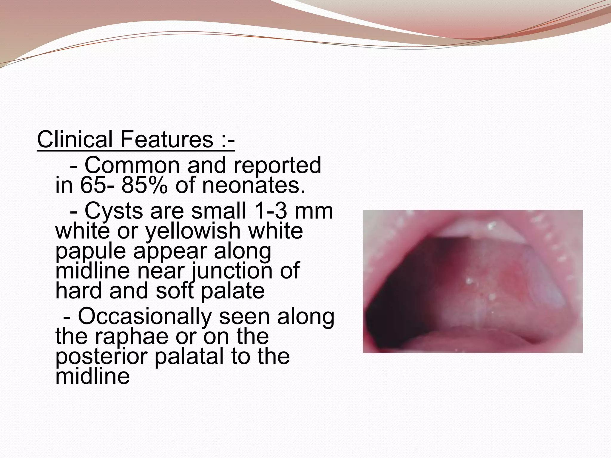
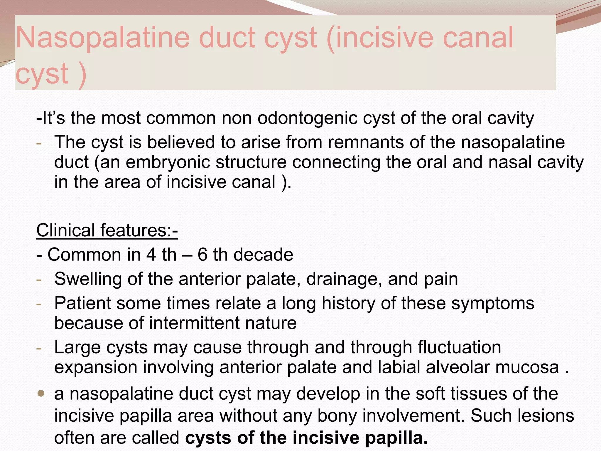
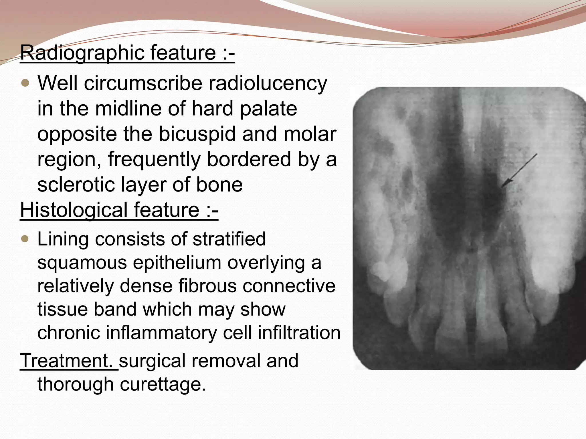
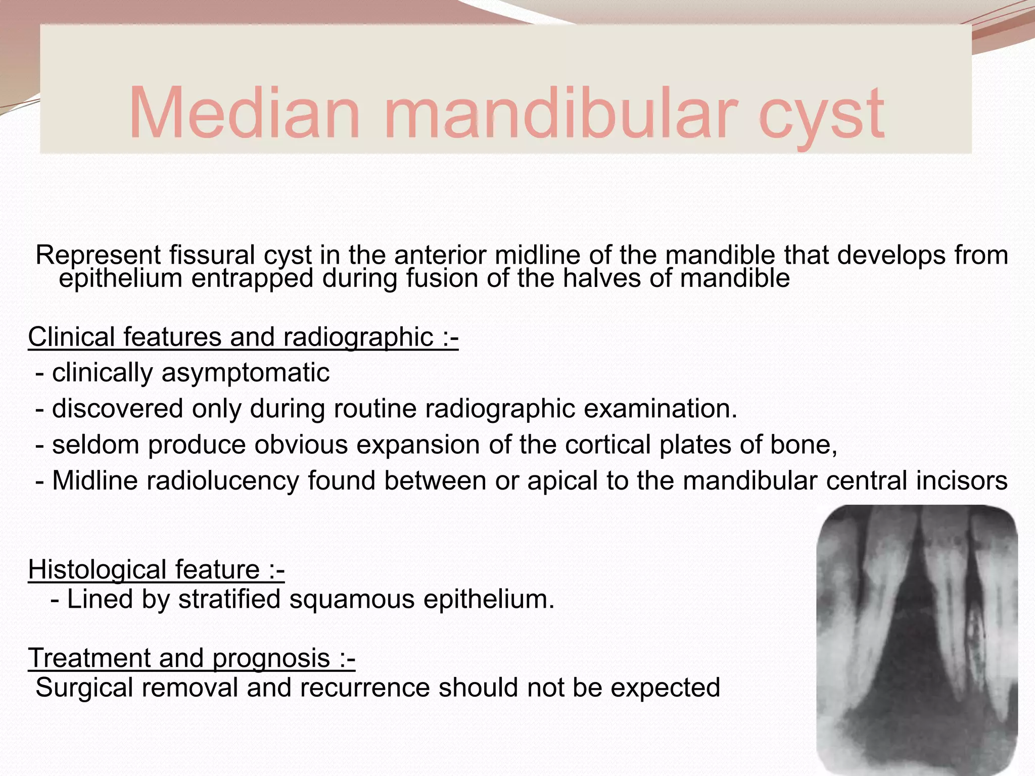
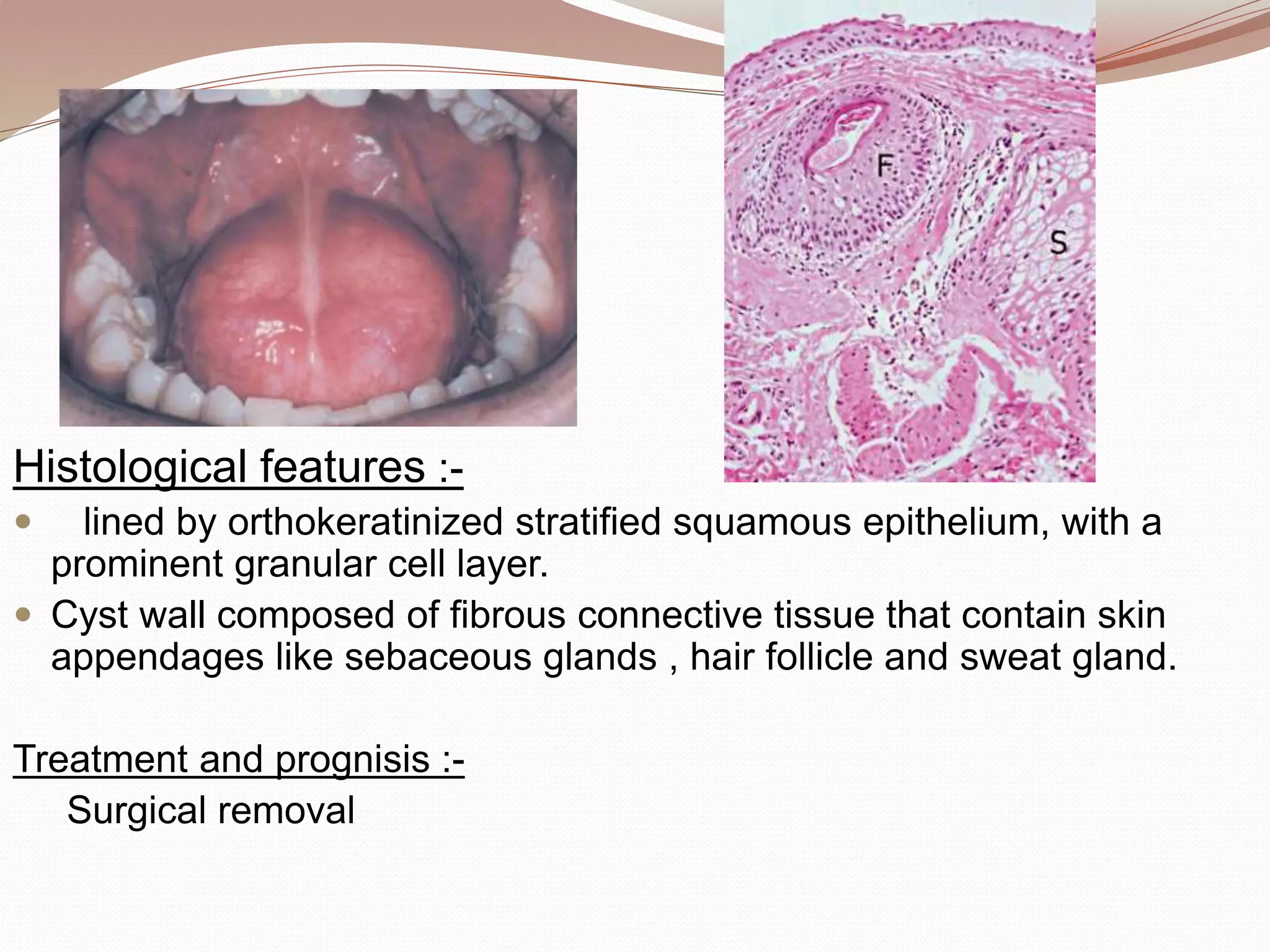
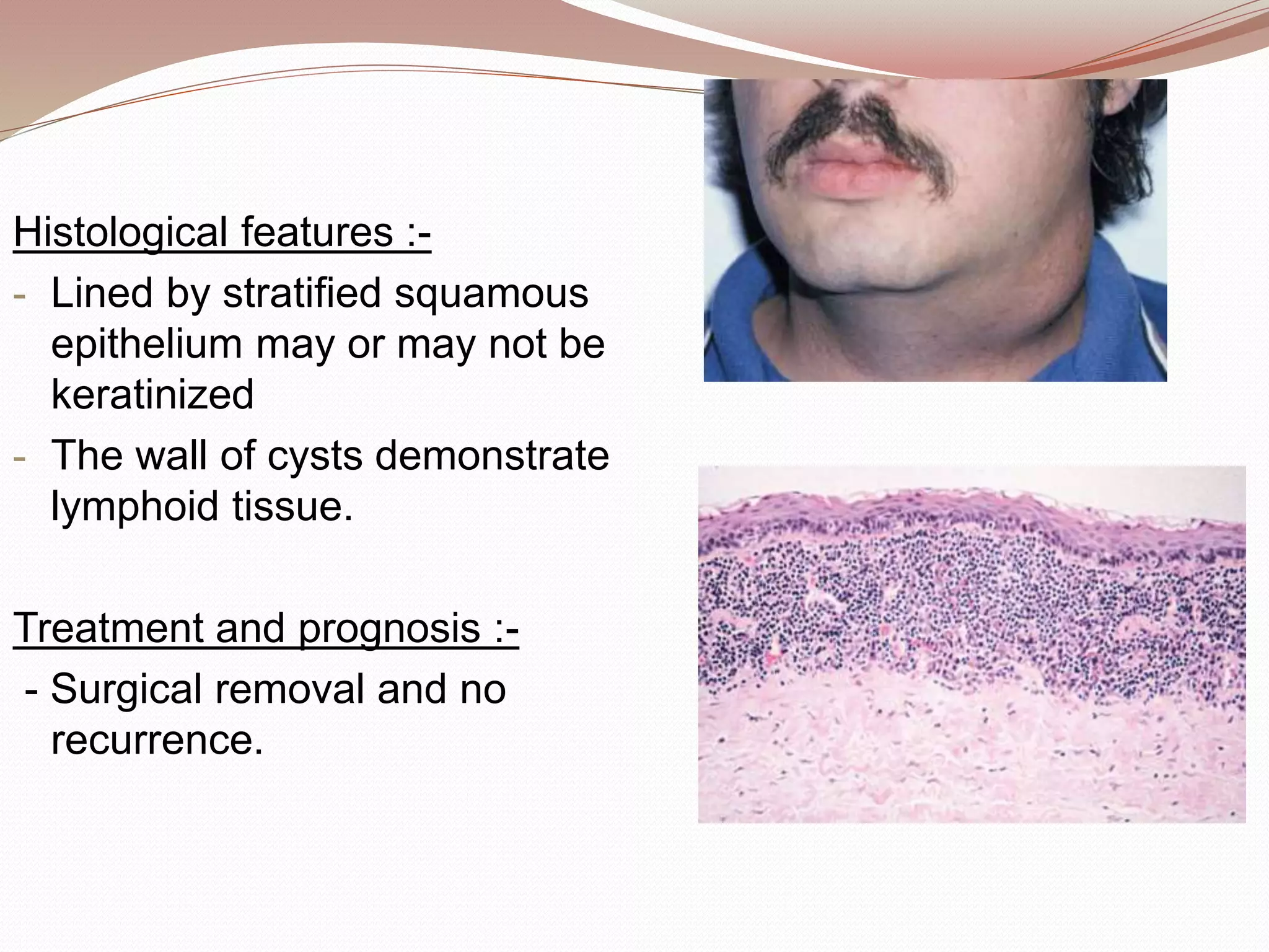
SlideShare a Scribd company logo
By
Dr. Surrendra Reddy
Cyst :- is a pathological cavity (often fluid filled) lined by
epithelium.
Fissural cysts are called so because they were
thought to arise from epithelium entrapped along
embryonal lines of fusion .
Regardless of origin, once cysts develop in the oral and
maxillofacial region ,they tend to slowly increase in size ,
possibly in response slightly elevated hydrostatic luminal
pressure.
 Various developmental cysts in oral and maxillofacial
region are :-
1. Palatal cyst of new born
2. Nasolabial cyst
3. Globullomaxillary cyst
4. Nasopalatine duct cyst
5. Median palatine cyst
6. Median mandibular cyst
7. Epidermoid cyst
8. Dermoid cyst
9. Thyroglossal duct cyst
10. Branchial cleft cyst
11. Oral lymphoepithelial cyst
Palatal cyst of new born (Epstein’s pearl
and Bohn’s nodule)
They are the inclusion cyst arise in one of two ways .
First , during formation of secondary palate small
islands of epithelium may entrapped below the
surface along the median palatal raphae and form
cyst.
Secondly this may arise from epithelial remnants
derived from the development of the minor salivary
gland of palate.
As originally described Epstein's pearl occurs along
medial palatal raphae arise from epithelium
entrapment . Bohn’s nodule are scattered over the
hard palate and soft palate arise from minor salivary
glands.
Clinical Features :-
- Common and reported
in 65- 85% of neonates.
- Cysts are small 1-3 mm
white or yellowish white
papule appear along
midline near junction of
hard and soft palate
- Occasionally seen along
the raphae or on the
posterior palatal to the
midline
Histological Features :-
- thin, stratified squamous epithelium cyst lining with a routine
fibrovascular connective tissue stroma, usually without an
inflammatory cell infiltrate.
- The cystic lumen is filled with degenerated keratin, usually
formed into concentric layers or onion rings and the epithelium lacks
rete processes.
Treatment and prognosis :-
- They are innocuous lesion and no treatment is required.
-They are self healing and rarely observable several weeks
after birth . Presumably the epithelium degenerates or cyst
rupture onto the mucosal surface and eliminate their keratin
content.
Nasolabial cyst
(Nasoalveolar cyst / klestadt’s cyst)
 The nasolabial cyst is a rare developmental cyst that occur
in upper lip lateral to the midline
 Origin is uncertain
 Two theories explain its pathogenesis,
one theory considers it to be fissural cyst arising from
epithelial remnants entrapped along the line of embryonic
fusion of maxillary medial nasal and lateral nasal process.
Second theory suggests that these cysts develop from
misplaced epithelium of nasolacrimal duct because of there
similar location and histology.
Clinical features :-
- Appears as swelling of upper lip lateral to the
midline , resulting in elevation of the ala of nose .
- Often elevates the mucosa of nasal vestibule
and obliterates the maxillary mucolabial fold.
- This expansion may result in nasal obstruction
or may interfere with wearing denture.
- Pain is uncommon unless the lesion is
secondarily infected .
- Cyst may rupture spontaneously and may drain
into oral cavity or nose.
- Commonly seen in adult with peak prevalence in 4 – 5 th
decade and more in females
- Pressure resorption of underlying bone can be noticed in
radiograph.
Histological features :-
- Cyst is lined by pseudo stratified columnar epithelium often
demonstrating the goblet cells and cilia . Areas of cuboidal
epithelium and squamous metaplasia are not unusual.
- Cyst wall composed of fibrous connective tissue with adjacent
skeletal muscle .
- Inflammatory changes seen if the cyst is secondarily infected.
Treatment and prognosis :-
-Complete surgical excision of cyst
via intra oral approach is treatment
of choice.
Nasopalatine duct cyst (incisive canal
cyst )
-It’s the most common non odontogenic cyst of the oral cavity
- The cyst is believed to arise from remnants of the nasopalatine
duct (an embryonic structure connecting the oral and nasal cavity
in the area of incisive canal ).
Clinical features:-
- Common in 4 th – 6 th decade
- Swelling of the anterior palate, drainage, and pain
- Patient some times relate a long history of these symptoms
because of intermittent nature
- Large cysts may cause through and through fluctuation
expansion involving anterior palate and labial alveolar mucosa .
 a nasopalatine duct cyst may develop in the soft tissues of the
incisive papilla area without any bony involvement. Such lesions
often are called cysts of the incisive papilla.
Radiological features :-
- Demonstrate well circumscribed
radiolucency in or near the
midline of anterior maxilla
between and apical to central
incisor.
- Lesion is round or oval with
sclerotic border.
- inverted pear shape due to
resistance of adjacent tooth or
heart shape as a result of
superimposition of nasal spine
- Lesion size will be less than 6
cm. and 6mm radiolucency is
regarded as normal foramen in
this area unless others
symptoms are present.
Histological features :-
- Epithelium of nasopalatine cyst is highly variable it
may be stratified squamous ,pseudo stratified
columnar, simple columnar , simple cuboidal.
- Because the nasopalatine duct cyst arises within the
incisive canal, moderate-sized nerves and small
muscular arteries and veins usually are found in the
wall of the cyst.
- Frequently inflammatory response is noted in cyst wall
and it will be chronic in nature and composed of
lymphocytes , plasma cells and histocytes
Treatment and prognosis :-
Surgical enucleation and biopsy
Median palatal (palatine) cyst
Fissural cysts develops from epithelium
entrapped along the embryonic line of
fusion of the lateral palatal shelves of the
maxilla.
Clinical features :-
- Firm and fluctuant swelling of midline of
hard palate posterior to palatine papilla.
- Affects young adults.
- Usually asymptomatic some times patient
complains of pain and expansion
- Average size of swelling is 2x2 cm.
- True median palatine cyst should exhibit
clinical enlargement of the palate
Radiographic feature :-
 Well circumscribe radiolucency
in the midline of hard palate
opposite the bicuspid and molar
region, frequently bordered by a
sclerotic layer of bone
Histological feature :-
 Lining consists of stratified
squamous epithelium overlying a
relatively dense fibrous connective
tissue band which may show
chronic inflammatory cell infiltration
Treatment. surgical removal and
thorough curettage.
 To differentiate the median palatal cyst from other cystic
lesions of the maxilla, Gingell and associates suggested the
following diagnostic criteria:
• Grossly appears symmetrical along the midline of the
hard palate
• Located posterior to the palatine papilla
• Appears ovoid or circular radiographically
• Not intimately associated with a nonvital tooth
• Does not communicate with the incisive canal
• Shows no microscopic evidence of large neurovascular
bundles, hyaline cartilage, or minor salivary glands in the
cyst wall
Globullomaxillary cyst
 It is a fissural cyst arises from epithelium entrapped
during the fusion of globular portion of median nasal
process with the maxillary process .
 cyst forms in the bone suture between the premaxilla and
maxilla,the incisive suture, (‘premaxilla-maxillary cyst’)
Clinical features :-
- Classically develop between maxillary lateral incisor and
cuspid teeth occasionally seen between central and
lateral incisor.
- As lesion grows there will be tipping of the tooth root.
Radiological feature :-
• an inverted, pear-shaped radiolucent area between
the roots of the lateral incisor and cuspid, usually
causing divergence of the roots of these teeth
Differential diagnosis:
 Periapical granuloma, apical periodontal cyst, lateral
periodontal cyst, odontogenic keratocyst, central giant
cell granuloma, calcifying odontogenic cyst, and
odontogenic myxoma
Histological features :-
 Histological globulomaxillary cyst lined by either
stratified squamous or ciliated columna epithelium
 the wall is made up of fibrous connective tissue,
usually showing inflammatory cell infiltration.
Treatment and prognosis :-
Surgical enucleation .
If the lesion can be related to an adjacent nonvital
tooth , then endodontic therapy may be appropriate.
Median mandibular cyst
Represent fissural cyst in the anterior midline of the mandible that develops from
epithelium entrapped during fusion of the halves of mandible
Clinical features and radiographic :-
- clinically asymptomatic
- discovered only during routine radiographic examination.
- seldom produce obvious expansion of the cortical plates of bone,
- Midline radiolucency found between or apical to the mandibular central incisors
Histological feature :-
- Lined by stratified squamous epithelium.
Treatment and prognosis :-
Surgical removal and recurrence should not be expected
Epidermal Inclusion Cyst
(Epidermal cyst, epidermoid cyst, epithelial cyst, keratin cyst, milia)
 Common cyst of skin lined by epidermis like epithelium
 Derived from the follicular infundibulum and called infundibular cyst.
 Arise after localized inflammation of hair follicle and represent nonneoplastic
proliferation of infundibular epithelium.
 Occasionally arise after traumatic implantation of epithelium.
Clinical features :-
 Common in acne prone areas of head neck and back region
 Usually affects before puberty unless they are associated with Gardner’s
syndrome
 Males are affected more than females
 Presents as nodular fluctuation subcutaneous lesion that may or may not
associated with inflammation and a non inflamed lesion is white to yellow in
color.
 Discharge of a foul-smelling cheese-like material is a common complaint.
 Less frequently, the cysts can become inflamed or infected, resulting in pain
and tenderness.
Milia (singular: milium) are tiny keratin-filled
cysts that resemble miniature epidermoid cysts
•When located orally, the cysts can
cause difficulty in feeding, swallowing, or
even speaking
•Epidermoid cysts appear as firm, round,
mobile, flesh-colored to yellow or white
subcutaneous nodules of variable size.
•A central pore or punctum is an
inconsistent finding that may adhere the
cyst to the overlying epidermis and from
which a thick cheesy material can
sometimes be expressed
Histological features :-
 Lined by stratified squamous epithelium
 A well developed granular cell layer is seen and the lumen is filled
with orthokeratin.
 Dystrophic calcification and reactive foreign body reaction are seen
associated with the cystic capsule.
 Pigmented epidermoid cysts may demonstrate melanin pigment in
the wall and a keratin mass.
 A surrounding infiltrate of melanocytes and melanophages may also
be observed.
Treatment and prognosis :-
- By conservative surgical excision and recurrence is uncommon .
- Malignant transformation has been reported but is exceedingly rare.
Dermoid cyst
 A hamartomatous tumor containing multiple sebaceous glands and almost
all skin adnexa, this may contain substances such as nails and dental,
cartilage like, and bone like structures.
 If limited to the skin or subcutaneous tissue, dermoid cysts are thin walled
tumors that contain different amounts of fatty masses; occasionally, they
contain horny masses and hairs.
 Origin : sequestration of skin and subsequent implantation of it along the
lines of embryonic closure.
Clinical features :-
- occur in individuals aged 10–30 years
- Commonly occur in midline of the floor of the mouth
- If cyst develops above geniohyoid muscle , a sublingual swelling may
displace the tongue towards the roof of mouth and causes dysphagia and
dysphonia or even dysponea.
- Submental swelling causing double chin appearance
- Slow growing painless.
- Presenting as a rubbery mass that frequently retains pitting after
application
Histological features :-
 lined by orthokeratinized stratified squamous epithelium, with a
prominent granular cell layer.
 Cyst wall composed of fibrous connective tissue that contain skin
appendages like sebaceous glands , hair follicle and sweat gland.
Treatment and prognisis :-
Surgical removal
Thyroglossal duct (tract) cyst
 The thyroglossal duct epithelium normally undergoes atrophy
however, remnants of this epithelium may persist and give rise
to Thyroglossal duct (tract) cyst.
Clinical features :-
- Develops in the midline and may occur anywhere from the
foramen caecum area of the tongue to the suprasternal notch.
- Common in first two decades .
- No sex prediliction.
- Most are smaller than 3 cm.
- Cyst is painless, fluctuant and movable swelling unless
secondarily infected.
- Lesion at the base of tongue may cause laryngeal obstruction.
 patients will have neck or throat pain, or dysphagia (difficulty in
swallowing).
- If cyst is attached to hyoid bone or
tongue , it will move vertically during
swallowing or protrusion of tongue.
Histological features :-
- Lined by columnar or stratified
squamous epithelium
- thyroid tissue may occur in the cyst
wall.
Treatment and prognosis :-
- Best treated by sistrunk procedure
and recurrence is 10 % in this
procedure .
Branchial cleft cyst
(Cervical lymphoepithelial cyst)
 Developmental cyst of lateral neck.
 The cyst develops from remnants of branchial cleft because it occurs in the
area of embryonic gill apparatus .
 95% of the cyst develops from second arch and 5% from 1st, 3rd & 4th
arches.
Clinical features :-
 Common in upper lateral neck along the anterior border of sternocleidomastoid
 Affects young adults of 20 – 40 yr
 Soft fluctuant mass of 1 – 10 cm in diameter.
 Tenderness and pain when secondarily infected.
 Anomalies from the first branchial arch comprise approximately 1% of branchial
cleft malformations and usually are found in close proximity to the parotid gland.
 Third-cleft and fourth-cleft anomalies are rare and may develop in the lower neck
or mediastinum.
Histological features :-
- Lined by stratified squamous
epithelium may or may not be
keratinized
- The wall of cysts demonstrate
lymphoid tissue.
Treatment and prognosis :-
- Surgical removal and no
recurrence.
 Uncommon cyst develop with in the oral lymphoid tissue.
 Similar to Branchial cleft cyst but smaller in size
Clinical features :-
- Appear as small submucosal mass less than 1 cm
- Cyst may feel firm or soft on palpation
- Overlying mucosa is smooth and nonulcerated.
- Lesion is typically white or yellow and often contain creamy
or cheesy keratinous material in the lumen.
- Usually asymptomatic pain is rare but occur due to trauma
- commonly occurs at floor of mouth of young people
Oral lymphoepithelial cyst
Histological features :-
- Cystic cavity lined by atropic
stratified squamous epithelium
with out rete ridges
- Epithelium is parakeratinized,
with desquamated epithelial
cells seen filling the cystic
lumen.
- Presence of lymphoid tissue in
the cyst wall .
Treatment and prognosis :-
Surgical excision and no
recurrence.

More Related Content

Similar to Developmental cysts.pptx

Cystic lesions in oral cavity
Cystic lesions in oral cavityCystic lesions in oral cavity
Cystic lesions in oral cavity
Saraah Gillani
 
CYSTS OF THE ORAL FACIAL REGION.pptx
CYSTS OF THE ORAL FACIAL REGION.pptxCYSTS OF THE ORAL FACIAL REGION.pptx
CYSTS OF THE ORAL FACIAL REGION.pptx
AngetileKasanga
 
Periapical cyst
Periapical  cystPeriapical  cyst
Periapical cyst
HagerMohammed12
 
Cysts of oral regions
Cysts of oral regionsCysts of oral regions
Cysts of oral regions
Naz Dizayee
 
Cysts in orofacial region
Cysts in orofacial regionCysts in orofacial region
Cysts in orofacial region
Mohammed Rhael
 
Soft tissue calcification of head and neck
Soft tissue calcification of head and neck Soft tissue calcification of head and neck
Soft tissue calcification of head and neck
Aravind Babudevan
 
Complications of sinusitis
Complications of sinusitisComplications of sinusitis
Complications of sinusitis
Sivendu P
 
Benign neck disease
Benign neck diseaseBenign neck disease
Benign neck disease
Shekhar Krishna Debnath
 
Non odontogenic cyst and pseudo cyst of the jaw- seminar 2- ORIGINAL.pptx
Non odontogenic cyst and pseudo cyst of the jaw- seminar 2- ORIGINAL.pptxNon odontogenic cyst and pseudo cyst of the jaw- seminar 2- ORIGINAL.pptx
Non odontogenic cyst and pseudo cyst of the jaw- seminar 2- ORIGINAL.pptx
ReshmaAmmu11
 
Radicular cyst or Periapical cyst
Radicular cyst or Periapical cystRadicular cyst or Periapical cyst
Radicular cyst or Periapical cyst
drabbasnaseem
 
Lab 6 developmental cyst
Lab 6 developmental cystLab 6 developmental cyst
Lab 6 developmental cyst
dina hameed
 
Non Odontogenic Cysts
Non Odontogenic CystsNon Odontogenic Cysts
Non Odontogenic Cysts
Mohammed Alawad
 
Neck surgery
Neck surgeryNeck surgery
Neck surgery
Laith AL-khalidy
 
Salivary Gland Diseases.pptx
Salivary Gland Diseases.pptxSalivary Gland Diseases.pptx
Salivary Gland Diseases.pptx
EidleMohamedsaed
 
CYSTS OF ORAL AND MAXILLOFACIAL REGION (2).ppt
CYSTS OF ORAL AND MAXILLOFACIAL REGION (2).pptCYSTS OF ORAL AND MAXILLOFACIAL REGION (2).ppt
CYSTS OF ORAL AND MAXILLOFACIAL REGION (2).ppt
Royal Dental College Library
 
Disease of external nose deviated nasal septum, fb in nose 02.05.16, dr.bini...
Disease of external nose deviated nasal septum, fb in nose  02.05.16, dr.bini...Disease of external nose deviated nasal septum, fb in nose  02.05.16, dr.bini...
Disease of external nose deviated nasal septum, fb in nose 02.05.16, dr.bini...
ophthalmgmcri
 
Cysts of Oral Cavity
Cysts of Oral Cavity Cysts of Oral Cavity
Cysts of Oral Cavity
KritiGarg25
 
Cyst
CystCyst
Cyst
Dr.Noreen
 
Neck mass-latest
Neck mass-latestNeck mass-latest
Neck mass-latest
Dennis Lee
 
Diseases of external nose
Diseases of external noseDiseases of external nose
Diseases of external nose
Karl Daniel, M.D.
 

Similar to Developmental cysts.pptx (20)

Cystic lesions in oral cavity
Cystic lesions in oral cavityCystic lesions in oral cavity
Cystic lesions in oral cavity
 
CYSTS OF THE ORAL FACIAL REGION.pptx
CYSTS OF THE ORAL FACIAL REGION.pptxCYSTS OF THE ORAL FACIAL REGION.pptx
CYSTS OF THE ORAL FACIAL REGION.pptx
 
Periapical cyst
Periapical  cystPeriapical  cyst
Periapical cyst
 
Cysts of oral regions
Cysts of oral regionsCysts of oral regions
Cysts of oral regions
 
Cysts in orofacial region
Cysts in orofacial regionCysts in orofacial region
Cysts in orofacial region
 
Soft tissue calcification of head and neck
Soft tissue calcification of head and neck Soft tissue calcification of head and neck
Soft tissue calcification of head and neck
 
Complications of sinusitis
Complications of sinusitisComplications of sinusitis
Complications of sinusitis
 
Benign neck disease
Benign neck diseaseBenign neck disease
Benign neck disease
 
Non odontogenic cyst and pseudo cyst of the jaw- seminar 2- ORIGINAL.pptx
Non odontogenic cyst and pseudo cyst of the jaw- seminar 2- ORIGINAL.pptxNon odontogenic cyst and pseudo cyst of the jaw- seminar 2- ORIGINAL.pptx
Non odontogenic cyst and pseudo cyst of the jaw- seminar 2- ORIGINAL.pptx
 
Radicular cyst or Periapical cyst
Radicular cyst or Periapical cystRadicular cyst or Periapical cyst
Radicular cyst or Periapical cyst
 
Lab 6 developmental cyst
Lab 6 developmental cystLab 6 developmental cyst
Lab 6 developmental cyst
 
Non Odontogenic Cysts
Non Odontogenic CystsNon Odontogenic Cysts
Non Odontogenic Cysts
 
Neck surgery
Neck surgeryNeck surgery
Neck surgery
 
Salivary Gland Diseases.pptx
Salivary Gland Diseases.pptxSalivary Gland Diseases.pptx
Salivary Gland Diseases.pptx
 
CYSTS OF ORAL AND MAXILLOFACIAL REGION (2).ppt
CYSTS OF ORAL AND MAXILLOFACIAL REGION (2).pptCYSTS OF ORAL AND MAXILLOFACIAL REGION (2).ppt
CYSTS OF ORAL AND MAXILLOFACIAL REGION (2).ppt
 
Disease of external nose deviated nasal septum, fb in nose 02.05.16, dr.bini...
Disease of external nose deviated nasal septum, fb in nose  02.05.16, dr.bini...Disease of external nose deviated nasal septum, fb in nose  02.05.16, dr.bini...
Disease of external nose deviated nasal septum, fb in nose 02.05.16, dr.bini...
 
Cysts of Oral Cavity
Cysts of Oral Cavity Cysts of Oral Cavity
Cysts of Oral Cavity
 
Cyst
CystCyst
Cyst
 
Neck mass-latest
Neck mass-latestNeck mass-latest
Neck mass-latest
 
Diseases of external nose
Diseases of external noseDiseases of external nose
Diseases of external nose
 

More from Lubna Nazneen

7. Oral mucosa.pptx
7. Oral mucosa.pptx7. Oral mucosa.pptx
7. Oral mucosa.pptx
Lubna Nazneen
 
Tumours of nerve tissue origin.pptx
Tumours of nerve tissue origin.pptxTumours of nerve tissue origin.pptx
Tumours of nerve tissue origin.pptx
Lubna Nazneen
 
Malignant Connective Tissue Tumors.pptx
Malignant Connective Tissue Tumors.pptxMalignant Connective Tissue Tumors.pptx
Malignant Connective Tissue Tumors.pptx
Lubna Nazneen
 
oral manifestations of HIV.pptx
oral manifestations of HIV.pptxoral manifestations of HIV.pptx
oral manifestations of HIV.pptx
Lubna Nazneen
 
Benign Connective Tissue Tumours.pptx
Benign Connective Tissue Tumours.pptxBenign Connective Tissue Tumours.pptx
Benign Connective Tissue Tumours.pptx
Lubna Nazneen
 
Candidiasis.pptx
Candidiasis.pptxCandidiasis.pptx
Candidiasis.pptx
Lubna Nazneen
 
6. Diff bw primary and permanent dentition.pptx
6. Diff bw primary and permanent dentition.pptx6. Diff bw primary and permanent dentition.pptx
6. Diff bw primary and permanent dentition.pptx
Lubna Nazneen
 
5. tongue.pptx
5. tongue.pptx5. tongue.pptx
5. tongue.pptx
Lubna Nazneen
 
4. mineralization.ppt
4. mineralization.ppt4. mineralization.ppt
4. mineralization.ppt
Lubna Nazneen
 
3. CELL JUNCTIONS.pptx
3. CELL JUNCTIONS.pptx3. CELL JUNCTIONS.pptx
3. CELL JUNCTIONS.pptx
Lubna Nazneen
 
2. Bone.pptx
2. Bone.pptx2. Bone.pptx
2. Bone.pptx
Lubna Nazneen
 
1. DEV OF FACE.pptx
1. DEV OF FACE.pptx1. DEV OF FACE.pptx
1. DEV OF FACE.pptx
Lubna Nazneen
 

More from Lubna Nazneen (12)

7. Oral mucosa.pptx
7. Oral mucosa.pptx7. Oral mucosa.pptx
7. Oral mucosa.pptx
 
Tumours of nerve tissue origin.pptx
Tumours of nerve tissue origin.pptxTumours of nerve tissue origin.pptx
Tumours of nerve tissue origin.pptx
 
Malignant Connective Tissue Tumors.pptx
Malignant Connective Tissue Tumors.pptxMalignant Connective Tissue Tumors.pptx
Malignant Connective Tissue Tumors.pptx
 
oral manifestations of HIV.pptx
oral manifestations of HIV.pptxoral manifestations of HIV.pptx
oral manifestations of HIV.pptx
 
Benign Connective Tissue Tumours.pptx
Benign Connective Tissue Tumours.pptxBenign Connective Tissue Tumours.pptx
Benign Connective Tissue Tumours.pptx
 
Candidiasis.pptx
Candidiasis.pptxCandidiasis.pptx
Candidiasis.pptx
 
6. Diff bw primary and permanent dentition.pptx
6. Diff bw primary and permanent dentition.pptx6. Diff bw primary and permanent dentition.pptx
6. Diff bw primary and permanent dentition.pptx
 
5. tongue.pptx
5. tongue.pptx5. tongue.pptx
5. tongue.pptx
 
4. mineralization.ppt
4. mineralization.ppt4. mineralization.ppt
4. mineralization.ppt
 
3. CELL JUNCTIONS.pptx
3. CELL JUNCTIONS.pptx3. CELL JUNCTIONS.pptx
3. CELL JUNCTIONS.pptx
 
2. Bone.pptx
2. Bone.pptx2. Bone.pptx
2. Bone.pptx
 
1. DEV OF FACE.pptx
1. DEV OF FACE.pptx1. DEV OF FACE.pptx
1. DEV OF FACE.pptx
 

Recently uploaded

micro teaching on communication m.sc nursing.pdf
micro teaching on communication m.sc nursing.pdfmicro teaching on communication m.sc nursing.pdf
micro teaching on communication m.sc nursing.pdf
Anurag Sharma
 
Identification and nursing management of congenital malformations .pptx
Identification and nursing management of congenital malformations .pptxIdentification and nursing management of congenital malformations .pptx
Identification and nursing management of congenital malformations .pptx
MGM SCHOOL/COLLEGE OF NURSING
 
Sex determination from mandible pelvis and skull
Sex determination from mandible pelvis and skullSex determination from mandible pelvis and skull
Sex determination from mandible pelvis and skull
ShashankRoodkee
 
Knee anatomy and clinical tests 2024.pdf
Knee anatomy and clinical tests 2024.pdfKnee anatomy and clinical tests 2024.pdf
Knee anatomy and clinical tests 2024.pdf
vimalpl1234
 
Chapter 11 Nutrition and Chronic Diseases.pptx
Chapter 11 Nutrition and Chronic Diseases.pptxChapter 11 Nutrition and Chronic Diseases.pptx
Chapter 11 Nutrition and Chronic Diseases.pptx
Earlene McNair
 
NVBDCP.pptx Nation vector borne disease control program
NVBDCP.pptx Nation vector borne disease control programNVBDCP.pptx Nation vector borne disease control program
NVBDCP.pptx Nation vector borne disease control program
Sapna Thakur
 
#cALL# #gIRLS# In Dehradun ꧁❤8107221448❤꧂#cALL# #gIRLS# Service In Dehradun W...
#cALL# #gIRLS# In Dehradun ꧁❤8107221448❤꧂#cALL# #gIRLS# Service In Dehradun W...#cALL# #gIRLS# In Dehradun ꧁❤8107221448❤꧂#cALL# #gIRLS# Service In Dehradun W...
#cALL# #gIRLS# In Dehradun ꧁❤8107221448❤꧂#cALL# #gIRLS# Service In Dehradun W...
chandankumarsmartiso
 
Aortic Association CBL Pilot April 19 – 20 Bern
Aortic Association CBL Pilot April 19 – 20 BernAortic Association CBL Pilot April 19 – 20 Bern
Aortic Association CBL Pilot April 19 – 20 Bern
suvadeepdas911
 
ANATOMY AND PHYSIOLOGY OF URINARY SYSTEM.pptx
ANATOMY AND PHYSIOLOGY OF URINARY SYSTEM.pptxANATOMY AND PHYSIOLOGY OF URINARY SYSTEM.pptx
ANATOMY AND PHYSIOLOGY OF URINARY SYSTEM.pptx
Swetaba Besh
 
CHEMOTHERAPY_RDP_CHAPTER 1_ANTI TB DRUGS.pdf
CHEMOTHERAPY_RDP_CHAPTER 1_ANTI TB DRUGS.pdfCHEMOTHERAPY_RDP_CHAPTER 1_ANTI TB DRUGS.pdf
CHEMOTHERAPY_RDP_CHAPTER 1_ANTI TB DRUGS.pdf
rishi2789
 
ARTHROLOGY PPT NCISM SYLLABUS AYURVEDA STUDENTS
ARTHROLOGY PPT NCISM SYLLABUS AYURVEDA STUDENTSARTHROLOGY PPT NCISM SYLLABUS AYURVEDA STUDENTS
ARTHROLOGY PPT NCISM SYLLABUS AYURVEDA STUDENTS
Dr. Vinay Pareek
 
Tests for analysis of different pharmaceutical.pptx
Tests for analysis of different pharmaceutical.pptxTests for analysis of different pharmaceutical.pptx
Tests for analysis of different pharmaceutical.pptx
taiba qazi
 
Non-respiratory Functions of the Lungs.pdf
Non-respiratory Functions of the Lungs.pdfNon-respiratory Functions of the Lungs.pdf
Non-respiratory Functions of the Lungs.pdf
MedicoseAcademics
 
THERAPEUTIC ANTISENSE MOLECULES .pptx
THERAPEUTIC ANTISENSE MOLECULES    .pptxTHERAPEUTIC ANTISENSE MOLECULES    .pptx
THERAPEUTIC ANTISENSE MOLECULES .pptx
70KRISHPATEL
 
The Electrocardiogram - Physiologic Principles
The Electrocardiogram - Physiologic PrinciplesThe Electrocardiogram - Physiologic Principles
The Electrocardiogram - Physiologic Principles
MedicoseAcademics
 
Physiology of Special Chemical Sensation of Taste
Physiology of Special Chemical Sensation of TastePhysiology of Special Chemical Sensation of Taste
Physiology of Special Chemical Sensation of Taste
MedicoseAcademics
 
Novas diretrizes da OMS para os cuidados perinatais de mais qualidade
Novas diretrizes da OMS para os cuidados perinatais de mais qualidadeNovas diretrizes da OMS para os cuidados perinatais de mais qualidade
Novas diretrizes da OMS para os cuidados perinatais de mais qualidade
Prof. Marcus Renato de Carvalho
 
CHEMOTHERAPY_RDP_CHAPTER 4_ANTI VIRAL DRUGS.pdf
CHEMOTHERAPY_RDP_CHAPTER 4_ANTI VIRAL DRUGS.pdfCHEMOTHERAPY_RDP_CHAPTER 4_ANTI VIRAL DRUGS.pdf
CHEMOTHERAPY_RDP_CHAPTER 4_ANTI VIRAL DRUGS.pdf
rishi2789
 
Local Advanced Lung Cancer: Artificial Intelligence, Synergetics, Complex Sys...
Local Advanced Lung Cancer: Artificial Intelligence, Synergetics, Complex Sys...Local Advanced Lung Cancer: Artificial Intelligence, Synergetics, Complex Sys...
Local Advanced Lung Cancer: Artificial Intelligence, Synergetics, Complex Sys...
Oleg Kshivets
 
CHEMOTHERAPY_RDP_CHAPTER 2 _LEPROSY.pdf1
CHEMOTHERAPY_RDP_CHAPTER 2 _LEPROSY.pdf1CHEMOTHERAPY_RDP_CHAPTER 2 _LEPROSY.pdf1
CHEMOTHERAPY_RDP_CHAPTER 2 _LEPROSY.pdf1
rishi2789
 

Recently uploaded (20)

micro teaching on communication m.sc nursing.pdf
micro teaching on communication m.sc nursing.pdfmicro teaching on communication m.sc nursing.pdf
micro teaching on communication m.sc nursing.pdf
 
Identification and nursing management of congenital malformations .pptx
Identification and nursing management of congenital malformations .pptxIdentification and nursing management of congenital malformations .pptx
Identification and nursing management of congenital malformations .pptx
 
Sex determination from mandible pelvis and skull
Sex determination from mandible pelvis and skullSex determination from mandible pelvis and skull
Sex determination from mandible pelvis and skull
 
Knee anatomy and clinical tests 2024.pdf
Knee anatomy and clinical tests 2024.pdfKnee anatomy and clinical tests 2024.pdf
Knee anatomy and clinical tests 2024.pdf
 
Chapter 11 Nutrition and Chronic Diseases.pptx
Chapter 11 Nutrition and Chronic Diseases.pptxChapter 11 Nutrition and Chronic Diseases.pptx
Chapter 11 Nutrition and Chronic Diseases.pptx
 
NVBDCP.pptx Nation vector borne disease control program
NVBDCP.pptx Nation vector borne disease control programNVBDCP.pptx Nation vector borne disease control program
NVBDCP.pptx Nation vector borne disease control program
 
#cALL# #gIRLS# In Dehradun ꧁❤8107221448❤꧂#cALL# #gIRLS# Service In Dehradun W...
#cALL# #gIRLS# In Dehradun ꧁❤8107221448❤꧂#cALL# #gIRLS# Service In Dehradun W...#cALL# #gIRLS# In Dehradun ꧁❤8107221448❤꧂#cALL# #gIRLS# Service In Dehradun W...
#cALL# #gIRLS# In Dehradun ꧁❤8107221448❤꧂#cALL# #gIRLS# Service In Dehradun W...
 
Aortic Association CBL Pilot April 19 – 20 Bern
Aortic Association CBL Pilot April 19 – 20 BernAortic Association CBL Pilot April 19 – 20 Bern
Aortic Association CBL Pilot April 19 – 20 Bern
 
ANATOMY AND PHYSIOLOGY OF URINARY SYSTEM.pptx
ANATOMY AND PHYSIOLOGY OF URINARY SYSTEM.pptxANATOMY AND PHYSIOLOGY OF URINARY SYSTEM.pptx
ANATOMY AND PHYSIOLOGY OF URINARY SYSTEM.pptx
 
CHEMOTHERAPY_RDP_CHAPTER 1_ANTI TB DRUGS.pdf
CHEMOTHERAPY_RDP_CHAPTER 1_ANTI TB DRUGS.pdfCHEMOTHERAPY_RDP_CHAPTER 1_ANTI TB DRUGS.pdf
CHEMOTHERAPY_RDP_CHAPTER 1_ANTI TB DRUGS.pdf
 
ARTHROLOGY PPT NCISM SYLLABUS AYURVEDA STUDENTS
ARTHROLOGY PPT NCISM SYLLABUS AYURVEDA STUDENTSARTHROLOGY PPT NCISM SYLLABUS AYURVEDA STUDENTS
ARTHROLOGY PPT NCISM SYLLABUS AYURVEDA STUDENTS
 
Tests for analysis of different pharmaceutical.pptx
Tests for analysis of different pharmaceutical.pptxTests for analysis of different pharmaceutical.pptx
Tests for analysis of different pharmaceutical.pptx
 
Non-respiratory Functions of the Lungs.pdf
Non-respiratory Functions of the Lungs.pdfNon-respiratory Functions of the Lungs.pdf
Non-respiratory Functions of the Lungs.pdf
 
THERAPEUTIC ANTISENSE MOLECULES .pptx
THERAPEUTIC ANTISENSE MOLECULES    .pptxTHERAPEUTIC ANTISENSE MOLECULES    .pptx
THERAPEUTIC ANTISENSE MOLECULES .pptx
 
The Electrocardiogram - Physiologic Principles
The Electrocardiogram - Physiologic PrinciplesThe Electrocardiogram - Physiologic Principles
The Electrocardiogram - Physiologic Principles
 
Physiology of Special Chemical Sensation of Taste
Physiology of Special Chemical Sensation of TastePhysiology of Special Chemical Sensation of Taste
Physiology of Special Chemical Sensation of Taste
 
Novas diretrizes da OMS para os cuidados perinatais de mais qualidade
Novas diretrizes da OMS para os cuidados perinatais de mais qualidadeNovas diretrizes da OMS para os cuidados perinatais de mais qualidade
Novas diretrizes da OMS para os cuidados perinatais de mais qualidade
 
CHEMOTHERAPY_RDP_CHAPTER 4_ANTI VIRAL DRUGS.pdf
CHEMOTHERAPY_RDP_CHAPTER 4_ANTI VIRAL DRUGS.pdfCHEMOTHERAPY_RDP_CHAPTER 4_ANTI VIRAL DRUGS.pdf
CHEMOTHERAPY_RDP_CHAPTER 4_ANTI VIRAL DRUGS.pdf
 
Local Advanced Lung Cancer: Artificial Intelligence, Synergetics, Complex Sys...
Local Advanced Lung Cancer: Artificial Intelligence, Synergetics, Complex Sys...Local Advanced Lung Cancer: Artificial Intelligence, Synergetics, Complex Sys...
Local Advanced Lung Cancer: Artificial Intelligence, Synergetics, Complex Sys...
 
CHEMOTHERAPY_RDP_CHAPTER 2 _LEPROSY.pdf1
CHEMOTHERAPY_RDP_CHAPTER 2 _LEPROSY.pdf1CHEMOTHERAPY_RDP_CHAPTER 2 _LEPROSY.pdf1
CHEMOTHERAPY_RDP_CHAPTER 2 _LEPROSY.pdf1
 

Developmental cysts.pptx

  • 2. Cyst :- is a pathological cavity (often fluid filled) lined by epithelium. Fissural cysts are called so because they were thought to arise from epithelium entrapped along embryonal lines of fusion . Regardless of origin, once cysts develop in the oral and maxillofacial region ,they tend to slowly increase in size , possibly in response slightly elevated hydrostatic luminal pressure.
  • 3.  Various developmental cysts in oral and maxillofacial region are :- 1. Palatal cyst of new born 2. Nasolabial cyst 3. Globullomaxillary cyst 4. Nasopalatine duct cyst 5. Median palatine cyst 6. Median mandibular cyst 7. Epidermoid cyst 8. Dermoid cyst 9. Thyroglossal duct cyst 10. Branchial cleft cyst 11. Oral lymphoepithelial cyst
  • 4. Palatal cyst of new born (Epstein’s pearl and Bohn’s nodule) They are the inclusion cyst arise in one of two ways . First , during formation of secondary palate small islands of epithelium may entrapped below the surface along the median palatal raphae and form cyst. Secondly this may arise from epithelial remnants derived from the development of the minor salivary gland of palate. As originally described Epstein's pearl occurs along medial palatal raphae arise from epithelium entrapment . Bohn’s nodule are scattered over the hard palate and soft palate arise from minor salivary glands.
  • 5. Clinical Features :- - Common and reported in 65- 85% of neonates. - Cysts are small 1-3 mm white or yellowish white papule appear along midline near junction of hard and soft palate - Occasionally seen along the raphae or on the posterior palatal to the midline
  • 6. Histological Features :- - thin, stratified squamous epithelium cyst lining with a routine fibrovascular connective tissue stroma, usually without an inflammatory cell infiltrate. - The cystic lumen is filled with degenerated keratin, usually formed into concentric layers or onion rings and the epithelium lacks rete processes. Treatment and prognosis :- - They are innocuous lesion and no treatment is required. -They are self healing and rarely observable several weeks after birth . Presumably the epithelium degenerates or cyst rupture onto the mucosal surface and eliminate their keratin content.
  • 7. Nasolabial cyst (Nasoalveolar cyst / klestadt’s cyst)  The nasolabial cyst is a rare developmental cyst that occur in upper lip lateral to the midline  Origin is uncertain  Two theories explain its pathogenesis, one theory considers it to be fissural cyst arising from epithelial remnants entrapped along the line of embryonic fusion of maxillary medial nasal and lateral nasal process. Second theory suggests that these cysts develop from misplaced epithelium of nasolacrimal duct because of there similar location and histology.
  • 8. Clinical features :- - Appears as swelling of upper lip lateral to the midline , resulting in elevation of the ala of nose . - Often elevates the mucosa of nasal vestibule and obliterates the maxillary mucolabial fold. - This expansion may result in nasal obstruction or may interfere with wearing denture. - Pain is uncommon unless the lesion is secondarily infected . - Cyst may rupture spontaneously and may drain into oral cavity or nose.
  • 9. - Commonly seen in adult with peak prevalence in 4 – 5 th decade and more in females - Pressure resorption of underlying bone can be noticed in radiograph. Histological features :- - Cyst is lined by pseudo stratified columnar epithelium often demonstrating the goblet cells and cilia . Areas of cuboidal epithelium and squamous metaplasia are not unusual. - Cyst wall composed of fibrous connective tissue with adjacent skeletal muscle . - Inflammatory changes seen if the cyst is secondarily infected. Treatment and prognosis :- -Complete surgical excision of cyst via intra oral approach is treatment of choice.
  • 10. Nasopalatine duct cyst (incisive canal cyst ) -It’s the most common non odontogenic cyst of the oral cavity - The cyst is believed to arise from remnants of the nasopalatine duct (an embryonic structure connecting the oral and nasal cavity in the area of incisive canal ). Clinical features:- - Common in 4 th – 6 th decade - Swelling of the anterior palate, drainage, and pain - Patient some times relate a long history of these symptoms because of intermittent nature - Large cysts may cause through and through fluctuation expansion involving anterior palate and labial alveolar mucosa .  a nasopalatine duct cyst may develop in the soft tissues of the incisive papilla area without any bony involvement. Such lesions often are called cysts of the incisive papilla.
  • 11. Radiological features :- - Demonstrate well circumscribed radiolucency in or near the midline of anterior maxilla between and apical to central incisor. - Lesion is round or oval with sclerotic border. - inverted pear shape due to resistance of adjacent tooth or heart shape as a result of superimposition of nasal spine - Lesion size will be less than 6 cm. and 6mm radiolucency is regarded as normal foramen in this area unless others symptoms are present.
  • 12. Histological features :- - Epithelium of nasopalatine cyst is highly variable it may be stratified squamous ,pseudo stratified columnar, simple columnar , simple cuboidal. - Because the nasopalatine duct cyst arises within the incisive canal, moderate-sized nerves and small muscular arteries and veins usually are found in the wall of the cyst. - Frequently inflammatory response is noted in cyst wall and it will be chronic in nature and composed of lymphocytes , plasma cells and histocytes Treatment and prognosis :- Surgical enucleation and biopsy
  • 13.
  • 14. Median palatal (palatine) cyst Fissural cysts develops from epithelium entrapped along the embryonic line of fusion of the lateral palatal shelves of the maxilla. Clinical features :- - Firm and fluctuant swelling of midline of hard palate posterior to palatine papilla. - Affects young adults. - Usually asymptomatic some times patient complains of pain and expansion - Average size of swelling is 2x2 cm. - True median palatine cyst should exhibit clinical enlargement of the palate
  • 15. Radiographic feature :-  Well circumscribe radiolucency in the midline of hard palate opposite the bicuspid and molar region, frequently bordered by a sclerotic layer of bone Histological feature :-  Lining consists of stratified squamous epithelium overlying a relatively dense fibrous connective tissue band which may show chronic inflammatory cell infiltration Treatment. surgical removal and thorough curettage.
  • 16.  To differentiate the median palatal cyst from other cystic lesions of the maxilla, Gingell and associates suggested the following diagnostic criteria: • Grossly appears symmetrical along the midline of the hard palate • Located posterior to the palatine papilla • Appears ovoid or circular radiographically • Not intimately associated with a nonvital tooth • Does not communicate with the incisive canal • Shows no microscopic evidence of large neurovascular bundles, hyaline cartilage, or minor salivary glands in the cyst wall
  • 17. Globullomaxillary cyst  It is a fissural cyst arises from epithelium entrapped during the fusion of globular portion of median nasal process with the maxillary process .  cyst forms in the bone suture between the premaxilla and maxilla,the incisive suture, (‘premaxilla-maxillary cyst’) Clinical features :- - Classically develop between maxillary lateral incisor and cuspid teeth occasionally seen between central and lateral incisor. - As lesion grows there will be tipping of the tooth root.
  • 18. Radiological feature :- • an inverted, pear-shaped radiolucent area between the roots of the lateral incisor and cuspid, usually causing divergence of the roots of these teeth
  • 19. Differential diagnosis:  Periapical granuloma, apical periodontal cyst, lateral periodontal cyst, odontogenic keratocyst, central giant cell granuloma, calcifying odontogenic cyst, and odontogenic myxoma Histological features :-  Histological globulomaxillary cyst lined by either stratified squamous or ciliated columna epithelium  the wall is made up of fibrous connective tissue, usually showing inflammatory cell infiltration. Treatment and prognosis :- Surgical enucleation . If the lesion can be related to an adjacent nonvital tooth , then endodontic therapy may be appropriate.
  • 20. Median mandibular cyst Represent fissural cyst in the anterior midline of the mandible that develops from epithelium entrapped during fusion of the halves of mandible Clinical features and radiographic :- - clinically asymptomatic - discovered only during routine radiographic examination. - seldom produce obvious expansion of the cortical plates of bone, - Midline radiolucency found between or apical to the mandibular central incisors Histological feature :- - Lined by stratified squamous epithelium. Treatment and prognosis :- Surgical removal and recurrence should not be expected
  • 21. Epidermal Inclusion Cyst (Epidermal cyst, epidermoid cyst, epithelial cyst, keratin cyst, milia)  Common cyst of skin lined by epidermis like epithelium  Derived from the follicular infundibulum and called infundibular cyst.  Arise after localized inflammation of hair follicle and represent nonneoplastic proliferation of infundibular epithelium.  Occasionally arise after traumatic implantation of epithelium. Clinical features :-  Common in acne prone areas of head neck and back region  Usually affects before puberty unless they are associated with Gardner’s syndrome  Males are affected more than females  Presents as nodular fluctuation subcutaneous lesion that may or may not associated with inflammation and a non inflamed lesion is white to yellow in color.  Discharge of a foul-smelling cheese-like material is a common complaint.  Less frequently, the cysts can become inflamed or infected, resulting in pain and tenderness.
  • 22. Milia (singular: milium) are tiny keratin-filled cysts that resemble miniature epidermoid cysts •When located orally, the cysts can cause difficulty in feeding, swallowing, or even speaking •Epidermoid cysts appear as firm, round, mobile, flesh-colored to yellow or white subcutaneous nodules of variable size. •A central pore or punctum is an inconsistent finding that may adhere the cyst to the overlying epidermis and from which a thick cheesy material can sometimes be expressed
  • 23. Histological features :-  Lined by stratified squamous epithelium  A well developed granular cell layer is seen and the lumen is filled with orthokeratin.  Dystrophic calcification and reactive foreign body reaction are seen associated with the cystic capsule.  Pigmented epidermoid cysts may demonstrate melanin pigment in the wall and a keratin mass.  A surrounding infiltrate of melanocytes and melanophages may also be observed. Treatment and prognosis :- - By conservative surgical excision and recurrence is uncommon . - Malignant transformation has been reported but is exceedingly rare.
  • 24. Dermoid cyst  A hamartomatous tumor containing multiple sebaceous glands and almost all skin adnexa, this may contain substances such as nails and dental, cartilage like, and bone like structures.  If limited to the skin or subcutaneous tissue, dermoid cysts are thin walled tumors that contain different amounts of fatty masses; occasionally, they contain horny masses and hairs.  Origin : sequestration of skin and subsequent implantation of it along the lines of embryonic closure. Clinical features :- - occur in individuals aged 10–30 years - Commonly occur in midline of the floor of the mouth - If cyst develops above geniohyoid muscle , a sublingual swelling may displace the tongue towards the roof of mouth and causes dysphagia and dysphonia or even dysponea. - Submental swelling causing double chin appearance - Slow growing painless. - Presenting as a rubbery mass that frequently retains pitting after application
  • 25. Histological features :-  lined by orthokeratinized stratified squamous epithelium, with a prominent granular cell layer.  Cyst wall composed of fibrous connective tissue that contain skin appendages like sebaceous glands , hair follicle and sweat gland. Treatment and prognisis :- Surgical removal
  • 26. Thyroglossal duct (tract) cyst  The thyroglossal duct epithelium normally undergoes atrophy however, remnants of this epithelium may persist and give rise to Thyroglossal duct (tract) cyst. Clinical features :- - Develops in the midline and may occur anywhere from the foramen caecum area of the tongue to the suprasternal notch. - Common in first two decades . - No sex prediliction. - Most are smaller than 3 cm. - Cyst is painless, fluctuant and movable swelling unless secondarily infected. - Lesion at the base of tongue may cause laryngeal obstruction.  patients will have neck or throat pain, or dysphagia (difficulty in swallowing).
  • 27. - If cyst is attached to hyoid bone or tongue , it will move vertically during swallowing or protrusion of tongue. Histological features :- - Lined by columnar or stratified squamous epithelium - thyroid tissue may occur in the cyst wall. Treatment and prognosis :- - Best treated by sistrunk procedure and recurrence is 10 % in this procedure .
  • 28. Branchial cleft cyst (Cervical lymphoepithelial cyst)  Developmental cyst of lateral neck.  The cyst develops from remnants of branchial cleft because it occurs in the area of embryonic gill apparatus .  95% of the cyst develops from second arch and 5% from 1st, 3rd & 4th arches. Clinical features :-  Common in upper lateral neck along the anterior border of sternocleidomastoid  Affects young adults of 20 – 40 yr  Soft fluctuant mass of 1 – 10 cm in diameter.  Tenderness and pain when secondarily infected.  Anomalies from the first branchial arch comprise approximately 1% of branchial cleft malformations and usually are found in close proximity to the parotid gland.  Third-cleft and fourth-cleft anomalies are rare and may develop in the lower neck or mediastinum.
  • 29. Histological features :- - Lined by stratified squamous epithelium may or may not be keratinized - The wall of cysts demonstrate lymphoid tissue. Treatment and prognosis :- - Surgical removal and no recurrence.
  • 30.  Uncommon cyst develop with in the oral lymphoid tissue.  Similar to Branchial cleft cyst but smaller in size Clinical features :- - Appear as small submucosal mass less than 1 cm - Cyst may feel firm or soft on palpation - Overlying mucosa is smooth and nonulcerated. - Lesion is typically white or yellow and often contain creamy or cheesy keratinous material in the lumen. - Usually asymptomatic pain is rare but occur due to trauma - commonly occurs at floor of mouth of young people Oral lymphoepithelial cyst
  • 31. Histological features :- - Cystic cavity lined by atropic stratified squamous epithelium with out rete ridges - Epithelium is parakeratinized, with desquamated epithelial cells seen filling the cystic lumen. - Presence of lymphoid tissue in the cyst wall . Treatment and prognosis :- Surgical excision and no recurrence.