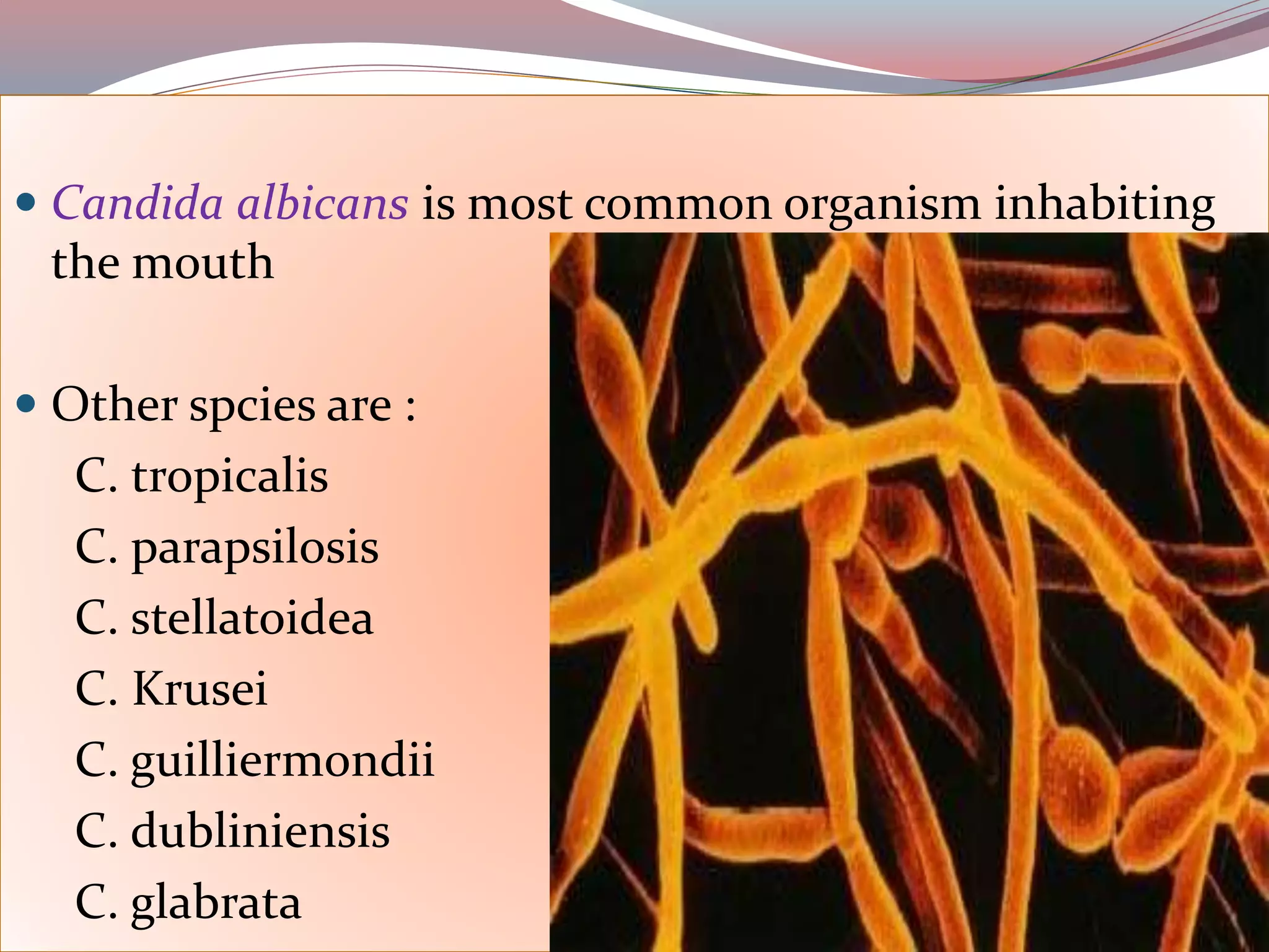
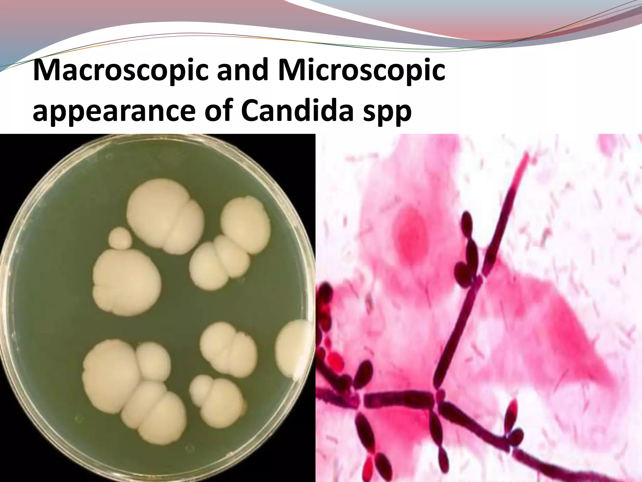
CANDIDIASIS
 Known as Candidosis, Moniliasis, Thrush
 ‘Candida’ white robes worn by the candidates for the Roman
Senate,
 ‘albicans’ (Latin word) to whiten.
 Thus ‘candida albicans’ literally translated can be defined as whitening white.
• Candidiasis refers to multiplicity of disease caused by yeast like fungus.
• Candida is the most common oral fungal infections in humans.
Introduction
 Candida albicans is most common organism inhabiting
the mouth
 Other spcies are :
C. tropicalis
C. parapsilosis
C. stellatoidea
C. Krusei
C. guilliermondii
C. dubliniensis
C. glabrata
CANDIDA:
-Normal flora
Exist in Mouth, Gastrointestinal tract
Vagina, skin in 20 % of normal Individuals
-Colonization increases with age, in pregnancy
-Important etiological agent presenting as opportunistic infection in
Diabetus and HIV patients
Macroscopic and Microscopic
appearance of Candida spp
FACTORS PRE-DISPOSING TO ORAL
CANDIDIASIS :-
Systemic factors :-
Physiologic:
Old age, Infancy, Pregnancy .
Endocrine:
Diabetes mellitus, hypothyroidism .
Nutritional Deficiencies :
Iron , Folate or vitamin B12 deficiency.
Malignancies :
Acute leukemia , agranulocytosis.
Immune defects , Immunosuppression , AIDS , Thymic
Aplasia , Corticosteroids
Local Factors:
• Xerostomia
-Sjogrens Syndrome, irradiation, drug therapy.
• Broad spectrum Antibiotics.
• Corticosteroids
• High carbohydrate diet
• Dentures
-change in environmental conditions, trauma, denture
usage, denture cleanliness.
• Smoking Tobacco
Classification
1. Acute Candidiasis
- Pseudomembranous type
- Atrophic type
2. Chronic Candidiasis
- Atrophic type
- Hypertrophic type
- Candida-associated angular chelitis
3. Systemic Candidiasis
- Candidal endocarditis
- Candidal meningitis
- Candidal septicaemia
4. Mucocutaneous Candidiasis
- Localised type
- Familial type
- Syndrome associated candidiasis
ACUTE PSEUDOMEMBRANEOUS CANDIDIASIS
(THRUSH )
 Best recognized form of candidiasis
 Development of white plaques
- Cottage Cheese or Curdled Milk resemblance
- composed of tangled masses of hyphae, yeasts,
desquamated epithelial cells and debris
- underlying mucosa appears normal or erythematous
 Occurs characteristically on
buccal mucosa, palate and
dorsal tongue
 Usually asymptomatic
 Patients may complains
burning sensation of mucosa or
unpleasant taste in mouth
 Oral thrush commonly occurs
among children, debilitated
elderly persons and AIDS
patients.
- Can be scraped off with tongue blade or by dry gauze
sponge
- May be initiated by broad spectrum antibiotics or
immune dysfunction
HISTOPATHOLOGY
 Hyperplastic epithelium with superficial necrotic and
desquamating parakeranitized layer.
 Hyperplastic epithelium is infiltrated by cadidal hyphae
and yeast cells along withPMN.
 Often there is separation between the superficial
pseudomembrane and the deeper layers of epithelium.
 The candidal hyphae often appears as a weakly basophilic
thread like structure.
 Lamina propria is infiltrated by chronic inflammatory cells.
ACUTE ATROPHIC
CANDIDIASIS:
( Erythematous Candidiasis)
 Also called “antibiotic sore mouth”
 Follows long course of broad spectrum
antibiotics
 Patients complains of pain & burning
sensation of mucosa
 Diffuse loss of filliform papillae of the
dorsal tongue resulting in a reddened
bald appearance of tongue
HISTOPATHOLOGY
 Thin, atrophic, non- keratinized epithelium with
occasional presence of candidal hyphae.
 Chronic inflammatory cell infiltration is seen in the
epithelium as well as in the lamina propria.
 Histologically these lesions resemble ‘oral thrush’
without the pseudomembrane.
CHRONIC ATROPHIC
CANDIDIASIS:
 Candida associated lesions
 Also known as denture sore mouth
 Characterized by varying degrees of
diffuse erythema and edema in
denture bearing areas.
- Usually in maxillary prostheses
 more often seen among females
than males.
 Usually asymptomatic
 It is regarded as secondary candidal infection of oral
tissues modified by continuous wearing of ill-fighting
dentures and associated poor oral hygiene.
 Histologically , shows presence of candidal hyphae
penetrating the keratin layer of host epithelium
ANGULAR CHEILITIS: -
 Also called Perleche
 Characterized by erythema,
fissuring and scaling of corners of
mouth
 Typically occurs along with
multifocal candidiasis
 It occurs at the angle of the mouth among persons
having deep commissural folds secondary to over closure
of mouth.
 It can also occur among persons with lip-licking habits,
denture wearing, or deficiency of riboflavin, vit B-12 and
folic acid deficiency,etc.
 Infection can be due to C.Albicans or Combined effect of
C.albicans + Staphylococcus Aureus
 Infrequently involves perioral skin , secondary to action
keeping skin moist ( in case of chronic lip licking ,
thumb sucking) – can be known as Cheilocandidiasis
CHRONIC HYPERPLASTIC
CANDIDIASIS
 Least common of all types
 Non scrapable
 It appears as a slightly elevated, indurated, persistent, white
plaque or patch on the oral mucosa that often resembles oral
leukoplakia. (candidal leukoplakia)
 The lesions could be bilateral and are mostly seen on the buccal
mucosa near the commissure.
 Some lesions may also develop over the tongue or the palate, etc.
 Homogeneous or speckled (nodular)
 Development of chronic hyperplastic candidiasis is often
favoured by certain conditions like smoking, denture wearing
and occlusal frictions.
 Histopathologically shows increased frequency of epithelial
dysplasia
 Diagnosis confirmed by demonstration of candidal hyphae
within the lesion and resolution of lesion after antifungal
therapy
HISTOPATHOLOGY
 Hyperplastic, acanthotic epithelium with
parakeratosis.
 Intercellular edema and PMN infitration sometimes
causing separation between different layers of the
epithelium.
 Microabscess formation in some cases.
 Atrophy of the epithelium with loss of keratin in the
clinically erythematous areas.
 Candidal hyphae invading the parakeratinized layer at
right angles to the surface.
.
 Epithelial displasia may be
present in some cases.
 Chronic inflammatory cell
infiltration in the underlying
connective tissue.
 PAS-stained sections best
demonstrate the presence of
candidal hyphae in the tissue.
Localised Mucocutaneous
Candidiasis
 This is characterised by
long standing and
persistent candidal
infections in the oral
cavity, skin, nails and
vaginal mucosa,etc.
Familial Mucocutaneous Candidiasis
 It is believed to be transmitted genetically as
autosomal recessive trait and most of the patients are
mildly affected.
 A traid of mucocutaneous candidiasis, thymoma and
myositis has been reported.
Candidiasis Endocrinopathy Syndrome
 Transmitted as autosomal recessive trait.
 Chronic oral candidiasis occuring mostly in the second
decade of life.
 Hypoparathyroidism, Addison’s disease, diabetes
mellitus and hypothyroidism.
Syndrome –associated candidiasis
 Severe candidiasis (both acute and chronic variety) are
well recognised opportunistic infections in
immunosuppressed patients, particularly those
suffering from AIDS.
 Depressed cell-mediated immunity is believed to be
the cause for the development of these lesions.
SYSTEMIC CANDIDIASIS
Candidal Endocarditis
 Patients who have undergone prosthetic heart valve
replacements and those who are using long time venous
catheters are at risk for developing candidal endocarditis.
 Clinically the patient often develops fever, dyspnoea,
edema and congestive cardiac failure, etc.
 Candidal growth in the valve may result in the
development of major venous embolism.
Candidal Meningitis
 Spread of candidal organisms into the brain results in
meningitis, which could be a consequence of oral
candidiasis and in such cases, the organism scan be
detected from the CSF.
 Patients often develop fever, headache, stiffness in the
body and hemiplegia.
 The condition is often fatal.
Candidal Septicaemia
 It occurs due to disseminated spread of candidal
organisms throughout the body and it can be
secondary to severe oral or oropharyngeal candidiasis.
 Clinically the patient often develops fever, chill,
nausea, vomiting, shock and coma, etc.
 The condition can be fatal if not treated in time.
Differential Diagnosis
 Chemical burns
 Mucous patch of syphilis
 Traumatic ulcers
 Leukoplakia
 Lichen planus
 Laborarory
 Saliva culture
 Biopsy and staining with PAS or Gomori methenamine
silver stain
 Serodiagnosis
 Imaging
 CT , or MRI to evaluate the submucosal extent
Investigation
 Other
 CBC with differential (eg, to evaluate for neutropenia or
lymphopenia)
 Immunologic testing
 Metabolic panel (eg, to evaluate for diabetes or malnutrition)
 HIV serostatus testing
Treatment
 Tropical and systemic administration of nystat is done
in conventional cases.
 In immunosuppressedpatients, systemic
administration of amphotericin-B and fluconazole
may be necessary.
 Removal of primary etiological factors and
improvement of oral hygiene is essential.
EXFOLIATIVE CYTOLOGY: -
 Candidal hyphae can also be demonstrated by exfoliative
cytology by PAS stain
 Hyphae are stained magenta color by PAS stain
RAPID DIAGNOSTIC TEST: - Gram’s staining
 10% - 20% KOH preparation used for rapid diagnosis
 KOH lyses background of epithelial cells allowing yeast
and hyphae to be seen
Easier Identification
Germ tube test
- Identifies C.albicans from other Candida species
- Majority of Diagnostic laboratories depend on this test
Emerging Methods for detection of
Candida Infections
 Molecular Methods
 PCR

Candidiasis.pptx

  • 1.
  • 2.
     Known asCandidosis, Moniliasis, Thrush  ‘Candida’ white robes worn by the candidates for the Roman Senate,  ‘albicans’ (Latin word) to whiten.  Thus ‘candida albicans’ literally translated can be defined as whitening white. • Candidiasis refers to multiplicity of disease caused by yeast like fungus. • Candida is the most common oral fungal infections in humans. Introduction
  • 3.
     Candida albicansis most common organism inhabiting the mouth  Other spcies are : C. tropicalis C. parapsilosis C. stellatoidea C. Krusei C. guilliermondii C. dubliniensis C. glabrata
  • 4.
    CANDIDA: -Normal flora Exist inMouth, Gastrointestinal tract Vagina, skin in 20 % of normal Individuals -Colonization increases with age, in pregnancy -Important etiological agent presenting as opportunistic infection in Diabetus and HIV patients
  • 5.
  • 6.
    FACTORS PRE-DISPOSING TOORAL CANDIDIASIS :- Systemic factors :- Physiologic: Old age, Infancy, Pregnancy . Endocrine: Diabetes mellitus, hypothyroidism . Nutritional Deficiencies : Iron , Folate or vitamin B12 deficiency. Malignancies : Acute leukemia , agranulocytosis. Immune defects , Immunosuppression , AIDS , Thymic Aplasia , Corticosteroids
  • 7.
    Local Factors: • Xerostomia -SjogrensSyndrome, irradiation, drug therapy. • Broad spectrum Antibiotics. • Corticosteroids • High carbohydrate diet • Dentures -change in environmental conditions, trauma, denture usage, denture cleanliness. • Smoking Tobacco
  • 8.
    Classification 1. Acute Candidiasis -Pseudomembranous type - Atrophic type 2. Chronic Candidiasis - Atrophic type - Hypertrophic type - Candida-associated angular chelitis 3. Systemic Candidiasis - Candidal endocarditis - Candidal meningitis - Candidal septicaemia 4. Mucocutaneous Candidiasis - Localised type - Familial type - Syndrome associated candidiasis
  • 9.
    ACUTE PSEUDOMEMBRANEOUS CANDIDIASIS (THRUSH)  Best recognized form of candidiasis  Development of white plaques - Cottage Cheese or Curdled Milk resemblance - composed of tangled masses of hyphae, yeasts, desquamated epithelial cells and debris - underlying mucosa appears normal or erythematous
  • 10.
     Occurs characteristicallyon buccal mucosa, palate and dorsal tongue  Usually asymptomatic  Patients may complains burning sensation of mucosa or unpleasant taste in mouth  Oral thrush commonly occurs among children, debilitated elderly persons and AIDS patients.
  • 11.
    - Can bescraped off with tongue blade or by dry gauze sponge - May be initiated by broad spectrum antibiotics or immune dysfunction
  • 12.
    HISTOPATHOLOGY  Hyperplastic epitheliumwith superficial necrotic and desquamating parakeranitized layer.  Hyperplastic epithelium is infiltrated by cadidal hyphae and yeast cells along withPMN.  Often there is separation between the superficial pseudomembrane and the deeper layers of epithelium.  The candidal hyphae often appears as a weakly basophilic thread like structure.  Lamina propria is infiltrated by chronic inflammatory cells.
  • 14.
    ACUTE ATROPHIC CANDIDIASIS: ( ErythematousCandidiasis)  Also called “antibiotic sore mouth”  Follows long course of broad spectrum antibiotics  Patients complains of pain & burning sensation of mucosa  Diffuse loss of filliform papillae of the dorsal tongue resulting in a reddened bald appearance of tongue
  • 15.
    HISTOPATHOLOGY  Thin, atrophic,non- keratinized epithelium with occasional presence of candidal hyphae.  Chronic inflammatory cell infiltration is seen in the epithelium as well as in the lamina propria.  Histologically these lesions resemble ‘oral thrush’ without the pseudomembrane.
  • 16.
    CHRONIC ATROPHIC CANDIDIASIS:  Candidaassociated lesions  Also known as denture sore mouth  Characterized by varying degrees of diffuse erythema and edema in denture bearing areas. - Usually in maxillary prostheses  more often seen among females than males.
  • 17.
     Usually asymptomatic It is regarded as secondary candidal infection of oral tissues modified by continuous wearing of ill-fighting dentures and associated poor oral hygiene.  Histologically , shows presence of candidal hyphae penetrating the keratin layer of host epithelium
  • 18.
    ANGULAR CHEILITIS: - Also called Perleche  Characterized by erythema, fissuring and scaling of corners of mouth  Typically occurs along with multifocal candidiasis
  • 19.
     It occursat the angle of the mouth among persons having deep commissural folds secondary to over closure of mouth.  It can also occur among persons with lip-licking habits, denture wearing, or deficiency of riboflavin, vit B-12 and folic acid deficiency,etc.  Infection can be due to C.Albicans or Combined effect of C.albicans + Staphylococcus Aureus  Infrequently involves perioral skin , secondary to action keeping skin moist ( in case of chronic lip licking , thumb sucking) – can be known as Cheilocandidiasis
  • 20.
    CHRONIC HYPERPLASTIC CANDIDIASIS  Leastcommon of all types  Non scrapable  It appears as a slightly elevated, indurated, persistent, white plaque or patch on the oral mucosa that often resembles oral leukoplakia. (candidal leukoplakia)  The lesions could be bilateral and are mostly seen on the buccal mucosa near the commissure.  Some lesions may also develop over the tongue or the palate, etc.
  • 21.
     Homogeneous orspeckled (nodular)  Development of chronic hyperplastic candidiasis is often favoured by certain conditions like smoking, denture wearing and occlusal frictions.  Histopathologically shows increased frequency of epithelial dysplasia  Diagnosis confirmed by demonstration of candidal hyphae within the lesion and resolution of lesion after antifungal therapy
  • 23.
    HISTOPATHOLOGY  Hyperplastic, acanthoticepithelium with parakeratosis.  Intercellular edema and PMN infitration sometimes causing separation between different layers of the epithelium.  Microabscess formation in some cases.  Atrophy of the epithelium with loss of keratin in the clinically erythematous areas.  Candidal hyphae invading the parakeratinized layer at right angles to the surface.
  • 24.
    .  Epithelial displasiamay be present in some cases.  Chronic inflammatory cell infiltration in the underlying connective tissue.  PAS-stained sections best demonstrate the presence of candidal hyphae in the tissue.
  • 25.
    Localised Mucocutaneous Candidiasis  Thisis characterised by long standing and persistent candidal infections in the oral cavity, skin, nails and vaginal mucosa,etc.
  • 26.
    Familial Mucocutaneous Candidiasis It is believed to be transmitted genetically as autosomal recessive trait and most of the patients are mildly affected.  A traid of mucocutaneous candidiasis, thymoma and myositis has been reported.
  • 27.
    Candidiasis Endocrinopathy Syndrome Transmitted as autosomal recessive trait.  Chronic oral candidiasis occuring mostly in the second decade of life.  Hypoparathyroidism, Addison’s disease, diabetes mellitus and hypothyroidism.
  • 28.
    Syndrome –associated candidiasis Severe candidiasis (both acute and chronic variety) are well recognised opportunistic infections in immunosuppressed patients, particularly those suffering from AIDS.  Depressed cell-mediated immunity is believed to be the cause for the development of these lesions.
  • 29.
  • 30.
    Candidal Endocarditis  Patientswho have undergone prosthetic heart valve replacements and those who are using long time venous catheters are at risk for developing candidal endocarditis.  Clinically the patient often develops fever, dyspnoea, edema and congestive cardiac failure, etc.  Candidal growth in the valve may result in the development of major venous embolism.
  • 31.
    Candidal Meningitis  Spreadof candidal organisms into the brain results in meningitis, which could be a consequence of oral candidiasis and in such cases, the organism scan be detected from the CSF.  Patients often develop fever, headache, stiffness in the body and hemiplegia.  The condition is often fatal.
  • 32.
    Candidal Septicaemia  Itoccurs due to disseminated spread of candidal organisms throughout the body and it can be secondary to severe oral or oropharyngeal candidiasis.  Clinically the patient often develops fever, chill, nausea, vomiting, shock and coma, etc.  The condition can be fatal if not treated in time.
  • 33.
    Differential Diagnosis  Chemicalburns  Mucous patch of syphilis  Traumatic ulcers  Leukoplakia  Lichen planus
  • 34.
     Laborarory  Salivaculture  Biopsy and staining with PAS or Gomori methenamine silver stain  Serodiagnosis  Imaging  CT , or MRI to evaluate the submucosal extent Investigation
  • 35.
     Other  CBCwith differential (eg, to evaluate for neutropenia or lymphopenia)  Immunologic testing  Metabolic panel (eg, to evaluate for diabetes or malnutrition)  HIV serostatus testing
  • 36.
    Treatment  Tropical andsystemic administration of nystat is done in conventional cases.  In immunosuppressedpatients, systemic administration of amphotericin-B and fluconazole may be necessary.  Removal of primary etiological factors and improvement of oral hygiene is essential.
  • 37.
    EXFOLIATIVE CYTOLOGY: - Candidal hyphae can also be demonstrated by exfoliative cytology by PAS stain  Hyphae are stained magenta color by PAS stain RAPID DIAGNOSTIC TEST: - Gram’s staining  10% - 20% KOH preparation used for rapid diagnosis  KOH lyses background of epithelial cells allowing yeast and hyphae to be seen
  • 38.
    Easier Identification Germ tubetest - Identifies C.albicans from other Candida species - Majority of Diagnostic laboratories depend on this test
  • 39.
    Emerging Methods fordetection of Candida Infections  Molecular Methods  PCR

Editor's Notes

  • #17 Note: Petechial haemorrhage can be seen iin denture areas
  • #35 periodic acid-Schiff (PAS), Gridley stain, or Gomori methenamine silver (GMS) stain is used
  • #36 periodic acid-Schiff (PAS), Gridley stain, or Gomori methenamine silver (GMS) stain is used