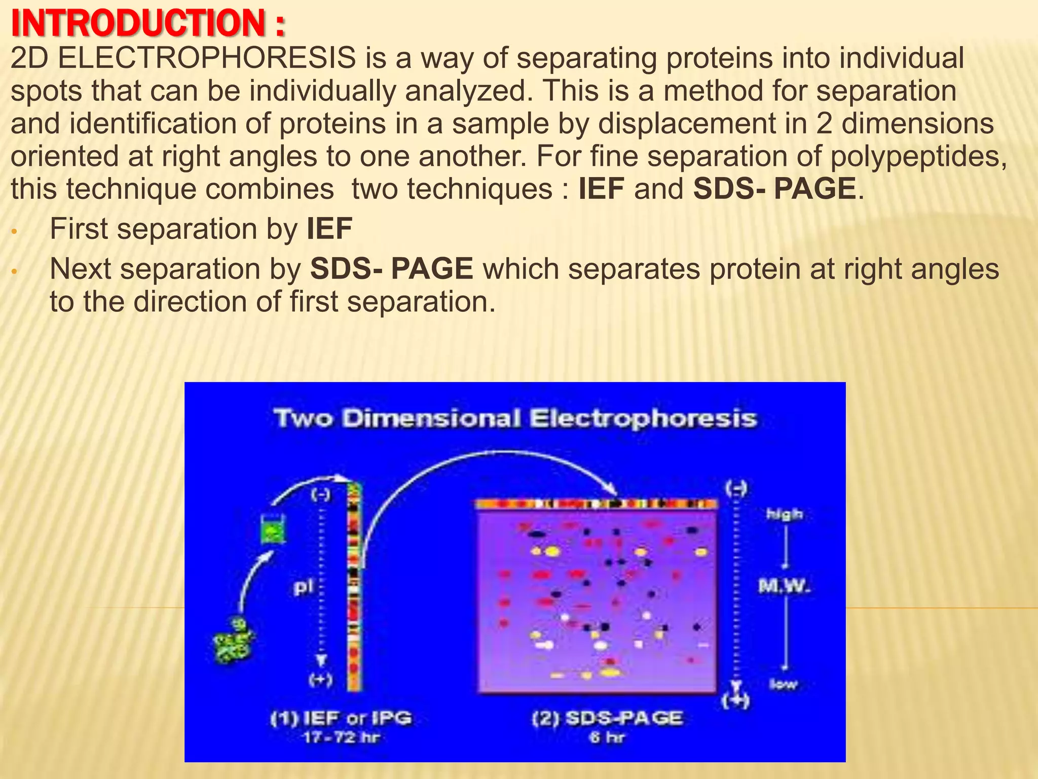
This document discusses electrophoresis, which separates molecules based on their charge and size. It describes the principles of electrophoresis, factors that affect separation, different support media used, techniques, detection methods, types of electrophoresis including zone electrophoresis and gel electrophoresis, applications, advantages, and disadvantages. Electrophoresis is used to analyze, identify, purify, and separate mixtures of charged biomolecules like proteins, nucleic acids, and other molecules.
This document discusses isoelectric focusing, a technique used to separate proteins based on their isoelectric point (PI). Proteins are subjected to an electric field within a pH gradient, which causes them to migrate to the point in the gradient where their net charge is zero (their PI). Different proteins have different PIs and will therefore migrate to distinct positions in the gel. Isoelectric focusing provides high resolution separation and is useful for research applications such as taxonomy, cytology and immunology.
This document discusses methods for separating proteins, focusing on electrophoresis techniques. It explains that proteins can be separated based on their size, charge, and binding properties to study each protein's individual structure and function. Electrophoresis methods separate proteins in an electric field based on factors like their molecular mass and charge. SDS-PAGE is described as denaturing proteins with SDS to give each one a uniform negative charge proportional to its size. Isoelectric focusing separates proteins based on their isoelectric point by using a pH gradient gel. Two-dimensional electrophoresis combines these methods to separate thousands of proteins based on both their isoelectric point and size.
Definition, factors affecting electrophoresis, classification of electrophoresis in general, Iso-electric focusing in detail, IEF and its types (based on ampholytes), step wise procedure of IEF process, Problems involved and their remedies, Capillary iso electric focusing and its types, detection of analytes explained in animation (so watch it in slide show mode), advantages and applications of CIEF.
This presentation contain the information about gel electrophoresis method , instruments & types.
Electrophoresis is a method through biological molecules are separated by applying an electric field.
Main purpose of this method is to determine the number , amount & mobility of biological component.
There are some internal & external factors that affects the process of electrophoresis.
The bio-molecules have charge on it & when we apply an electric field , the charge particles move to the opposite cathode. In this way, charge particles are separated
There are 3 types of gels that use in this process .
In this buffers are also used which provide ions that carry a current.
Capillary electrophoresis and application by Dr. Anurag YadavDr Anurag Yadav
Dr. Anurag Yadav presented on capillary electrophoresis. Capillary electrophoresis is a technique used to separate molecules like amino acids, peptides, proteins, DNA fragments, and drugs based on their charge and size. It involves applying a high voltage to a thin capillary filled with buffer, causing molecules to separate as they migrate through the capillary at different speeds. Detection is usually done through ultraviolet absorption, refractive index changes, or fluorescence near the end of the capillary. Capillary electrophoresis provides high resolution, requires only small sample volumes, and can separate a wide range of biomolecules.
Gel electrophoresis is a method used to separate macromolecules like DNA, RNA, and proteins based on their size and charge. It uses a gel as a medium for separation under an electric field. Smaller molecules move faster through the gel towards the positive electrode. The separated molecules can then be visualized and identified. Gel electrophoresis is more efficient at separation than paper electrophoresis and allows for separation of a wider range of molecule sizes.
This presentation provides an overview of electrophoresis techniques. It defines electrophoresis as a separation technique where solutes migrate through a conductive medium under an applied electric field. It describes how charges migrate based on their size and charge. It then discusses the different forms of electrophoresis, focusing on capillary zone electrophoresis and gel electrophoresis. For capillary electrophoresis, it explains the concepts of electrophoretic mobility, electroosmotic mobility, and their roles in solute migration. It also outlines the basic instrumentation and processes involved like injection, separation, and detection. For gel electrophoresis, it discusses how it separates biomolecules like DNA and proteins based on size and provides examples of its applications.
Electrophoresis is an analytical technique that separates and analyzes ionized analytes using their migration in an electric field. Charged molecules are separated as they migrate through a porous medium like agarose or polyacrylamide gel under the influence of an electric field generated between electrodes. The direction of migration depends on the charge, with positively charged ions or DNA fragments migrating toward the negative cathode and negatively charged molecules toward the positive anode. Different electrophoresis methods like agarose gel electrophoresis and polyacrylamide gel electrophoresis are used depending on the size of DNA fragments to be separated.
Gel electrophoresis is a technique used to separate macromolecules like proteins and nucleic acids based on size, charge, or conformation. It works by applying an electric field to push molecules through a gel containing small pores. Smaller molecules will travel farther through the gel than larger ones. After electrophoresis, the gel can be stained to make the separated molecules visible, then used for various applications like DNA fingerprinting in forensics, mapping cellular components in biochemistry, and determining purity in protein analysis.
Gel electrophoresis is a technique used to separate molecules like proteins and nucleic acids based on their size and charge. It works by applying an electric current to a gel, which causes negatively charged molecules to migrate toward the positive electrode at rates dependent on their size and charge. There are different types of gels used like agarose, polyacrylamide, and starch. Agarose is commonly used for separating DNA fragments, while polyacrylamide is used for separating smaller molecules like proteins and nucleic acids. The separated molecules can then be visualized by staining the gel. Gel electrophoresis is widely applied in areas like determining protein purity and molecular weights.
Technique combining ideas of isoelectric points and electric fields. It gives good separation with a high resolution compared to any other method. Proteins are separated in a pH gradient according to their isoelectric points, with proteins migrating towards the cathode or anode depending on whether their pI is below or above the pH at that point in the gradient. Applications include separation and identification of serum proteins in clinical settings and separation of proteins, enzymes, and other biomolecules in research.
Isoelectric focusing is a technique used to separate proteins based on their isoelectric point. It involves creating an immobilized pH gradient using carrier ampholytes within an acrylamide gel. When an electric current is applied, proteins will migrate within the gel until they reach the point where they carry no net charge and stop, allowing separation based on subtle differences in pI. The key steps are preparation of the IEF gel, addition of ampholytes to generate the pH gradient, running electrophoresis to allow protein migration, and staining to visualize the separated protein bands.
This document provides an overview of isoelectric focusing (IEF). IEF separates proteins in a gel according to their isoelectric point (pI), which is the pH at which a protein has no net charge. During IEF, proteins migrate through an immobilized pH gradient generated by ampholytes until they reach the pH that matches their pI and cease moving. IEF provides high resolution separation and is useful for research applications such as taxonomy, cytology, and immunology.
Two-dimensional gel electrophoresis (2D-GE) separates proteins based on two properties - isoelectric focusing (IEF) according to isoelectric point (pI) in the first dimension, and SDS-PAGE according to molecular weight in the second dimension. This allows for high resolution separation of complex protein mixtures. Key steps include sample preparation to solubilize and stabilize proteins, IEF to separate by pI, equilibration then SDS-PAGE to separate by size, and visualization techniques like autoradiography or mass spectrometry for analysis.
Electrophoresis is an electrokinetic process which separates charged particles in a fluid using a field of electrical charge. It is most often used in life sciences to separate protein molecules or DNA and can be achieved through several different procedures depending on the type and size of the molecules. The procedures differ in some ways but all need a source for the electrical charge, a support medium and a buffer solution. Electrophoresis is used in laboratories for the separation of molecules based on size, density and purity.An electric field is applied to molecules and as they are electrically charged themselves it results in a force acting upon them. The greater the charge of the molecule the greater the force applied by the electrical field and therefore the further through the support medium the molecule will move relative to its mass.
Some example applications of electrophoresis include DNA and RNA analysis as well as protein electrophoresis which is a medical procedure used to analyse and separate the molecules found in a fluid sample (most commonly blood and urine samples).Different types of gels are usually used as the support medium for electrophoresis and this may be in slab or tube form depending on which is more beneficial. Gel slabs enable many samples to be run simultaneously and so are frequently used in laboratories. However, tube gels give a better resolution of the results so are often chosen for protein electrophoresis.
Agarose gel is commonly used for electrophoresis of DNA. It has a large pore structure allowing larger molecules to move easily but it is not suitable for sequencing smaller molecules.
Polyacrylamide gel electrophoresis (PAGE) has a clearer resolution than agarose gel making it more suitable for quantitative analysis. This makes it possible to identify how proteins bind to DNA. It can also be used to develop an understanding of how bacteria is becoming resistant to antibiotics through plasmid analysis.
2D gel electrophoresis is a widely used technique in molecular biology and biochemistry to separate and analyze complex mixtures of proteins. It combines two dimensions of separation, isoelectric focusing (IEF), and sodium dodecyl sulfate polyacrylamide gel electrophoresis (SDS-PAGE), to achieve high-resolution separation of proteins based on their isoelectric point (pI) and molecular weight.
Here is a step-by-step overview of the 2D gel electrophoresis process:
1. Sample Preparation: The first step involves extracting proteins from the biological sample of interest. The sample can be a cell lysate, tissue extract, or any other protein-containing mixture. The proteins are typically solubilized and denatured using a lysis buffer containing detergents and denaturing agents.
2. Isoelectric Focusing (IEF): The next step is to perform the first dimension separation, which separates proteins based on their pI. In IEF, proteins are loaded onto an immobilized pH gradient (IPG) gel strip or a strip of carrier ampholytes with a pH gradient. An electric field is applied across the strip, causing the proteins to migrate toward their respective pI, where they become electrically neutral and stop moving. The separation occurs in a tube gel or a flat gel format.
3. Equilibration: After the completion of IEF, the IPG strip is equilibrated to prepare it for the second dimension separation. This involves treating the strip with reducing and alkylating agents to ensure proper SDS-PAGE separation and to prevent protein aggregation.
4. SDS-PAGE: In the second dimension, the equilibrated IPG strip is placed on top of an SDS-PAGE gel, which is typically a polyacrylamide gel with a concentration gradient. The proteins are separated based on their molecular weight as an electric field is applied across the gel. SDS, a detergent, denatures the proteins and imparts a negative charge to them, allowing for separation based on size. The proteins migrate through the gel, with smaller proteins moving faster and larger proteins moving more slowly.
5. Visualization and Analysis: After the electrophoresis run, the proteins are typically stained using specific dyes, such as Coomassie Brilliant Blue or silver stain, to visualize the protein bands. The gel can be scanned or photographed for documentation and further analysis. Advanced techniques like mass spectrometry can be used to identify individual proteins within the gel spots/bands.
Overall, 2D gel electrophoresis allows researchers to obtain a two-dimensional map of the protein composition within a sample, facilitating the detection of differences in protein expression, post-translational modifications, and protein-protein interactions. It has been a valuable tool in various fields, including proteomics, biomedical research, and biomarker discovery.
This document provides information about electrophoresis. It discusses different types of electrophoretic techniques including slab electrophoresis, capillary electrophoresis, capillary zone electrophoresis, capillary gel electrophoresis, capillary isotachophoresis, and micellar electrokinetic chromatography. It also covers principles, instrumentation, applications in areas like DNA analysis and vaccine analysis.
Electrophoresis is a technique used to separate charged particles like proteins by using an electric field to migrate them through a medium like filter paper or gel based on factors like their size and charge. It can be used to separate plasma proteins, lipoproteins, and immunoglobulins into distinct bands. The document discusses several electrophoresis techniques including paper, gel, SDS-PAGE, isoelectric focusing, and immunoelectrophoresis and how they are used to separate biomolecules.
Chromatofocusing is a protein separation technique that uses ion exchange resins and buffers with changing pH to separate proteins based on their isoelectric point. As the buffer pH passes through a protein's pI, the protein loses its charge and elutes from the resin. Chromatofocusing provides high resolution separation of proteins that have similar pI values. However, some proteins may aggregate at high concentrations and clog the resin. Isoelectric focusing uses immobilized pH gradients in gels to separate proteins based on their pI through electrophoresis. Two-dimensional electrophoresis separates proteins first by pI using isoelectric focusing, then by molecular weight using SDS-PAGE to provide high resolution separation and identification of
Electrophoresis is a technique used to separate charged particles such as proteins, nucleic acids, and other macromolecules. It works by applying an electric field to move these particles through a medium such as a gel or paper based on their size and charge. There are different types of electrophoresis including gel electrophoresis, paper electrophoresis, capillary electrophoresis, and isoelectric focusing which separates particles based on their isoelectric point. Electrophoresis has many applications in fields like forensics, molecular biology, genetics, and biochemistry to analyze proteins, DNA, RNA, and other biomolecules.
This document discusses capillary electrophoresis, a technique for separating charged molecules. It can separate proteins, peptides, amino acids, nucleic acids, and other molecules. Capillary electrophoresis works by applying an electric field across a thin capillary tube, which causes different molecules to migrate through the buffer at different rates based on their charge and size. It provides high separation efficiency using only small sample volumes. The document outlines the basic components and process of capillary electrophoresis.
Poly acrylamide gel electrophoresis (page)Dharmesh Gol
This document provides an overview of polyacrylamide gel electrophoresis (PAGE). It describes how PAGE uses an electric field to separate charged biomolecules like proteins and nucleic acids based on their size and charge. The document discusses the principles and instrumentation of electrophoresis. It explains how polyacrylamide gels are made and the procedures for PAGE. Different types of PAGE like SDS-PAGE and native PAGE are also summarized.
INTRODUCTION, DEFINATION OF ELECTROPHORESIS, ELECTROPHORESIS PRINCIPLE, TYPES OF ELECTROPHORESIS, FREE ELECTROPHORESIS, ZONE ELECTROPHORESIS,PAPER ELECTROPHORESIS, WORKING OF PAPER ELECTROPHORESIS, PROCEDURE FOR PAPER ELECTROPHORESIS, VISUALISATION, FACTORS AFFECTING SEPARATION OF MOLECULES, APPLICATIONS, working of paper electrophoresis ,procedure for paper electrophoresis ,visualisation ,factors affecting separation of molecules ,applications ,forensics ,dna fingerprinting ,molecular biology ,microbiology information about the organisms ,biochemistry mapping of cellular components ,paper electrophoresis is also used in study of sic ,hemoglobin abnormalities ,separation of blood clotting factors ,serum plasma proteins from blood sample ,used in separation and identification of alkaloids ,used for testing water samples ,toxicity of water ,drug industry to determine presence of illelgal drUGS
This document provides an overview of isoelectric focusing (IEF), a technique used to separate amphoteric molecules like proteins based on their isoelectric point (pI). IEF involves subjecting proteins to an electric field within a pH gradient formed in a polyacrylamide gel. Proteins will migrate within the gel until they reach the point where they are electrically neutral (pI) and stop migrating. Different proteins have different pIs and so become separated. Key steps include establishing a stable pH gradient using ampholytes, loading protein samples mixed with loading buffer, running the gel at increasing voltages, and then fixing and staining to visualize separated protein bands. IEF can also be used in the first dimension
This presentation consist of all information regarding sodium dodecyl sulphate PAGE. The general information regarding electrophoresis and its main types are also included in this presentation.
Electrophoresis-PAPER ELECTROPHORESIS,GEL ELCTROPHORESIS, PAGE-SDS AND NON-SDSAmrutha Hari
The document discusses different electrophoresis techniques including paper electrophoresis, gel electrophoresis, polyacrylamide gel electrophoresis (PAGE), sodium dodecyl sulfate–polyacrylamide gel electrophoresis (SDS-PAGE), and non-SDS PAGE. It describes the basic principles, apparatus, procedures, applications and advantages/disadvantages of each technique. Paper electrophoresis is a simple method that uses filter paper as the support medium while gel electrophoresis uses gels like agarose or polyacrylamide as the support medium. SDS-PAGE and PAGE are variations that separate molecules based on their molecular weight or size.
This document provides an overview of electrophoresis techniques. It defines electrophoresis as the migration of charged particles in an electric field, which allows for separation based on differences in charge-to-size ratios. Several electrophoresis methods are described, including capillary electrophoresis, paper electrophoresis, gel electrophoresis, and SDS-PAGE. Capillary electrophoresis provides high-speed separation of small sample volumes, while SDS-PAGE is used to determine protein molecular weights. Electrophoresis has applications in biochemistry, clinical analysis, environmental analysis and other fields.
Electrophoresis is a method used to separate charged molecules such as proteins and nucleic acids. It works by applying an electric field to encourage the migration of molecules towards the positively or negatively charged electrode, depending on their own charge. The document discusses the principle of electrophoresis, different types such as paper and zone electrophoresis, and factors that affect separation like charge, size, electric field strength, and buffer composition. It also outlines some applications in clinical testing, forensics, and environmental analysis.
This document summarizes techniques for exploring and analyzing proteins, including concentrating purified proteins using lyophilization or ultrafiltration, separating proteins using electrophoresis or mass spectrometry, and identifying proteins using mass spectrometry. Electrophoresis techniques like SDS-PAGE and 2D gels separate proteins based on size and charge, allowing visualization and quantification of purified proteins. Mass spectrometry further identifies proteins by correlating detected ion masses with known protein standards. These techniques provide a quantitative evaluation of protein purification schemes.
The document provides answers to questions about various topics in biochemistry and molecular biology. It discusses:
1) How the charge distribution of a protein changes between pH 0-14.
2) The differences between differential, rate zonal, and isopycnic centrifugation methods.
3) The advantages of a stacking gel and the principles of 2D gel electrophoresis and staining methods.
4) Features of ESI and MALDI-TOF mass spectrometry and how it can be used to analyze phosphorylated proteins.
5) How to further purify a protein of interest using gel filtration chromatography after ion exchange chromatography.
6) The principles of CD spectroscopy and the
Electrophoresis is a technique used to separate charged molecules like proteins and nucleic acids based on their size and charge. It involves applying an electric field to move molecules through a medium like agarose gel or capillary. Shorter/less charged molecules move faster and are separated from longer/more charged molecules. It is used in laboratories and clinical settings to analyze biological samples and diagnose conditions.
Thank you for the detailed presentation on electrophoresis. I appreciate you taking the time to explain the key concepts and techniques. Please let me know if you have any other questions.
Gel electrophoresis is a method to separate biomolecules like proteins, nucleic acids, and lipids based on their charge and size. During gel electrophoresis, an electric current is applied across a gel, causing negatively charged molecules to migrate toward the positive electrode and positively charged molecules to migrate toward the negative electrode. Smaller molecules migrate faster through the gel than larger molecules. Factors like the charge, size, and shape of molecules, as well as the electric current, gel composition, and buffer used, determine how far each type of molecule will migrate through the gel. Gel electrophoresis has applications in separating DNA, RNA, proteins, and other biomolecules.
Gel electrophoresis is a method to separate biomolecules like DNA, RNA, and proteins based on their size and charge. During gel electrophoresis, charged molecules are placed in wells in a gel and an electric current is applied, causing them to migrate through the gel at different rates depending on their size and charge. Larger molecules migrate more slowly through the gel pores than smaller molecules. This allows separation of molecules by size. Common gels used include agarose and polyacrylamide. Samples can be visualized after electrophoresis using dyes like ethidium bromide or stains. Gel electrophoresis has applications in DNA sequencing, forensic analysis, and medical research.
This document discusses various types of electrophoresis techniques. It begins by explaining that electrophoresis involves charged molecules like proteins and nucleic acids migrating in response to an electrical field. It then discusses different gel types like agarose and polyacrylamide gels that are used as supporting matrices. It also covers techniques like SDS-PAGE, isoelectric focusing, two-dimensional gel electrophoresis, and capillary electrophoresis. The document provides details on how each technique separates molecules based on properties like size, charge, and isoelectric point.
Electrophoresis is a technique used to separate charged molecules such as proteins and nucleic acids. It works by applying an electric field to move these molecules through a buffer or gel based on their size and charge. There are several types of electrophoresis including gel electrophoresis, which separates molecules based on size using a gel matrix, and capillary electrophoresis, which uses narrow capillaries to achieve high resolution separations. Electrophoresis has many applications in areas like biochemistry, genetics, and medicine.
it is a short ppt. on Electrophoresis. Which gives it's defination, Types of electrophoresis ; Like:- Zone electrophoresis, Isoelectric focussing, Immunoelectrophoresis. and their images.
This document discusses two electrophoresis techniques: moving boundary electrophoresis and isoelectric focusing electrophoresis. Moving boundary electrophoresis separates charged molecules in a free solution without a supporting medium by measuring the speed of the boundary between solutions under an applied electric current. Isoelectric focusing electrophoresis separates molecules based on their isoelectric point, which is the pH where a molecule has no net charge, by migrating molecules through a pH gradient gel until they reach the pH matching their isoelectric point. The document covers the principles, instrumentation, factors affecting separation, advantages, disadvantages and applications of these two electrophoresis techniques.
It contains 2-3 acetyl groups per glucose unit and its adsorption capacity is less than that of paper.
It gives sharper bands.
Provides a good background for staining glycoproteins.
ADVANTAGE:
No tailing of proteins or hydrophilic materials.
Available in wide range of particle size and layer thickness.
Give sharp bands and offer good resolution.
High voltage can be applied which will enhance the resolution.
This document summarizes the process of 2-D gel electrophoresis used to separate proteins. It begins with an introduction explaining that 2-D gel electrophoresis couples isoelectric focusing in the first dimension based on isoelectric point and SDS-PAGE in the second dimension based on molecular mass. The document then covers the principles, processes, and techniques involved in isoelectric focusing, SDS-PAGE, blotting, and concludes by discussing the applications and references for 2-D gel electrophoresis.
BIOTRANSFORMATION OF NON-STEROID COMPOUNDS.pptxdrpvczback
BIOTRANSFORMATION OF NON-STEROID COMPOUNDS
BIOTRANSFORMATION
Oxidation
Reduction
Hydrolysis
Isomerization
Condensation
Formation of new carbon bond
Introduction of functional group
Agarose Gel Electrophoresis
Estimate the size of molecules
Agarose in AGE
Gel Loading Buffer
Nucleic Acid Stain
Factor affecting mobility of DNA
Factor affecting Resolution
Smearing
Atomic absorption spectrometry is a technique used to determine the concentration of chemical elements in solution. It works by vaporizing the elements in a flame or graphite furnace and measuring how much light of a specific wavelength is absorbed, which indicates the concentration. Key components of an atomic absorption spectrometer include a light source, atomizer such as a flame or furnace, monochromator, detector, and display. Flame atomic absorption is used for higher concentrations while graphite furnace atomic absorption can detect trace levels. Potential interferences must also be considered and addressed.
Spectrofluorometry
what cause fluorescence
Quantification
Instrumentation
Effect of Solvent, Temperature and pH
Application
Energy change of excited molecules Molecular Spectra
Fate of Excited State
LASER
Colorimeter and Spectrophotometer
Electromagnetic Radiation
EMR
THE ELECTROMAGNETIC SPECTRUM
Interaction of e.m.r. with Matter
Molecular Spectra
Spectrophotometry
Principles OF Spectrophotometry
Introduction
Types of Transcription
factors involves in different Polymerase initiation complex
Structure of transcription factor
Role of transcription factor
Significance
RNA transport
Multiple classes of RNA are exported from the nucleus
Transportation through nuclear pore complex.
Ribosomal subunits are assembled in the nucleolus and exported by exportin 1
tRNAs are exported by a dedicated exportin
Messenger RNAs are exported from the nucleus as RNA-protein complexes
hnRNPs move from sites of processing to NPCs
Precursors to microRNAs are exported from the nucleus and processed in the cytoplasm
RNA Editing
Discovery of RNA Editing in Trypanosome Mitochondria
real functional genes
RNA EDITING IN KINETOPLAST OF TRYPANOSOMES.
Guide RNA (gRNA)
Guide RNAs Direct Editing in Trypanosomes
Editing is catalyzed by a multiprotein complex
Other Systems with RNA Editing
RECOMBINATION MECHANISM
PROKARYOTIC AND EUKARYOTIC CELLS
RECOMBINATION
MITOTIC AND MEIOTIC RECOMBINATION
CLASSES OF RECOMBINATION
HOMOLOGOUS RECOMBINATION
DOUBLE-STRAND BREAK MODEL
DNA RECOMBINATION
Recombination
Breaking and rejoining of two parental DNA molecules to produce new DNA molecules
Types of recombination
Definition of recombination
Gene Conversion – Characteristics
Holliday model
Holliday junction cleavage
This document discusses protein targeting in eukaryotic cells. It explains that each organelle has a distinct set of proteins that allow it to perform specific functions. There is a complex system that moves newly synthesized proteins from the site of synthesis to their proper destination. Proteins contain targeting sequences that direct them to the correct organelle, such as receptors to the plasma membrane or DNA polymerase to the nucleus. Targeting can occur co-translationally as the protein is synthesized on ER-bound ribosomes or post-translationally after cytosolic synthesis. The targeting sequences help distinguish the destination but are sometimes cleaved off later.
The document provides information about protein synthesis and processing. It begins with an overview of the topics to be covered, including ribosome formation, initiation and elongation factors, termination, the genetic code, tRNA aminoacylation, aminoacyl-tRNA synthetases, translational proofreading, inhibitors, and post-translational modifications. It then discusses the machinery of protein synthesis, including transcription, the genetic code, RNA, tRNA identity, aminoacyl-tRNA synthetases, aminoacylation of tRNA, and the ribosome. The mechanisms of initiation, elongation, and termination are explained in detail.
BIOSYNTHESIS OF PYRIMIDINES
SALVAGE PATHWAY
DE NOVO PATHWAY
SYNTHESIS OF OTHER PYRIMIDINE NUCLEOTIDES
IMPORTANCE
Essential building blocks of nucleic acids
Biologically very important heterocycles
Used in anti-biotics, used as anti-bacterial and anti-fungal also
Derivatives of pyrimidine also possess good anti-viral properties
MECHANISM OF TRANSCRIPTION prashant.pptxdrpvczback
MECHANISM OF TRANSCRIPTION
Transcription is the first step of gene expression, in which a particular segment of DNA is copied into RNA by the enzyme RNA polymerase. During transcription, a DNA sequence is read by RNA polymerase, which produces a complementary RNA strand.
Professional air quality monitoring systems provide immediate, on-site data for analysis, compliance, and decision-making.
Monitor common gases, weather parameters, particulates.
ESR spectroscopy in liquid food and beverages.pptxPRIYANKA PATEL
With increasing population, people need to rely on packaged food stuffs. Packaging of food materials requires the preservation of food. There are various methods for the treatment of food to preserve them and irradiation treatment of food is one of them. It is the most common and the most harmless method for the food preservation as it does not alter the necessary micronutrients of food materials. Although irradiated food doesn’t cause any harm to the human health but still the quality assessment of food is required to provide consumers with necessary information about the food. ESR spectroscopy is the most sophisticated way to investigate the quality of the food and the free radicals induced during the processing of the food. ESR spin trapping technique is useful for the detection of highly unstable radicals in the food. The antioxidant capability of liquid food and beverages in mainly performed by spin trapping technique.
The binding of cosmological structures by massless topological defectsSérgio Sacani
Assuming spherical symmetry and weak field, it is shown that if one solves the Poisson equation or the Einstein field
equations sourced by a topological defect, i.e. a singularity of a very specific form, the result is a localized gravitational
field capable of driving flat rotation (i.e. Keplerian circular orbits at a constant speed for all radii) of test masses on a thin
spherical shell without any underlying mass. Moreover, a large-scale structure which exploits this solution by assembling
concentrically a number of such topological defects can establish a flat stellar or galactic rotation curve, and can also deflect
light in the same manner as an equipotential (isothermal) sphere. Thus, the need for dark matter or modified gravity theory is
mitigated, at least in part.
This presentation explores a brief idea about the structural and functional attributes of nucleotides, the structure and function of genetic materials along with the impact of UV rays and pH upon them.
Nucleophilic Addition of carbonyl compounds.pptxSSR02
Nucleophilic addition is the most important reaction of carbonyls. Not just aldehydes and ketones, but also carboxylic acid derivatives in general.
Carbonyls undergo addition reactions with a large range of nucleophiles.
Comparing the relative basicity of the nucleophile and the product is extremely helpful in determining how reversible the addition reaction is. Reactions with Grignards and hydrides are irreversible. Reactions with weak bases like halides and carboxylates generally don’t happen.
Electronic effects (inductive effects, electron donation) have a large impact on reactivity.
Large groups adjacent to the carbonyl will slow the rate of reaction.
Neutral nucleophiles can also add to carbonyls, although their additions are generally slower and more reversible. Acid catalysis is sometimes employed to increase the rate of addition.
Comparing Evolved Extractive Text Summary Scores of Bidirectional Encoder Rep...University of Maribor
Slides from:
11th International Conference on Electrical, Electronics and Computer Engineering (IcETRAN), Niš, 3-6 June 2024
Track: Artificial Intelligence
https://www.etran.rs/2024/en/home-english/
Deep Behavioral Phenotyping in Systems Neuroscience for Functional Atlasing a...Ana Luísa Pinho
Functional Magnetic Resonance Imaging (fMRI) provides means to characterize brain activations in response to behavior. However, cognitive neuroscience has been limited to group-level effects referring to the performance of specific tasks. To obtain the functional profile of elementary cognitive mechanisms, the combination of brain responses to many tasks is required. Yet, to date, both structural atlases and parcellation-based activations do not fully account for cognitive function and still present several limitations. Further, they do not adapt overall to individual characteristics. In this talk, I will give an account of deep-behavioral phenotyping strategies, namely data-driven methods in large task-fMRI datasets, to optimize functional brain-data collection and improve inference of effects-of-interest related to mental processes. Key to this approach is the employment of fast multi-functional paradigms rich on features that can be well parametrized and, consequently, facilitate the creation of psycho-physiological constructs to be modelled with imaging data. Particular emphasis will be given to music stimuli when studying high-order cognitive mechanisms, due to their ecological nature and quality to enable complex behavior compounded by discrete entities. I will also discuss how deep-behavioral phenotyping and individualized models applied to neuroimaging data can better account for the subject-specific organization of domain-general cognitive systems in the human brain. Finally, the accumulation of functional brain signatures brings the possibility to clarify relationships among tasks and create a univocal link between brain systems and mental functions through: (1) the development of ontologies proposing an organization of cognitive processes; and (2) brain-network taxonomies describing functional specialization. To this end, tools to improve commensurability in cognitive science are necessary, such as public repositories, ontology-based platforms and automated meta-analysis tools. I will thus discuss some brain-atlasing resources currently under development, and their applicability in cognitive as well as clinical neuroscience.
Remote Sensing and Computational, Evolutionary, Supercomputing, and Intellige...University of Maribor
Slides from talk:
Aleš Zamuda: Remote Sensing and Computational, Evolutionary, Supercomputing, and Intelligent Systems.
11th International Conference on Electrical, Electronics and Computer Engineering (IcETRAN), Niš, 3-6 June 2024
Inter-Society Networking Panel GRSS/MTT-S/CIS Panel Session: Promoting Connection and Cooperation
https://www.etran.rs/2024/en/home-english/
2. INTRODUCTION :
2D ELECTROPHORESIS is a way of separating proteins into individual
spots that can be individually analyzed. This is a method for separation
and identification of proteins in a sample by displacement in 2 dimensions
oriented at right angles to one another. For fine separation of polypeptides,
this technique combines two techniques : IEF and SDS- PAGE.
• First separation by IEF
• Next separation by SDS- PAGE which separates protein at right angles
to the direction of first separation.
3. PRINCIPLE :
1)ISO ELECTRIC FOCUSING:
Technique for separating proteins or
peptides, on the basis of their isoelectric
point (pI). IEF works because in an electric
field molecules in a pH gradient will
migrate towards their pI. The protein will
arrive at the point where the pH gradient is
equal to its pI. There, being uncharged, it
will stop migrating.
2)SDS- PAGE:
Sodium dodecyl sulfate (SDS) is used to
linearize proteins and to negatively charge
the proteins. The binding of SDS to the
polypeptide chain imparts an even
distribution of charge per unit mass. As a
result, negatively charged proteins will
migrate towards the positive electrode and
will be fractionated by approximate size
during electrophoresis. This procedure is
called SDS-PAGE.
6. 1)Sample Preparation
2)IEF: IEF uses a mixture of
ampholytes, when an electric field is
applied the ampholytes move to a
position in the gel where their net charge
is zero and in the process they set up a
pH gradient. Proteins also move down
the pH gradient until they reach a pH
where they have no net charge, their
isoelectric point.
3)SDS-PAGE: Separates protein which
migrates through a medium when
subjected to an electric field from anode
to cathode terminal. Molecules flow at
different rates depending upon the
molecular size of the protein. SDS-
coated large proteins migrate slowly
through the gel matrix and small proteins
migrate quickly through the matrix. The
nearer the band to the well, the larger
the molecular size of the protein.
4)Analysis:
Spot analysis
Mass spectrometry
PROCEDURE:
7. BIOLOGICAL APPLICATIONS:
The analysis involves the systematic
separation, identification, and quantitation of
many proteins simultaneously from a single
sample.
It has the ability to detect post- translational
modifications.
On the basis of above applications we can use
2D-ELECTROPHORESIS for the fulfilment of -
cell differentiation, detection of disease
markers, therapy monitoring, drug discovery,
cancer research, purity checks.
REFERENCE:
www.boundless.com/textbooks