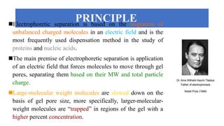
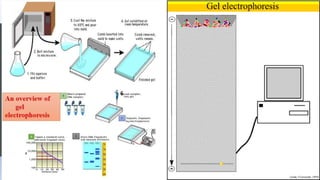
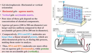
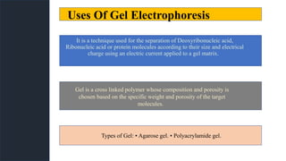
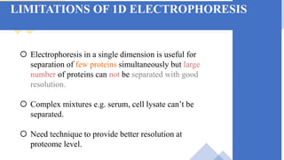
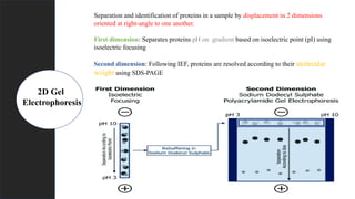
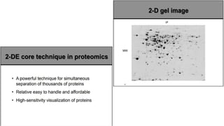
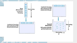
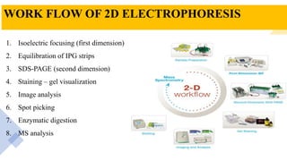
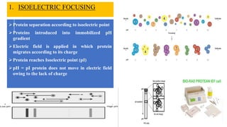
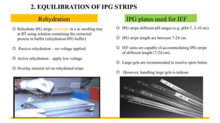
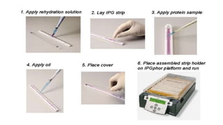
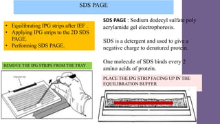
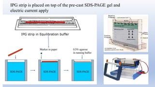
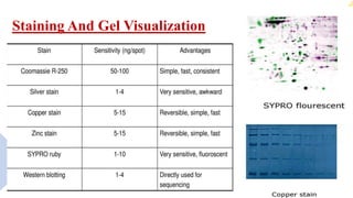
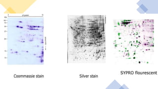
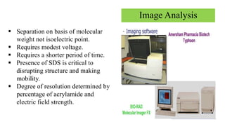
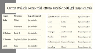
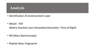
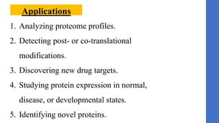
2 D GEL ELECTROPHORESIS
Presented by:
ISHWAR PATIL
REGULATORY TOXICOLOGY
M.S.PHARM/2022-24/RT/10
LEARNING OBJECTIVES
• Understanding of gel electrophoresis and its uses.
• Types of gel electrophoresis
• Limitations of gel electrophoresis.
• Understanding of 2D gel electrophoresis
• Steps involved in 2D gel electrophoresis
• Its applications.
2D GEL
ELECTROPHORESIS
PRINCIPLE
Electrophoretic separation is based on the migration of
unbalanced charged molecules in an electric field and is the
most frequently used dispensation method in the study of
proteins and nucleic acids.
The main premise of electrophoretic separation is application
of an electric field that forces molecules to move through gel
pores, separating them based on their MW and total particle
charge.
Large-molecular weight molecules are slowed down on the
basis of gel pore size, more specifically, larger-molecular-
weight molecules are “trapped” in regions of the gel with a
higher percent concentration.
• Gel electrophoresis -Horizontal or vertical
orientation
• Horizontal gels - agarose matrix.
• Vertical gels -acrylamide matrix.
• Pore sizes of these gels depend on the
concentration of chemical components.
• Agarose gel pores (100 to 500 nm diameter) are
larger and less uniform compared to that of
acrylamide gel pores (10 to 200 nm in diameter).
• Comparatively, DNA and RNA molecules are
larger than a linear strand of protein, which are
often denatured prior to, or during this process,
making them easier to analyze.
• Thus, DNA and RNA molecules are more often
run on agarose gels (horizontally), while proteins
are run on acrylamide gels (vertically).
Uses Of Gel Electrophoresis
It is a technique used for the separation of Deoxyribonucleic acid,
Ribonucleic acid or protein molecules according to their size and electrical
charge using an electric current applied to a gel matrix.
Gel is a cross linked polymer whose composition and porosity is
chosen based on the specific weight and porosity of the target
molecules.
Types of Gel: ▪ Agarose gel. ▪ Polyacrylamide gel.
 Electrophoresis in a single dimension is useful for
separation of few proteins simultaneously but large
number of proteins can not be separated with good
resolution.
 Complex mixtures e.g. serum, cell lysate can’t be
separated.
 Need technique to provide better resolution at
proteome level.
LIMITATIONS OF 1D ELECTROPHORESIS
Separation and identification of proteins in a sample by displacement in 2 dimensions
oriented at right-angle to one another.
First dimension: Separates proteins pH on gradient based on isoelectric point (pI) using
isoelectric focusing
Second dimension: Following IEF, proteins are resolved according to their molecular
weight using SDS-PAGE
2D Gel
Electrophoresis
WORK FLOW OF 2D ELECTROPHORESIS
1. Isoelectric focusing (first dimension)
2. Equilibration of IPG strips
3. SDS-PAGE (second dimension)
4. Staining – gel visualization
5. Image analysis
6. Spot picking
7. Enzymatic digestion
8. MS analysis
Protein separation according to isoelectric point
Proteins introduced into immobilized pH
gradient
Electric field is applied in which protein
migrates according to its charge
Protein reaches Isoelectric point (pI)
pH = pI protein does not move in electric field
owing to the lack of charge
1. ISOELECTRIC FOCUSING
Rehydration
 Rehydrate IPG strips overnight in a re swelling tray
at RT using solution containing the extracted
protein in buffer (rehydration/IPG buffer)
 Passive rehydration – no voltage applied
 Active rehydration – apply low voltage
 Overlay mineral oil on rehydrated strips
IPG plates used for IEF
 IPG strips different pH ranges (e.g. pH4-7, 3-10 etc)
 IPG strips length are between 7-24 cm.
 IEF units are capable of accommodating IPG strips
of different length (7-24 cm).
 Large gels are recommended to resolve spots better.
 However, handling large gels is tedious
2. EQUILIBRATION OF IPG STRIPS
SDS PAGE
• Equilibrating IPG strips after IEF .
• Applying IPG strips to the 2D SDS
PAGE.
• Performing SDS PAGE.
REMOVE THE IPG STRIPS FROM THE TRAY
SDS PAGE : Sodium dodecyl sulfate poly
acrylamide gel electrophoresis.
SDS is a detergent and used to give a
negative charge to denatured protein.
One molecule of SDS binds every 2
amino acids of protein.
PLACE THE IPG STRIP FACING UP IN THE
EQUILIBRATION BUFFER
IPG strip is placed on top of the pre-cast SDS-PAGE gel and
electric current apply
Staining And Gel Visualization
Image Analysis
 Separation on basis of molecular
weight not isoelectric point.
 Requires modest voltage.
 Requires a shorter period of time.
 Presence of SDS is critical to
disrupting structure and making
mobility.
 Degree of resolution determined by
percentage of acrylamide and
electric field strength.
Analysis
• Identification of eluted proteins spot
• MALDI - TOF
(Matrix Assisted Laser Desorption/Ionization -Time of flight)
• MS (Mass Spectroscopy)
• Peptide Mass Fingerprint
Applications
1. Analyzing proteome profiles.
2. Detecting post- or co-translational
modifications.
3. Discovering new drug targets.
4. Studying protein expression in normal,
disease, or developmental states.
5. Identifying novel proteins.
 A. Drabik and A. Bodzon-Kułakowska ; J. Silberring ; GEL
ELECTROPHORESIS; Proteomic Profiling and Analytical
Chemistry; Proteomic Profiling and Analytical Chemistry.
http://dx.doi.org/10.1016/B978-0-444-63688-1.00007-0 2016
Elsevier
 Sameh Magdeldin , Shymaa Enany , Yutaka Yoshida , Bo Xu ,
Ying Zhang , Zam Zureena , Ilambarthi Lokamani , Eishin
Yaoita and Tadashi Yamamoto; Basics and recent advances of
two dimensional- polyacrylamide gel electrophoresis;
Magdeldinetal. Clinical Proteomics 2014
References
THANK YOU

2D Gel Electrophoresis

  • 1.
    2 D GELELECTROPHORESIS Presented by: ISHWAR PATIL REGULATORY TOXICOLOGY M.S.PHARM/2022-24/RT/10
  • 2.
    LEARNING OBJECTIVES • Understandingof gel electrophoresis and its uses. • Types of gel electrophoresis • Limitations of gel electrophoresis. • Understanding of 2D gel electrophoresis • Steps involved in 2D gel electrophoresis • Its applications.
  • 3.
  • 4.
    PRINCIPLE Electrophoretic separation isbased on the migration of unbalanced charged molecules in an electric field and is the most frequently used dispensation method in the study of proteins and nucleic acids. The main premise of electrophoretic separation is application of an electric field that forces molecules to move through gel pores, separating them based on their MW and total particle charge. Large-molecular weight molecules are slowed down on the basis of gel pore size, more specifically, larger-molecular- weight molecules are “trapped” in regions of the gel with a higher percent concentration.
  • 6.
    • Gel electrophoresis-Horizontal or vertical orientation • Horizontal gels - agarose matrix. • Vertical gels -acrylamide matrix. • Pore sizes of these gels depend on the concentration of chemical components. • Agarose gel pores (100 to 500 nm diameter) are larger and less uniform compared to that of acrylamide gel pores (10 to 200 nm in diameter). • Comparatively, DNA and RNA molecules are larger than a linear strand of protein, which are often denatured prior to, or during this process, making them easier to analyze. • Thus, DNA and RNA molecules are more often run on agarose gels (horizontally), while proteins are run on acrylamide gels (vertically).
  • 7.
    Uses Of GelElectrophoresis It is a technique used for the separation of Deoxyribonucleic acid, Ribonucleic acid or protein molecules according to their size and electrical charge using an electric current applied to a gel matrix. Gel is a cross linked polymer whose composition and porosity is chosen based on the specific weight and porosity of the target molecules. Types of Gel: ▪ Agarose gel. ▪ Polyacrylamide gel.
  • 8.
     Electrophoresis ina single dimension is useful for separation of few proteins simultaneously but large number of proteins can not be separated with good resolution.  Complex mixtures e.g. serum, cell lysate can’t be separated.  Need technique to provide better resolution at proteome level. LIMITATIONS OF 1D ELECTROPHORESIS
  • 9.
    Separation and identificationof proteins in a sample by displacement in 2 dimensions oriented at right-angle to one another. First dimension: Separates proteins pH on gradient based on isoelectric point (pI) using isoelectric focusing Second dimension: Following IEF, proteins are resolved according to their molecular weight using SDS-PAGE 2D Gel Electrophoresis
  • 12.
    WORK FLOW OF2D ELECTROPHORESIS 1. Isoelectric focusing (first dimension) 2. Equilibration of IPG strips 3. SDS-PAGE (second dimension) 4. Staining – gel visualization 5. Image analysis 6. Spot picking 7. Enzymatic digestion 8. MS analysis
  • 13.
    Protein separation accordingto isoelectric point Proteins introduced into immobilized pH gradient Electric field is applied in which protein migrates according to its charge Protein reaches Isoelectric point (pI) pH = pI protein does not move in electric field owing to the lack of charge 1. ISOELECTRIC FOCUSING
  • 14.
    Rehydration  Rehydrate IPGstrips overnight in a re swelling tray at RT using solution containing the extracted protein in buffer (rehydration/IPG buffer)  Passive rehydration – no voltage applied  Active rehydration – apply low voltage  Overlay mineral oil on rehydrated strips IPG plates used for IEF  IPG strips different pH ranges (e.g. pH4-7, 3-10 etc)  IPG strips length are between 7-24 cm.  IEF units are capable of accommodating IPG strips of different length (7-24 cm).  Large gels are recommended to resolve spots better.  However, handling large gels is tedious 2. EQUILIBRATION OF IPG STRIPS
  • 16.
    SDS PAGE • EquilibratingIPG strips after IEF . • Applying IPG strips to the 2D SDS PAGE. • Performing SDS PAGE. REMOVE THE IPG STRIPS FROM THE TRAY SDS PAGE : Sodium dodecyl sulfate poly acrylamide gel electrophoresis. SDS is a detergent and used to give a negative charge to denatured protein. One molecule of SDS binds every 2 amino acids of protein. PLACE THE IPG STRIP FACING UP IN THE EQUILIBRATION BUFFER
  • 17.
    IPG strip isplaced on top of the pre-cast SDS-PAGE gel and electric current apply
  • 18.
    Staining And GelVisualization
  • 20.
    Image Analysis  Separationon basis of molecular weight not isoelectric point.  Requires modest voltage.  Requires a shorter period of time.  Presence of SDS is critical to disrupting structure and making mobility.  Degree of resolution determined by percentage of acrylamide and electric field strength.
  • 22.
    Analysis • Identification ofeluted proteins spot • MALDI - TOF (Matrix Assisted Laser Desorption/Ionization -Time of flight) • MS (Mass Spectroscopy) • Peptide Mass Fingerprint
  • 23.
    Applications 1. Analyzing proteomeprofiles. 2. Detecting post- or co-translational modifications. 3. Discovering new drug targets. 4. Studying protein expression in normal, disease, or developmental states. 5. Identifying novel proteins.
  • 24.
     A. Drabikand A. Bodzon-Kułakowska ; J. Silberring ; GEL ELECTROPHORESIS; Proteomic Profiling and Analytical Chemistry; Proteomic Profiling and Analytical Chemistry. http://dx.doi.org/10.1016/B978-0-444-63688-1.00007-0 2016 Elsevier  Sameh Magdeldin , Shymaa Enany , Yutaka Yoshida , Bo Xu , Ying Zhang , Zam Zureena , Ilambarthi Lokamani , Eishin Yaoita and Tadashi Yamamoto; Basics and recent advances of two dimensional- polyacrylamide gel electrophoresis; Magdeldinetal. Clinical Proteomics 2014 References
  • 25.