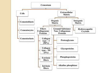
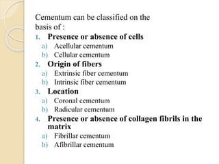
CEMENTUM
PERIDONTOLOGY
By Safura Ijaz
Batch’24 Dental Technology
CONTENTS
 INTRODUCTION
 PHYSICAL CHARACTERISTICS
 COMPOSITION
 FUNCTIONS
 CLASSIFICATION
 CEMENTO-ENAMEL JUNCTION (CEJ)
 CEMENTO-DENTINAL JUNCTION (CDJ)
 CEMENTUM RESORPTION AND REPAIR
 ABNORMALITIES OF CEMENTUM
INTRODUCTION
“Cementum is avascular, mineralized connective
tissue that forms the outer covering of the anatomic
root.”
PHYSICAL
CHARACTERISTICS
 Hardness < Dentin
 Light yellow in color
 Permeability of cellular cementum is greater
than Acellular cementum and this
permeability diminishes with age.
 Thinnest on the coronal half of the root i.e. 16
to 60 μm.
 Thickest in the apical 3rd and in the furcation
area i.e. ≤ 150 to 200 µm.
 Thicker in distal surfaces than in mesial
surfaces because of mesial drift.
COMPOSITION
Cementum
Cells
Cementoblasts
Cementocytes
Cementoclasts
Extracellular
Matrix
Organic
Matrix
Fibers
Collagenous
Proteins
Collagen
type – I
(90%)
Collagen
type – III
(5%)
Extrinsic
fibers
Intrinsic
fibers
Ground Substance
Non- Collagenous
Proteins
Proteoglycans
Glycoproteins
Phosphoproteins
Alkaline phosphtase
Inorganic
Matrix
Hydroxyapatite
Crystals
SOURCES OF COLLAGEN
FIBERS
There are 02 main sources of collagen fibers:
1. Extrinsic fibers: “Fibers that belong to
the principal fibers of
PDL (Sharpey’s fibers)
and formed by fibroblasts.”
2. Intrinsic fibers: “Fibers that belong to the
cementum matrix and
formed by cementoblasts.”
FUNCTIONS OF
CEMENTUM
Followings are the functions of cementum
 Anchorage : Provides a medium for the
attachment of collagen fibers that bind tooth
to the alveolar bone
 Adaptation: In response to tooth wear and
movement.
 Repair : Associated with repair of
periodontal tissues and damage to roots
such as fractures and resorptions.
CLASSIFICATION OF
CEMENTUM
Cementum can be classified on the
basis of :
1. Presence or absence of cells
a) Acellular cementum
b) Cellular cementum
2. Origin of fibers
a) Extrinsic fiber cementum
b) Intrinsic fiber cementum
3. Location
a) Coronal cementum
b) Radicular cementum
4. Presence or absence of collagen fibrils in the
matrix
a) Fibrillar cementum
b) Afibrillar cementum
Acellular cementum Cellular cementum
Forms before tooth reaches occlusal plane -
first formed cementum / primary cementum
Forms after tooth reach
plane i.e. secondary ce
Absent Present (Cementocytes
Cervical 3rd or half of the root
i.e. coronal portion of the root
Apical portion of the roo
furcation area
mation Slower Faster
n More calcified Less calcified
nt of collagen Regularly arranged Irregularly arranged
lines More i.e. closer together Sparse i.e. wide apart
bers and
cation
More and completely calcified Less and completely or
calcified
Thin cementum i.e. 30 to 230 µm Thick cementum i.e.10
Anchorage Adaptation and Repair
SCHROEDER’S
CLASSIFICATION
On the basis of presence or absence of cells
and collagen fibers in matrix, location and
origin of fibers, SCHROEDER classified
cementum into 05 different types.
1) Acellular afibrillar cementum
2) Acellular extrinsic fiber cementum
3) Cellular mixed stratified cementum
4) Cellular intrinsic fiber cementum
5) Intermediate cementum
Acellular
Afibrillar
Cementum
(AAC)
Acellular
Extrinsic Fiber
Cementum
(AEFC)
Cellular Mixed
Stratified
Cementum
(AMSC)
Cellular
Intrinsic Fiber
Cementum
(AIFC)
Absent Densely packed
bundles of
Sharpey’s fibers
Extrinsic and
intrinsic fibers
Intrinsic fibers
from the
cementum matrix
Absent Absent Present Present C
o
Cementoblasts Fibroblasts and
cementoblasts
Fibroblasts and
cementoblasts
Cementoblasts
Coronal cementum
i.e. CEJ
Cervical 3rd of the
root and may
extend farther
apically
Apical 3rd of the
root and in
furcation area
Resorption
lacunae
C
j
1 – 15 µm 30 – 230 µm 100 – 1000 µm
CEMENTO-ENAMEL
JUNCTION
PATTERNS OF CEJ
TYPE – 1 : In about 60% - 65% cases cementum overlaps the
enamel.
TYPE – 2 : In about 30% cases edge-to-edge butt joint exists.
TYPE – 3 : In about 5% - 10% cases cementum and enamel fails to
meet which cause accentuated sensitivity because of
exposed dentin.
CEMENTO-DENTINAL
JUNCTION
“The terminal apical
area of cementum
where it joins the
internal root dentin is
called CDJ.”
It is 2 – 3 µm wide
its width remains
same i.e. stable with
age.
During RCT,
obturating material
should be at CDJ.
CEMENTUM
RESORPTION AND REPAIR
“It is the process by which old cementum is removed
and replaced by new cementum.”
Cells responsible for resorption of cementum:-
These cells are found adjacent to the cementum
that is undergoing active resorption.
 Multinucleated giant cells
 Large mononuclear macrophages
Cells responsible for repair of cementum:-
 Cementoblasts
 Cells rest of Malassez
 Growth factors
Members of transforming growth factors super-family
Platelet derived growth factor
Insulin like growth factors
Enamel matrix derivatives
CAUSES OF CEMENTUM
RESORPTION
Local Factors Systemic Factors
 Trauma from occlusion
 Orthodontic movement
 Pressure from malaligned
erupting teeth
 Cysts and tumors
 Teeth without functional
antagonist
 Embedded teeth
 Replanted and transplanted
teeth
 Periapical and periodontal
disease
 Calcium deficiency
 Hypothyroidism
 Paget’s disease
 Hereditary fibrous
osteodystrophy
ABNORMALITIES OF
CEMENTUM
Abnormalities of cementum can be:
Cemental Aplasia “Absence of cellular cementum.”
Cemental Hypoplasia “Paucity of cellular cementum.”
Cementum Hyperplasia
Or
Hypercementosis
“Excessive deposition of cementum.”
• It is largely an age related phenomena.
• It can be localized to one tooth or
generalized i.e. affect the entire dentition.
Causes :-
• Loss of antagonist
• Inflammation of the root
• Trauma to the root
Treatment:-
• It itself does not need treatment.
• It could pose a problem if an affected
tooth requires extraction.
• In a multi-rooted tooth, sectioning of the
tooth may be required before extraction.
ANKYLOSIS
“Fusion of cementum and the alveolar bone
with the obliteration of PDL is called
ankylosis.”
Causes of ankylosis:-
 Cemental resorption
 Chronic apical inflammation
 Tooth replantation
 Occlusal trauma
ANKYLOSIS
EXPOSURE OF CEMENTUM TO
THE ORAL ENVIRONMENT
Cementum becomes exposed to the oral
environment in cases of:
 Gingival recession
 Loss of attachment in resulting in pocket
formation
This results in:
 Periodontal diseases
 Cementum caries
because cementum is sufficiently
permeable to be penetrated by organic
substances, inorganic ions, and bacteria.
cementum.pptx

cementum.pptx

  • 1.
  • 2.
    CONTENTS  INTRODUCTION  PHYSICALCHARACTERISTICS  COMPOSITION  FUNCTIONS  CLASSIFICATION  CEMENTO-ENAMEL JUNCTION (CEJ)  CEMENTO-DENTINAL JUNCTION (CDJ)  CEMENTUM RESORPTION AND REPAIR  ABNORMALITIES OF CEMENTUM
  • 3.
  • 4.
    “Cementum is avascular,mineralized connective tissue that forms the outer covering of the anatomic root.”
  • 5.
  • 6.
     Hardness <Dentin  Light yellow in color  Permeability of cellular cementum is greater than Acellular cementum and this permeability diminishes with age.  Thinnest on the coronal half of the root i.e. 16 to 60 μm.  Thickest in the apical 3rd and in the furcation area i.e. ≤ 150 to 200 µm.  Thicker in distal surfaces than in mesial surfaces because of mesial drift.
  • 7.
  • 8.
    Cementum Cells Cementoblasts Cementocytes Cementoclasts Extracellular Matrix Organic Matrix Fibers Collagenous Proteins Collagen type – I (90%) Collagen type– III (5%) Extrinsic fibers Intrinsic fibers Ground Substance Non- Collagenous Proteins Proteoglycans Glycoproteins Phosphoproteins Alkaline phosphtase Inorganic Matrix Hydroxyapatite Crystals
  • 9.
    SOURCES OF COLLAGEN FIBERS Thereare 02 main sources of collagen fibers: 1. Extrinsic fibers: “Fibers that belong to the principal fibers of PDL (Sharpey’s fibers) and formed by fibroblasts.” 2. Intrinsic fibers: “Fibers that belong to the cementum matrix and formed by cementoblasts.”
  • 10.
  • 11.
    Followings are thefunctions of cementum  Anchorage : Provides a medium for the attachment of collagen fibers that bind tooth to the alveolar bone  Adaptation: In response to tooth wear and movement.  Repair : Associated with repair of periodontal tissues and damage to roots such as fractures and resorptions.
  • 12.
  • 13.
    Cementum can beclassified on the basis of : 1. Presence or absence of cells a) Acellular cementum b) Cellular cementum 2. Origin of fibers a) Extrinsic fiber cementum b) Intrinsic fiber cementum 3. Location a) Coronal cementum b) Radicular cementum 4. Presence or absence of collagen fibrils in the matrix a) Fibrillar cementum b) Afibrillar cementum
  • 15.
    Acellular cementum Cellularcementum Forms before tooth reaches occlusal plane - first formed cementum / primary cementum Forms after tooth reach plane i.e. secondary ce Absent Present (Cementocytes Cervical 3rd or half of the root i.e. coronal portion of the root Apical portion of the roo furcation area mation Slower Faster n More calcified Less calcified nt of collagen Regularly arranged Irregularly arranged lines More i.e. closer together Sparse i.e. wide apart bers and cation More and completely calcified Less and completely or calcified Thin cementum i.e. 30 to 230 µm Thick cementum i.e.10 Anchorage Adaptation and Repair
  • 16.
    SCHROEDER’S CLASSIFICATION On the basisof presence or absence of cells and collagen fibers in matrix, location and origin of fibers, SCHROEDER classified cementum into 05 different types. 1) Acellular afibrillar cementum 2) Acellular extrinsic fiber cementum 3) Cellular mixed stratified cementum 4) Cellular intrinsic fiber cementum 5) Intermediate cementum
  • 17.
    Acellular Afibrillar Cementum (AAC) Acellular Extrinsic Fiber Cementum (AEFC) Cellular Mixed Stratified Cementum (AMSC) Cellular IntrinsicFiber Cementum (AIFC) Absent Densely packed bundles of Sharpey’s fibers Extrinsic and intrinsic fibers Intrinsic fibers from the cementum matrix Absent Absent Present Present C o Cementoblasts Fibroblasts and cementoblasts Fibroblasts and cementoblasts Cementoblasts Coronal cementum i.e. CEJ Cervical 3rd of the root and may extend farther apically Apical 3rd of the root and in furcation area Resorption lacunae C j 1 – 15 µm 30 – 230 µm 100 – 1000 µm
  • 19.
  • 20.
    PATTERNS OF CEJ TYPE– 1 : In about 60% - 65% cases cementum overlaps the enamel. TYPE – 2 : In about 30% cases edge-to-edge butt joint exists. TYPE – 3 : In about 5% - 10% cases cementum and enamel fails to meet which cause accentuated sensitivity because of exposed dentin.
  • 21.
  • 22.
    “The terminal apical areaof cementum where it joins the internal root dentin is called CDJ.” It is 2 – 3 µm wide its width remains same i.e. stable with age. During RCT, obturating material should be at CDJ.
  • 23.
  • 24.
    “It is theprocess by which old cementum is removed and replaced by new cementum.” Cells responsible for resorption of cementum:- These cells are found adjacent to the cementum that is undergoing active resorption.  Multinucleated giant cells  Large mononuclear macrophages Cells responsible for repair of cementum:-  Cementoblasts  Cells rest of Malassez  Growth factors Members of transforming growth factors super-family Platelet derived growth factor Insulin like growth factors Enamel matrix derivatives
  • 25.
    CAUSES OF CEMENTUM RESORPTION LocalFactors Systemic Factors  Trauma from occlusion  Orthodontic movement  Pressure from malaligned erupting teeth  Cysts and tumors  Teeth without functional antagonist  Embedded teeth  Replanted and transplanted teeth  Periapical and periodontal disease  Calcium deficiency  Hypothyroidism  Paget’s disease  Hereditary fibrous osteodystrophy
  • 26.
  • 27.
    Abnormalities of cementumcan be: Cemental Aplasia “Absence of cellular cementum.” Cemental Hypoplasia “Paucity of cellular cementum.” Cementum Hyperplasia Or Hypercementosis “Excessive deposition of cementum.” • It is largely an age related phenomena. • It can be localized to one tooth or generalized i.e. affect the entire dentition. Causes :- • Loss of antagonist • Inflammation of the root • Trauma to the root Treatment:- • It itself does not need treatment. • It could pose a problem if an affected tooth requires extraction. • In a multi-rooted tooth, sectioning of the tooth may be required before extraction.
  • 28.
    ANKYLOSIS “Fusion of cementumand the alveolar bone with the obliteration of PDL is called ankylosis.” Causes of ankylosis:-  Cemental resorption  Chronic apical inflammation  Tooth replantation  Occlusal trauma
  • 29.
  • 30.
    EXPOSURE OF CEMENTUMTO THE ORAL ENVIRONMENT Cementum becomes exposed to the oral environment in cases of:  Gingival recession  Loss of attachment in resulting in pocket formation This results in:  Periodontal diseases  Cementum caries because cementum is sufficiently permeable to be penetrated by organic substances, inorganic ions, and bacteria.