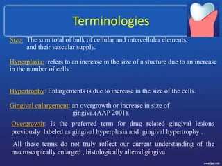
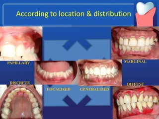
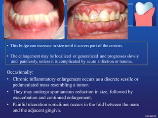
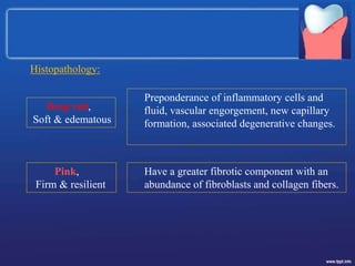
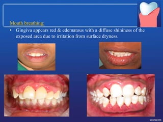
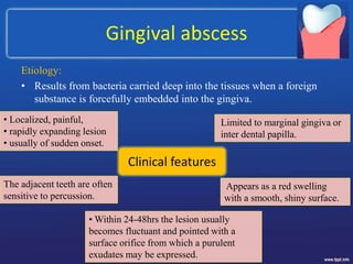
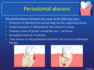
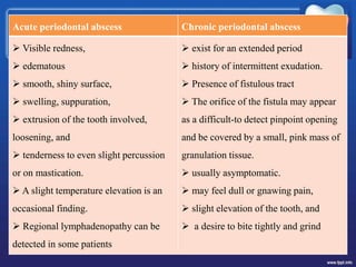
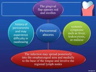
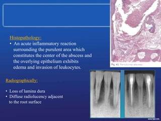
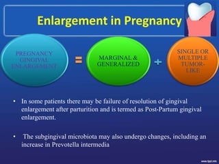
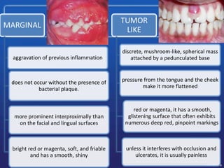
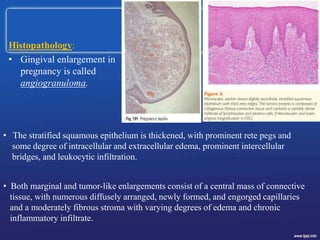
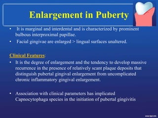
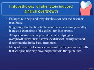
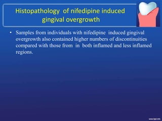
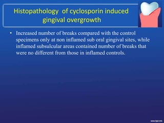
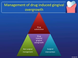
GINGIVAL ENLARGEMENT
Dr.D.Navya,MDS
Introduction
• Clinically apparent enlargement of the papillary and marginal
gingiva is a common finding in otherwise healthy humans and
other mammals.
• Swelling ("tumor") is one of the five cardinal symptoms of
inflammation.
• Affected gingival tissues are soft in consistency, generally
more or less erythematous, and bleed without exception when
gently probed.
Terminologies
Size: The sum total of bulk of cellular and intercellular elements,
and their vascular supply.
Hyperplasia: refers to an increase in the size of a stucture due to an increase
in the number of cells
Hypertrophy: Enlargements is due to increase in the size of the cells.
Gingival enlargement: an overgrowth or increase in size of
gingiva.(AAP 2001).
Overgrowth: Is the preferred term for drug related gingival lesions
previously labeled as gingival hyperplasia and gingival hypertrophy .
All these terms do not truly reflect our current understanding of the
macroscopically enlarged , histologically altered gingiva.
According to location & distribution
GENERALIZED
LOCALIZED
PAPILLARY
DISCRETE DIFFUSE
MARGINAL
According to etiologic factors &
pathologic changes
I. Inflammatory enlargement
A. Chronic
B. Acute
II. Drug-induced enlargement
III. Enlargements associated with systemic diseases
A. Conditioned enlargement
1. Pregnancy
2. Puberty
3. Vitamin C deficiency
4. Plasma cell gingivitis
5. Nonspecific conditioned enlargement (pyogenic granuloma)
B. Systemic diseases causing gingival enlargement
1. Leukemia
2. Granulomatous diseases (Wegener's granulomatosis, sarcoidosis)
IV. Neoplastic enlargement (gingival tumors)
A. Benign tumors
B. Malignant tumors
V. False enlargement
According to degree of gingival enlargement
Grade Description
0 No signs of gingival enlargement
I Enlargement confined to
interdental papilla
II Enlargement involves
papilla and marginal gingiva
III Enlargement covers
three quarters or more of the crown
Chronic inflammatory enlargement
Clinical features:
• Slight ballooning of the interdental
papilla and/or the marginal gingiva.
• In the early stages it produces a life
preserver-shaped bulge around the
involved teeth.
Inflammatory enlargements commonly are a secondary
complication to any of the other types of enlargement,
creating a combined gingival enlargement.
Etiology:
• Prolonged exposure to dental plaque, poor oral hygiene,
Irritation by anatomic abnormalities, improper restorative
and orthodontic appliances
Occasionally:
• Chronic inflammatory enlargement occurs as a discrete sessile or
pedunculated mass resembling a tumor.
• They may undergo spontaneous reduction in size, followed by
exacerbation and continued enlargement.
• Painful ulceration sometimes occurs in the fold between the mass
and the adjacent gingiva.
• This bulge can increase in size until it covers part of the crowns.
• The enlargement may be localized or generalized and progresses slowly
and painlessly, unless it is complicated by acute infection or trauma.
Preponderance of inflammatory cells and
fluid, vascular engorgement, new capillary
formation, associated degenerative changes.
Have a greater fibrotic component with an
abundance of fibroblasts and collagen fibers.
Deep red,
Soft & edematous
Pink,
Firm & resilient
Histopathology:
Mouth breathing:
• Gingiva appears red & edematous with a diffuse shininess of the
exposed area due to irritation from surface dryness.
Gingival abscess
Etiology:
• Results from bacteria carried deep into the tissues when a foreign
substance is forcefully embedded into the gingiva.
Clinical features
• Localized, painful,
• rapidly expanding lesion
• usually of sudden onset.
Limited to marginal gingiva or
inter dental papilla.
Appears as a red swelling
with a smooth, shiny surface.
• Within 24-48hrs the lesion usually
becomes fluctuant and pointed with a
surface orifice from which a purulent
exudates may be expressed.
The adjacent teeth are often
sensitive to percussion.
Periodontal abscess
Periodontal abscess formation may occur in the following ways:
• Extension of infection from pocket deep into the supporting tissues.
• Lateral extension of inflammation into connective tissue.
• Tortuous course of pocket around the root – cul-de-sac.
• Incomplete removal of calculus.
• After trauma or with perforation of lateral wall of root in endodontic
therapy.
Acute periodontal abscess Chronic periodontal abscess
 Visible redness,
 edematous
 smooth, shiny surface,
 swelling, suppuration,
 extrusion of the tooth involved,
loosening, and
 tenderness to even slight percussion
or on mastication.
 A slight temperature elevation is an
occasional finding.
 Regional lymphadenopathy can be
detected in some patients
 exist for an extended period
 history of intermittent exudation.
 Presence of fistulous tract
 The orifice of the fistula may appear
as a difficult-to detect pinpoint opening
and be covered by a small, pink mass of
granulation tissue.
 usually asymptomatic.
 may feel dull or gnawing pain,
 slight elevation of the tooth, and
 a desire to bite tightly and grind
Pericoronal
abscess
The infection may spread posteriorly
into the oropharyngeal area and medially
to the base of the tongue and involve the
regional lymph nodes
systemic
symptoms
such as fever,
leukocytosis,
or malaise
The gingival
flap appears red
and swollen
history of
pericoronitis
and may
experience
difficulty in
swallowing
Histopathology:
• An acute inflammatory reaction
surrounding the purulent area which
constitutes the center of the abscess and
the overlying epithelium exhibits
edema and invasion of leukocytes.
Radiographically:
• Loss of lamina dura
• Diffuse radiolucency adjacent
to the root surface
Idiopathic gingival enlargement
• Idiopathic Gingival enlargement is a rare condition of undetermined
cause, may present as a specific entity in an isolated form or
associated with several uncommon syndromes or genetic disorders.
1. Hereditary gingival fibromatosis:
a) Zimmerman- Laband
syndrome
b) Rutherfurd syndrome
c) Neurofibromatosis type 1
2. Lysomal storage disorders:
a) Hurler syndrome
b) Hunter syndrome
c) Cowden’s syndrome
3. Vascular disorders:
a) Sturge-Weber’s syndrome
b) Klippel-Trenaunay
syndrome
4. Disorders associated with
dental anamolies:
a) Wilson syndrome
b) Goltz syndrome
c) Regional odontodysplasia
Etiology:
• Some cases have a hereditary basis found that
mode of inheritance to be autosomal dominant or
recessive.
Clinical features:
• Enlargement usually begins with eruption of teeth
and may regress after extraction.
• The overgrowth may result in functional and
esthetic concern, create diastemas, impede or
delay tooth eruption and create changes in the
facial appearance as a result of lip protrusion.
• Gingiva is pale pink, firm and leathery in
consistency and has a characteristic minutely
pebbeled surface.
Histopathology:
• Epithelium is thickened and acanthotic with elongated rete pegs.
• Shows a bulbous increase in the amount of connective tissue
consisting of densely arranged collagen bundles and numerous
fibroblasts, relatively avascular.
• Also exhibits an accumulation of elastic and oxytalan fibers.
• The hallmark of Hereditary gingival fibromatosis is the
accumulation of excess ECM where TGF upregulation promotes
excess ECM synthesis and Matrix metalloproteases (MMP)
downregulation inhibits ECM degradation.
Enlargements associated with
Systemic conditions
Conditioned Enlargements:
• 1) Hormonal :
a) Pregnancy
b) Puberty
• 2)Nutritional:Vitamin C deficiency
• 3) Allergic: Allergic stomatitis
a) Systemic diseases causing gingival
enlargement: leukemia,
granulomatous diseases.
b) Neoplastic enlargements (gingival
tumors)
1) Magnification of existing
inflammation initiated by
dental plaque.
2) Manifestation of the systemic
disease independent of the
inflammatory status of the
gingiva.
Enlargement in Pregnancy
• In some patients there may be failure of resolution of gingival
enlargement after parturition and is termed as Post-Partum gingival
enlargement.
• The subgingival microbiota may also undergo changes, including an
increase in Prevotella intermedia
PREGNANCY
GINGIVAL
ENLARGEMENT
MARGINAL &
GENERALIZED
SINGLE OR
MULTIPLE
TUMOR-
LIKE
MARGINAL
aggravation of previous inflammation
does not occur without the presence of
bacterial plaque.
more prominent interproximally than
on the facial and lingual surfaces
bright red or magenta, soft, and friable
and has a smooth, shiny
TUMOR
LIKE
discrete, mushroom-like, spherical mass
attached by a pedunculated base
pressure from the tongue and the cheek
make it more flattened
red or magenta, it has a smooth,
glistening surface that often exhibits
numerous deep red, pinpoint markings
unless it interferes with occlusion and
ulcerates, it is usually painless
Histopathology:
• Gingival enlargement in
pregnancy is called
angiogranuloma.
• The stratified squamous epithelium is thickened, with prominent rete pegs and
some degree of intracellular and extracellular edema, prominent intercellular
bridges, and leukocytic infiltration.
• Both marginal and tumor-like enlargements consist of a central mass of connective
tissue, with numerous diffusely arranged, newly formed, and engorged capillaries
and a moderately fibrous stroma with varying degrees of edema and chronic
inflammatory infiltrate.
Enlargement in Puberty
• It is marginal and interdental and is characterized by prominent
bulbous interproximal papillae.
• Facial gingivae are enlarged > lingual surfaces unaltered.
Clinical Features:
• It is the degree of enlargement and the tendency to develop massive
recurrence in the presence of relatively scant plaque deposits that
distinguish pubertal gingival enlargement from uncomplicated
chronic inflammatory gingival enlargement.
• Association with clinical parameters has implicated
Capnocytophaga species in the initiation of pubertal gingivitis
• Nakagawa et al 1994, have reported that hormonal changes coincide
with an increase in the proportion of Prevotella intermedia and
Prevotella nigrescens.
Histopathology:
• The microscopic picture is that of chronic inflammation with
prominent edema and associated degenerative changes.
Enlargement in Vitamin-C Deficiency
• Acute vitamin C deficiency does not of itself cause gingival
inflammation, but it does cause hemorrhage, collagen degeneration,
and edema of the gingival connective tissue.
• These changes modify the response of the gingiva to plaque to the
extent that the normal defensive delimiting reaction is inhibited,
and  the extent of the inflammation is exaggerated.
• The combined effect of acute vitamin C deficiency and
inflammation produces the massive gingival enlargement in Scurvy.
Clinical Features:
• Gingival enlargement in vitamin C deficiency is marginal; the
gingiva is bluish red, soft, and friable and has a smooth, shiny
surface.
• Hemorrhage, occurring either spontaneously or on slight
provocation, and surface necrosis with pseudomembrane formation
are common features.
Histopathology:
• The gingiva has a chronic inflammatory cellular infiltration with a
superficial acute response.
• There are scattered areas of hemorrhage, with engorged capillaries.
Marked diffuse edema, collagen degeneration, and scarcity of
collagen fibrils or fibroblasts are striking findings.
Allergic stomatitis
Plasma cell gingivitis
Also termed as
• Atypical gingivitis and Plasma cell gingivostomatitis  if generalized
• Plasma cell granuloma  if localized
Etiology:
• Thought to be allergic in origin
Clinical Features:
• The gingiva appears red, friable, and sometimes granular and bleeds
easily; usually it does not induce a loss of attachment.
• This lesion is located in the oral aspect of the attached gingiva and
therefore differs from plaque-induced gingivitis.
Pathogenesis:
• The large number of plasma cells may represent an autoimmune
reaction or an alteration of blood flow imposing congestive
vasodilation.
Histopathology:
• The oral epithelium shows spongiosis and infiltration with
inflammatory cells; ultra structurally there are signs of damage in the
lower spinous layers and the basal layers.
• The underlying connective tissue contains a dense infiltrate of plasma
cells that also extends to the oral epithelium, inducing a dissecting type
of injury.
Pyogenic granuloma
Clinical Features:
• The lesion is discrete spherical,
tumor-like mass with a pedunculated
attachment to a flattened, keloid like
enlargement with a broad base.
• It is bright red or purple and either friable
or firm, depending on its duration; in the
majority of cases it presents with surface
ulceration and purulent exudation.
• The lesion tends to involute spontaneously to become a
fibroepithelial papilloma or persists relatively unchanged for years.
• Histopathology:
• Pyogenic granuloma appears as a mass of
granulation tissue with chronic inflammatory
cellular infiltration.
• Endothelial proliferation and the formation of
numerous vascular spaces are the prominent
features.
• The surface epithelium is atrophic in some areas
and hyperplastic in others. Surface ulceration
and exudation are common features.
SYSTEMIC DISEASES CAUSING
GINGIVAL ENLARGEMENT
LEUKEMIA:
• Leukemias are uniformly fatal diseases of unknown etiology
characterized by excessive and abnormal proliferation of primitive
white blood cells and their precursors with infiltration into the
various tissues of the body, especially bone marrow, spleen, and
lymph nodes.
• Pallor, hyperplasia of the gingiva, spontaneous hemorrhage,
petechiae and ulceration showed a higher incidence of occurrence in
the acute forms compared to the chronic forms.
• Osgood (1935) noted that swelling of the gingiva was one of the
most constant features in monocytic Leukemias.
Clinical Features:
• It may appear as a diffuse enlargement of the gingival mucosa, an
oversized extension of the marginal gingiva, or a discrete tumor-
like inter-proximal mass.
• Color - gingiva is generally bluish red and
has a shiny surface.
• Consistency - is moderately firm,
but there is a tendency toward
friability and hemorrhage, occurring
either spontaneously or on slight irritation.
• Acute painful necrotizing ulcerative inflammatory involvement
sometimes occurs in the crevice formed at the junction of the
enlarged gingiva and the contiguous tooth surfaces.
Histopathology:
• Various degrees of chronic inflammation with mature
leukocytes and areas of connective tissue infiltrated with a
dense mass of immature and proliferating leukocytes, the
specific nature of which varies with the type of leukemia.
• Engorged capillaries, edematous and degenerated
connective tissue, and epithelium with various degrees of
leukocytic infiltration and edema are found.
• Isolated surface areas of acute necrotizing inflammation
with a pseudomembranous meshwork of fibrin, necrotic
epithelial cells, polymorphonuclear neutrophils (PMNs),
and bacteria are often seen.
Wegener's Granulomatosis
• The cause is unknown, but the condition is considered an
immunologically mediated tissue injury (Robbins et al 1989).
• Wegener's granulomatosis is a rare disease characterized by acute
granulomatous necrotizing lesions of the respiratory tract, including
nasal and oral defects.
• Renal lesions develop, and acute necrotizing vasculitis affects the
blood vessels.
• The initial manifestations may involve the oro-facial region and
include oral mucosal ulceration, gingival enlargement, abnormal
tooth mobility, exfoliation of teeth, and delayed healing response.
Clinical Features:
• The granulomatous papillary enlargement is reddish purple and
bleeds easily on stimulation.
Histopathology:
• Chronic inflammation occurs with scattered giant cells and foci of
acute inflammation and micro abscesses covered by a thin
acanthotic epithelium.
• Vascular changes have not been described, probably owing to the
small size of the gingival blood vessels.
Neoplastic enlargement
(Gingival tumors)
Benign tumors of the gingiva
• “Epulis” is a generic term used clinically to designate discrete
tumors and tumor like masses of the gingiva.
• It serves to locate the tumor but not to describe it.
• Most lesions referred to as “epulis” are inflammatory rather than
neoplastic.
Fibroma
Clinical features:
• Fibromas of the gingiva arise from the gingival connective tissue or
from the periodontal ligament.
• They are slow growing, spherical tumors that t end to be firm and
nodular but may be soft and vascular, usually pedunculated.
Histopathology:
• Fibromas are composed of bundles of
well-formed collagen fibers with a
scattering of fibrocytes and a variable
vascularity.
 Giant cell fibroma and peripheral
ossifying fibroma are other variants.
Haemangioma
• These are usually hamartoms rather than true tumors and are
sometimes part of a wide spread developmental defect
Microscopy:
• Capillary haemangiomas:
These consists of a mass of fine capillaries or
imperforate rosettes of endothelial cells,
covered by squamous epithelium
• Cavernous haemangiomas:
These consists of dilated, thin walled, blood filled
vessels or sinusoids covered by squamous epithelium.
Management:
If a haemangioma needs to be removed, cryotherapy is
probably the treatment of choice because of the risk of
serious haemorrhage
Squamous cell carcinoma
• Because of its proximity to the teeth and periodontium, the tumor
can mimic tooth related benign inflammatory conditions which can
lead to misdiagnosis.
False enlargements
Drug induced gingival enlargement
• 3 drugs are most frequently implicated:
a) Phenytoin b) Cyclosporine c) Calcium channel blockers
Risk factors
• A variety of risk factors for drug induced gingival overgrowth
have been identified
1) Age and other demographic factors
2) Drug variables
3) Concomitant medication
4) Periodontal variables and
5) Genetic factors.
Anticonvulsants
• Fibroblasts from a phenytoin-induced gingival overgrowth
show increased synthesis of sulfated glycosaminoglycans in
vitro. Phenytoin may induce a decrease in collagen
degradation as a result of the production of an inactive
fibroblastic collagenase.
• Mechanism of action:
• Phenytoin is a drug with high plasma protein binding and the
free phenytoin concentrations are better correlated with both
efficacy and toxicity.
• A reduced plasma protein binding favors tissue distribution,
particularly to those tissues with high phenytoin binding
affinity such as the gingival fibroblasts
• Phenytoin is not only responsible for the initiation of
enlargement of the gingival tissue, but has also been noted to
interfere with folic acid absorption, thereby leading to a
significant decrease in folate levels
• The latter hypothesis was that folic acid supplementation in
patients with high phenytoin levels led to a significant
regression of gingival enlargement.
• Clinical features:
• In the absence of gingivitis, is usually firm, pink, and
somewhat pebbly.
• In severe cases, teeth surfaces are completely covered with
gingival overgrowth.
Immunosuppressants
• Another major side effect associated with cyclosporine is
increased blood pressure. Calcium channel blockers, such as
nifedipine, are usually the drugs of choice to control this
problem.
• Mechanism of action:
• Its exact mechanism of action is not well known, but it appears
to selectively and reversibly inhibit helper T cells, which play
a role in cellular and humoral immune responses.
• Clinical features:
• Cyclosporin-induced gingival overgrowth commences as a papillary
enlargement which is more pronounced on the labial aspects of the gingiva
than the palatal or lingual surfaces
• This gives the gingival tissues a lobulated appearance.
• Overgrowth is restricted to the width of attached gingiva.
• Cyclosporin induced gingival overgrowth has not been reported in
edentulous subjects
• They bleed readily on probing and are generally more hyperaemic than the
gingival tissue from phenytoin-induced gingival overgrowth.
Calcium Channel Blockers
• It has been speculated that some alteration to Ca++ metabolism
is involved, but others have suggested that nifedipine may act
indirectly by stimulating either production of IL-2 by T cells or
metabolites of testosterone.
Drug-induced Alterations in Gingival
Connective Tissue Homeostasis
• Collagen production from gingival fibroblasts is controlled by the
co-ordination of transcripted and post-transplation collagen
regulatory mechanisms, including intracellular degradation.
• The latter is controlled by synthesis and release of metalloproteinase
and tissue inhibitors of metalloproteinase (TIMPs).
• Histometric analysis of phenytoin-induced gingival overgrowth has
shown that the lesion is characterized by an increase in "normal
growth" (i.e. the ratio of cells to matrix remains the same) as
opposed to a cellular hypertrophy or hyperplasia (hence the accepted
term gingival overgrowth
Alteration in connective tissue
metabolism
• Overproduction of collagen by gingival fibroblasts in phenytoin-
induced gingival enlargement involves an increased steady state
level of collagen mRNA and not a decrease in collagen degradation
• Early in vitro studies showed that phenytoin-induced gingival
overgrowth may be more related to a lack of collagen breakdown as
opposed to an increase in collagen production
Histopathology, Ultra structural
Factors and Inflammatory Changes
• Potential drug related differences have been described only
when immune histochemical staining techniques or electron
microscopy have been employed.
• Drug induced gingival overgrowth is associated with
1. Thickening of epithelium &
2. Elongated rete pegs extending deep into the underlying
connective tissue.
• This phenomenon is related to expansion of the stratum
spinosum and is associated with increased mitotic activity
within the epithelial layer.
Histopathology of phenytoin induced
gingival overgrowth
• Enlarged rete pegs and irregularities at or near the basement
membrane.
• Suggesting that the fibrotic transformation is accompanied by
increased extensions of the epithelium into stroma.
• All specimens from the phenytoin induced gingival
overgrowth individuals showed evidence of disruptions and
discontinuities in the basal membrane.
• Many of these breaks are accompanied by the presence of cells
that we speculate may have migrated from the epithelium.
Histopathology of nifedipine induced
gingival overgrowth
• Samples from individuals with nifedipine induced gingival
overgrowth also contained higher numbers of discontinuities
compared with those from in both inflamed and less inflamed
regions.
Histopathology of cyclosporin induced
gingival overgrowth
• Increased number of breaks compared with the control
specimens only at non inflamed sub oral gingival sites, while
inflamed subsulcular areas contained number of breaks that
were no different from those in inflamed controls.
Management of drug induced gingival
overgrowth
Drug
substitution
Non surgical
management
Drug
induced
gingival
overgrowth
Surgical
intervention
Drug substitution
• Phenytoin  Vigabartin, Lomatrigine, Gabapentin, Sulthiame
and Topiramate.
• Cyclosporine  reduction in the dose is beneficial,
Tacrolimus, Rapamycin and Mycophenolate mofetil.
• Nifedipine  Verapamil, Amlodipine and Felodipine.
• Primary aim is to reduce inflammatory component.
• Professional teeth cleaning: Scaling and root planing
• Systemic antibiotics: Azithromycin and Metranidazole
• Folic acid mouth wash: 1mg/ml reduced recurrence of phenytoin
induced gingival overgrowth.
• Full mouth disinfection: An anti infective regime combining non
surgical mechanical therapy and a chemotherapeutic approach.
Non surgical
Management
• Current surgical management includes:
• Gingivoplasty /Gingivectomy-
1) Scalpel
2) Electrosurgery
3) Laser
4) Chemical
• Overgrowth flap surgery
Surgical
Intervention
THANK YOU

3.gingival enlargement.ppt

  • 1.
  • 2.
    Introduction • Clinically apparentenlargement of the papillary and marginal gingiva is a common finding in otherwise healthy humans and other mammals. • Swelling ("tumor") is one of the five cardinal symptoms of inflammation. • Affected gingival tissues are soft in consistency, generally more or less erythematous, and bleed without exception when gently probed.
  • 3.
    Terminologies Size: The sumtotal of bulk of cellular and intercellular elements, and their vascular supply. Hyperplasia: refers to an increase in the size of a stucture due to an increase in the number of cells Hypertrophy: Enlargements is due to increase in the size of the cells. Gingival enlargement: an overgrowth or increase in size of gingiva.(AAP 2001). Overgrowth: Is the preferred term for drug related gingival lesions previously labeled as gingival hyperplasia and gingival hypertrophy . All these terms do not truly reflect our current understanding of the macroscopically enlarged , histologically altered gingiva.
  • 4.
    According to location& distribution GENERALIZED LOCALIZED PAPILLARY DISCRETE DIFFUSE MARGINAL
  • 5.
    According to etiologicfactors & pathologic changes I. Inflammatory enlargement A. Chronic B. Acute II. Drug-induced enlargement III. Enlargements associated with systemic diseases A. Conditioned enlargement 1. Pregnancy 2. Puberty 3. Vitamin C deficiency 4. Plasma cell gingivitis 5. Nonspecific conditioned enlargement (pyogenic granuloma)
  • 6.
    B. Systemic diseasescausing gingival enlargement 1. Leukemia 2. Granulomatous diseases (Wegener's granulomatosis, sarcoidosis) IV. Neoplastic enlargement (gingival tumors) A. Benign tumors B. Malignant tumors V. False enlargement
  • 7.
    According to degreeof gingival enlargement Grade Description 0 No signs of gingival enlargement I Enlargement confined to interdental papilla II Enlargement involves papilla and marginal gingiva III Enlargement covers three quarters or more of the crown
  • 8.
    Chronic inflammatory enlargement Clinicalfeatures: • Slight ballooning of the interdental papilla and/or the marginal gingiva. • In the early stages it produces a life preserver-shaped bulge around the involved teeth. Inflammatory enlargements commonly are a secondary complication to any of the other types of enlargement, creating a combined gingival enlargement. Etiology: • Prolonged exposure to dental plaque, poor oral hygiene, Irritation by anatomic abnormalities, improper restorative and orthodontic appliances
  • 9.
    Occasionally: • Chronic inflammatoryenlargement occurs as a discrete sessile or pedunculated mass resembling a tumor. • They may undergo spontaneous reduction in size, followed by exacerbation and continued enlargement. • Painful ulceration sometimes occurs in the fold between the mass and the adjacent gingiva. • This bulge can increase in size until it covers part of the crowns. • The enlargement may be localized or generalized and progresses slowly and painlessly, unless it is complicated by acute infection or trauma.
  • 10.
    Preponderance of inflammatorycells and fluid, vascular engorgement, new capillary formation, associated degenerative changes. Have a greater fibrotic component with an abundance of fibroblasts and collagen fibers. Deep red, Soft & edematous Pink, Firm & resilient Histopathology:
  • 11.
    Mouth breathing: • Gingivaappears red & edematous with a diffuse shininess of the exposed area due to irritation from surface dryness.
  • 12.
    Gingival abscess Etiology: • Resultsfrom bacteria carried deep into the tissues when a foreign substance is forcefully embedded into the gingiva. Clinical features • Localized, painful, • rapidly expanding lesion • usually of sudden onset. Limited to marginal gingiva or inter dental papilla. Appears as a red swelling with a smooth, shiny surface. • Within 24-48hrs the lesion usually becomes fluctuant and pointed with a surface orifice from which a purulent exudates may be expressed. The adjacent teeth are often sensitive to percussion.
  • 13.
    Periodontal abscess Periodontal abscessformation may occur in the following ways: • Extension of infection from pocket deep into the supporting tissues. • Lateral extension of inflammation into connective tissue. • Tortuous course of pocket around the root – cul-de-sac. • Incomplete removal of calculus. • After trauma or with perforation of lateral wall of root in endodontic therapy.
  • 14.
    Acute periodontal abscessChronic periodontal abscess  Visible redness,  edematous  smooth, shiny surface,  swelling, suppuration,  extrusion of the tooth involved, loosening, and  tenderness to even slight percussion or on mastication.  A slight temperature elevation is an occasional finding.  Regional lymphadenopathy can be detected in some patients  exist for an extended period  history of intermittent exudation.  Presence of fistulous tract  The orifice of the fistula may appear as a difficult-to detect pinpoint opening and be covered by a small, pink mass of granulation tissue.  usually asymptomatic.  may feel dull or gnawing pain,  slight elevation of the tooth, and  a desire to bite tightly and grind
  • 15.
    Pericoronal abscess The infection mayspread posteriorly into the oropharyngeal area and medially to the base of the tongue and involve the regional lymph nodes systemic symptoms such as fever, leukocytosis, or malaise The gingival flap appears red and swollen history of pericoronitis and may experience difficulty in swallowing
  • 16.
    Histopathology: • An acuteinflammatory reaction surrounding the purulent area which constitutes the center of the abscess and the overlying epithelium exhibits edema and invasion of leukocytes. Radiographically: • Loss of lamina dura • Diffuse radiolucency adjacent to the root surface
  • 17.
    Idiopathic gingival enlargement •Idiopathic Gingival enlargement is a rare condition of undetermined cause, may present as a specific entity in an isolated form or associated with several uncommon syndromes or genetic disorders. 1. Hereditary gingival fibromatosis: a) Zimmerman- Laband syndrome b) Rutherfurd syndrome c) Neurofibromatosis type 1 2. Lysomal storage disorders: a) Hurler syndrome b) Hunter syndrome c) Cowden’s syndrome 3. Vascular disorders: a) Sturge-Weber’s syndrome b) Klippel-Trenaunay syndrome 4. Disorders associated with dental anamolies: a) Wilson syndrome b) Goltz syndrome c) Regional odontodysplasia
  • 18.
    Etiology: • Some caseshave a hereditary basis found that mode of inheritance to be autosomal dominant or recessive. Clinical features: • Enlargement usually begins with eruption of teeth and may regress after extraction. • The overgrowth may result in functional and esthetic concern, create diastemas, impede or delay tooth eruption and create changes in the facial appearance as a result of lip protrusion. • Gingiva is pale pink, firm and leathery in consistency and has a characteristic minutely pebbeled surface.
  • 19.
    Histopathology: • Epithelium isthickened and acanthotic with elongated rete pegs. • Shows a bulbous increase in the amount of connective tissue consisting of densely arranged collagen bundles and numerous fibroblasts, relatively avascular. • Also exhibits an accumulation of elastic and oxytalan fibers. • The hallmark of Hereditary gingival fibromatosis is the accumulation of excess ECM where TGF upregulation promotes excess ECM synthesis and Matrix metalloproteases (MMP) downregulation inhibits ECM degradation.
  • 20.
    Enlargements associated with Systemicconditions Conditioned Enlargements: • 1) Hormonal : a) Pregnancy b) Puberty • 2)Nutritional:Vitamin C deficiency • 3) Allergic: Allergic stomatitis a) Systemic diseases causing gingival enlargement: leukemia, granulomatous diseases. b) Neoplastic enlargements (gingival tumors) 1) Magnification of existing inflammation initiated by dental plaque. 2) Manifestation of the systemic disease independent of the inflammatory status of the gingiva.
  • 21.
    Enlargement in Pregnancy •In some patients there may be failure of resolution of gingival enlargement after parturition and is termed as Post-Partum gingival enlargement. • The subgingival microbiota may also undergo changes, including an increase in Prevotella intermedia PREGNANCY GINGIVAL ENLARGEMENT MARGINAL & GENERALIZED SINGLE OR MULTIPLE TUMOR- LIKE
  • 22.
    MARGINAL aggravation of previousinflammation does not occur without the presence of bacterial plaque. more prominent interproximally than on the facial and lingual surfaces bright red or magenta, soft, and friable and has a smooth, shiny TUMOR LIKE discrete, mushroom-like, spherical mass attached by a pedunculated base pressure from the tongue and the cheek make it more flattened red or magenta, it has a smooth, glistening surface that often exhibits numerous deep red, pinpoint markings unless it interferes with occlusion and ulcerates, it is usually painless
  • 23.
    Histopathology: • Gingival enlargementin pregnancy is called angiogranuloma. • The stratified squamous epithelium is thickened, with prominent rete pegs and some degree of intracellular and extracellular edema, prominent intercellular bridges, and leukocytic infiltration. • Both marginal and tumor-like enlargements consist of a central mass of connective tissue, with numerous diffusely arranged, newly formed, and engorged capillaries and a moderately fibrous stroma with varying degrees of edema and chronic inflammatory infiltrate.
  • 24.
    Enlargement in Puberty •It is marginal and interdental and is characterized by prominent bulbous interproximal papillae. • Facial gingivae are enlarged > lingual surfaces unaltered. Clinical Features: • It is the degree of enlargement and the tendency to develop massive recurrence in the presence of relatively scant plaque deposits that distinguish pubertal gingival enlargement from uncomplicated chronic inflammatory gingival enlargement. • Association with clinical parameters has implicated Capnocytophaga species in the initiation of pubertal gingivitis
  • 25.
    • Nakagawa etal 1994, have reported that hormonal changes coincide with an increase in the proportion of Prevotella intermedia and Prevotella nigrescens. Histopathology: • The microscopic picture is that of chronic inflammation with prominent edema and associated degenerative changes.
  • 26.
    Enlargement in Vitamin-CDeficiency • Acute vitamin C deficiency does not of itself cause gingival inflammation, but it does cause hemorrhage, collagen degeneration, and edema of the gingival connective tissue. • These changes modify the response of the gingiva to plaque to the extent that the normal defensive delimiting reaction is inhibited, and  the extent of the inflammation is exaggerated. • The combined effect of acute vitamin C deficiency and inflammation produces the massive gingival enlargement in Scurvy.
  • 27.
    Clinical Features: • Gingivalenlargement in vitamin C deficiency is marginal; the gingiva is bluish red, soft, and friable and has a smooth, shiny surface. • Hemorrhage, occurring either spontaneously or on slight provocation, and surface necrosis with pseudomembrane formation are common features. Histopathology: • The gingiva has a chronic inflammatory cellular infiltration with a superficial acute response. • There are scattered areas of hemorrhage, with engorged capillaries. Marked diffuse edema, collagen degeneration, and scarcity of collagen fibrils or fibroblasts are striking findings.
  • 28.
  • 29.
    Plasma cell gingivitis Alsotermed as • Atypical gingivitis and Plasma cell gingivostomatitis  if generalized • Plasma cell granuloma  if localized Etiology: • Thought to be allergic in origin Clinical Features: • The gingiva appears red, friable, and sometimes granular and bleeds easily; usually it does not induce a loss of attachment. • This lesion is located in the oral aspect of the attached gingiva and therefore differs from plaque-induced gingivitis.
  • 30.
    Pathogenesis: • The largenumber of plasma cells may represent an autoimmune reaction or an alteration of blood flow imposing congestive vasodilation. Histopathology: • The oral epithelium shows spongiosis and infiltration with inflammatory cells; ultra structurally there are signs of damage in the lower spinous layers and the basal layers. • The underlying connective tissue contains a dense infiltrate of plasma cells that also extends to the oral epithelium, inducing a dissecting type of injury.
  • 31.
    Pyogenic granuloma Clinical Features: •The lesion is discrete spherical, tumor-like mass with a pedunculated attachment to a flattened, keloid like enlargement with a broad base. • It is bright red or purple and either friable or firm, depending on its duration; in the majority of cases it presents with surface ulceration and purulent exudation. • The lesion tends to involute spontaneously to become a fibroepithelial papilloma or persists relatively unchanged for years.
  • 32.
    • Histopathology: • Pyogenicgranuloma appears as a mass of granulation tissue with chronic inflammatory cellular infiltration. • Endothelial proliferation and the formation of numerous vascular spaces are the prominent features. • The surface epithelium is atrophic in some areas and hyperplastic in others. Surface ulceration and exudation are common features.
  • 33.
    SYSTEMIC DISEASES CAUSING GINGIVALENLARGEMENT LEUKEMIA: • Leukemias are uniformly fatal diseases of unknown etiology characterized by excessive and abnormal proliferation of primitive white blood cells and their precursors with infiltration into the various tissues of the body, especially bone marrow, spleen, and lymph nodes. • Pallor, hyperplasia of the gingiva, spontaneous hemorrhage, petechiae and ulceration showed a higher incidence of occurrence in the acute forms compared to the chronic forms. • Osgood (1935) noted that swelling of the gingiva was one of the most constant features in monocytic Leukemias.
  • 34.
    Clinical Features: • Itmay appear as a diffuse enlargement of the gingival mucosa, an oversized extension of the marginal gingiva, or a discrete tumor- like inter-proximal mass. • Color - gingiva is generally bluish red and has a shiny surface. • Consistency - is moderately firm, but there is a tendency toward friability and hemorrhage, occurring either spontaneously or on slight irritation. • Acute painful necrotizing ulcerative inflammatory involvement sometimes occurs in the crevice formed at the junction of the enlarged gingiva and the contiguous tooth surfaces.
  • 35.
    Histopathology: • Various degreesof chronic inflammation with mature leukocytes and areas of connective tissue infiltrated with a dense mass of immature and proliferating leukocytes, the specific nature of which varies with the type of leukemia. • Engorged capillaries, edematous and degenerated connective tissue, and epithelium with various degrees of leukocytic infiltration and edema are found. • Isolated surface areas of acute necrotizing inflammation with a pseudomembranous meshwork of fibrin, necrotic epithelial cells, polymorphonuclear neutrophils (PMNs), and bacteria are often seen.
  • 36.
    Wegener's Granulomatosis • Thecause is unknown, but the condition is considered an immunologically mediated tissue injury (Robbins et al 1989). • Wegener's granulomatosis is a rare disease characterized by acute granulomatous necrotizing lesions of the respiratory tract, including nasal and oral defects. • Renal lesions develop, and acute necrotizing vasculitis affects the blood vessels. • The initial manifestations may involve the oro-facial region and include oral mucosal ulceration, gingival enlargement, abnormal tooth mobility, exfoliation of teeth, and delayed healing response.
  • 37.
    Clinical Features: • Thegranulomatous papillary enlargement is reddish purple and bleeds easily on stimulation. Histopathology: • Chronic inflammation occurs with scattered giant cells and foci of acute inflammation and micro abscesses covered by a thin acanthotic epithelium. • Vascular changes have not been described, probably owing to the small size of the gingival blood vessels.
  • 38.
    Neoplastic enlargement (Gingival tumors) Benigntumors of the gingiva • “Epulis” is a generic term used clinically to designate discrete tumors and tumor like masses of the gingiva. • It serves to locate the tumor but not to describe it. • Most lesions referred to as “epulis” are inflammatory rather than neoplastic.
  • 39.
    Fibroma Clinical features: • Fibromasof the gingiva arise from the gingival connective tissue or from the periodontal ligament. • They are slow growing, spherical tumors that t end to be firm and nodular but may be soft and vascular, usually pedunculated. Histopathology: • Fibromas are composed of bundles of well-formed collagen fibers with a scattering of fibrocytes and a variable vascularity.  Giant cell fibroma and peripheral ossifying fibroma are other variants.
  • 41.
    Haemangioma • These areusually hamartoms rather than true tumors and are sometimes part of a wide spread developmental defect
  • 42.
    Microscopy: • Capillary haemangiomas: Theseconsists of a mass of fine capillaries or imperforate rosettes of endothelial cells, covered by squamous epithelium • Cavernous haemangiomas: These consists of dilated, thin walled, blood filled vessels or sinusoids covered by squamous epithelium. Management: If a haemangioma needs to be removed, cryotherapy is probably the treatment of choice because of the risk of serious haemorrhage
  • 43.
    Squamous cell carcinoma •Because of its proximity to the teeth and periodontium, the tumor can mimic tooth related benign inflammatory conditions which can lead to misdiagnosis.
  • 44.
  • 45.
    Drug induced gingivalenlargement • 3 drugs are most frequently implicated: a) Phenytoin b) Cyclosporine c) Calcium channel blockers
  • 46.
    Risk factors • Avariety of risk factors for drug induced gingival overgrowth have been identified 1) Age and other demographic factors 2) Drug variables 3) Concomitant medication 4) Periodontal variables and 5) Genetic factors.
  • 47.
    Anticonvulsants • Fibroblasts froma phenytoin-induced gingival overgrowth show increased synthesis of sulfated glycosaminoglycans in vitro. Phenytoin may induce a decrease in collagen degradation as a result of the production of an inactive fibroblastic collagenase.
  • 48.
    • Mechanism ofaction: • Phenytoin is a drug with high plasma protein binding and the free phenytoin concentrations are better correlated with both efficacy and toxicity. • A reduced plasma protein binding favors tissue distribution, particularly to those tissues with high phenytoin binding affinity such as the gingival fibroblasts
  • 49.
    • Phenytoin isnot only responsible for the initiation of enlargement of the gingival tissue, but has also been noted to interfere with folic acid absorption, thereby leading to a significant decrease in folate levels • The latter hypothesis was that folic acid supplementation in patients with high phenytoin levels led to a significant regression of gingival enlargement.
  • 50.
    • Clinical features: •In the absence of gingivitis, is usually firm, pink, and somewhat pebbly. • In severe cases, teeth surfaces are completely covered with gingival overgrowth.
  • 51.
    Immunosuppressants • Another majorside effect associated with cyclosporine is increased blood pressure. Calcium channel blockers, such as nifedipine, are usually the drugs of choice to control this problem. • Mechanism of action: • Its exact mechanism of action is not well known, but it appears to selectively and reversibly inhibit helper T cells, which play a role in cellular and humoral immune responses.
  • 52.
    • Clinical features: •Cyclosporin-induced gingival overgrowth commences as a papillary enlargement which is more pronounced on the labial aspects of the gingiva than the palatal or lingual surfaces • This gives the gingival tissues a lobulated appearance. • Overgrowth is restricted to the width of attached gingiva. • Cyclosporin induced gingival overgrowth has not been reported in edentulous subjects • They bleed readily on probing and are generally more hyperaemic than the gingival tissue from phenytoin-induced gingival overgrowth.
  • 53.
    Calcium Channel Blockers •It has been speculated that some alteration to Ca++ metabolism is involved, but others have suggested that nifedipine may act indirectly by stimulating either production of IL-2 by T cells or metabolites of testosterone.
  • 54.
    Drug-induced Alterations inGingival Connective Tissue Homeostasis • Collagen production from gingival fibroblasts is controlled by the co-ordination of transcripted and post-transplation collagen regulatory mechanisms, including intracellular degradation. • The latter is controlled by synthesis and release of metalloproteinase and tissue inhibitors of metalloproteinase (TIMPs). • Histometric analysis of phenytoin-induced gingival overgrowth has shown that the lesion is characterized by an increase in "normal growth" (i.e. the ratio of cells to matrix remains the same) as opposed to a cellular hypertrophy or hyperplasia (hence the accepted term gingival overgrowth
  • 55.
    Alteration in connectivetissue metabolism • Overproduction of collagen by gingival fibroblasts in phenytoin- induced gingival enlargement involves an increased steady state level of collagen mRNA and not a decrease in collagen degradation • Early in vitro studies showed that phenytoin-induced gingival overgrowth may be more related to a lack of collagen breakdown as opposed to an increase in collagen production
  • 56.
    Histopathology, Ultra structural Factorsand Inflammatory Changes • Potential drug related differences have been described only when immune histochemical staining techniques or electron microscopy have been employed. • Drug induced gingival overgrowth is associated with 1. Thickening of epithelium & 2. Elongated rete pegs extending deep into the underlying connective tissue. • This phenomenon is related to expansion of the stratum spinosum and is associated with increased mitotic activity within the epithelial layer.
  • 57.
    Histopathology of phenytoininduced gingival overgrowth • Enlarged rete pegs and irregularities at or near the basement membrane. • Suggesting that the fibrotic transformation is accompanied by increased extensions of the epithelium into stroma. • All specimens from the phenytoin induced gingival overgrowth individuals showed evidence of disruptions and discontinuities in the basal membrane. • Many of these breaks are accompanied by the presence of cells that we speculate may have migrated from the epithelium.
  • 58.
    Histopathology of nifedipineinduced gingival overgrowth • Samples from individuals with nifedipine induced gingival overgrowth also contained higher numbers of discontinuities compared with those from in both inflamed and less inflamed regions.
  • 59.
    Histopathology of cyclosporininduced gingival overgrowth • Increased number of breaks compared with the control specimens only at non inflamed sub oral gingival sites, while inflamed subsulcular areas contained number of breaks that were no different from those in inflamed controls.
  • 60.
    Management of druginduced gingival overgrowth Drug substitution Non surgical management Drug induced gingival overgrowth Surgical intervention
  • 61.
    Drug substitution • Phenytoin Vigabartin, Lomatrigine, Gabapentin, Sulthiame and Topiramate. • Cyclosporine  reduction in the dose is beneficial, Tacrolimus, Rapamycin and Mycophenolate mofetil. • Nifedipine  Verapamil, Amlodipine and Felodipine.
  • 62.
    • Primary aimis to reduce inflammatory component. • Professional teeth cleaning: Scaling and root planing • Systemic antibiotics: Azithromycin and Metranidazole • Folic acid mouth wash: 1mg/ml reduced recurrence of phenytoin induced gingival overgrowth. • Full mouth disinfection: An anti infective regime combining non surgical mechanical therapy and a chemotherapeutic approach. Non surgical Management
  • 63.
    • Current surgicalmanagement includes: • Gingivoplasty /Gingivectomy- 1) Scalpel 2) Electrosurgery 3) Laser 4) Chemical • Overgrowth flap surgery Surgical Intervention
  • 64.