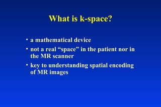
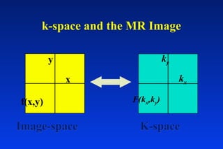
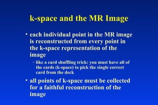
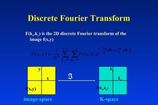
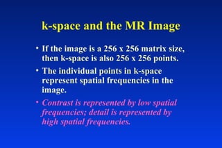
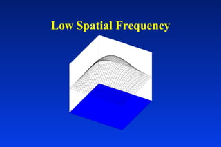
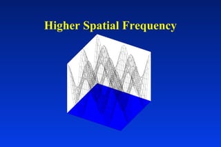
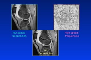
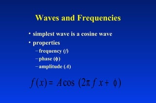
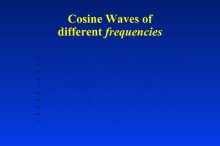
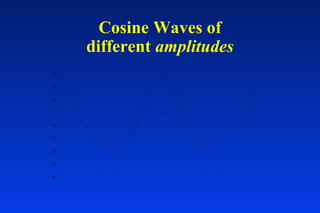
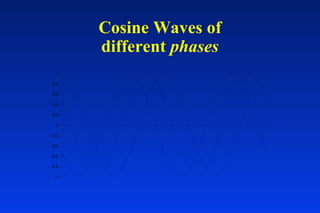
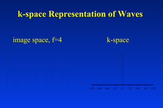
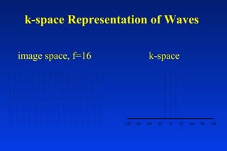
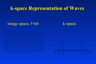
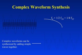
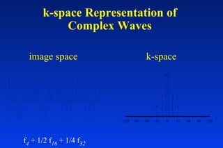
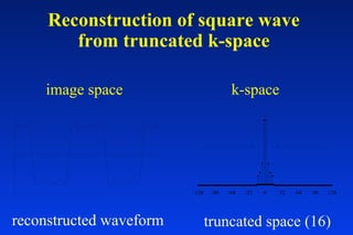
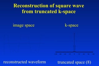
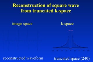
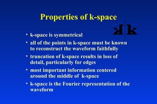
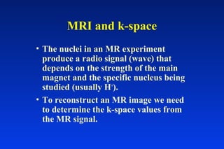
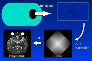
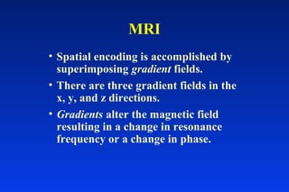
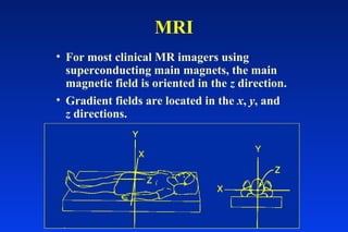
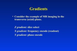
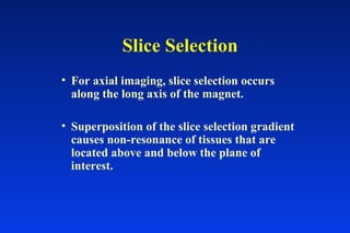
Topics
• spatial encoding - part 1
K-space, the path to MRI.K-space, the path to MRI.
ENTER IF YOU DAREENTER IF YOU DARE
What is k-space?
• a mathematical device
• not a real “space” in the patient nor in
the MR scanner
• key to understanding spatial encoding
of MR images
k-space and the MR Image
x
y
f(x,y)
kx
ky
K-spaceK-space
F(kx,ky)
Image-spaceImage-space
k-space and the MR Image
• each individual point in the MR image
is reconstructed from every point in
the k-space representation of the
image
– like a card shuffling trick: you must have all of
the cards (k-space) to pick the single correct
card from the deck
• all points of k-space must be collected
for a faithful reconstruction of the
image
Discrete Fourier Transform
F(kx,ky) is the 2D discrete Fourier transform of the
image f(x,y)
x
y
f(x,y)
kx
ky
ℑ
K-space
F(kx,ky)
f x y
N
F k k e
xk yk
kk
x y
j
N
x j
N
yNN
yx
( , ) ( , )=
+






=
−
=
−
∑∑
1
2
2 2
0
1
0
1 π π
image-space
k-space and the MR Image
• If the image is a 256 x 256 matrix size,
then k-space is also 256 x 256 points.
• The individual points in k-space
represent spatial frequencies in the
image.
• Contrast is represented by low spatial
frequencies; detail is represented by
high spatial frequencies.
Low Spatial Frequency
Higher Spatial Frequency
low spatial
frequencies
high spatial
frequencies
all
frequencies
Spatial Frequencies
• low frequency = contrast
• high frequency = detail
• The most abrupt change occurs at an
edge. Images of edges contain the
highest spatial frequencies.
Waves and Frequencies
• simplest wave is a cosine wave
• properties
–frequency (f)
–phase (φ)
–amplitude (A)
f x A f x( ) cos ( )= +2π φ
Cosine Waves of
different frequencies
-1
-0.8
-0.6
-0.4
-0.2
0
0.2
0.4
0.6
0.8
1
Cosine Waves of
different amplitudes
-4
-3
-2
-1
0
1
2
3
4
Cosine Waves of
different phases
-1
-0.8
-0.6
-0.4
-0.2
0
0.2
0.4
0.6
0.8
1
k-space Representation of Waves
image space, f=4 k-space
-128 -96 -64 -32 0 32 64 96 128
k-space Representation of Waves
image space, f=16 k-space
-128 -96 -64 -32 0 32 64 96 128
k-space Representation of Waves
image space, f=64 k-space
-128 -96 -64 -32 0 32 64 96 128
Complex Waveform Synthesis
f4 + 1/2 f16 + 1/4 f32
Complex waveforms can be
synthesized by adding simple
waves together.
k-space Representation of
Complex Waves
f4 + 1/2 f16 + 1/4 f32
-128 -96 -64 -32 0 32 64 96 128
image space k-space
k-space Representation of
Complex Waves
“square” wave
image space k-space
-128 -96 -64 -32 0 32 64 96 128
Reconstruction of square wave
from truncated k-space
truncated space (16)
image space k-space
-128 -96 -64 -32 0 32 64 96 128
reconstructed waveform
Reconstruction of square wave
from truncated k-space
truncated space (8)
image space k-space
-128 -96 -64 -32 0 32 64 96 128
reconstructed waveform
Reconstruction of square wave
from truncated k-space
truncated space (240)
image space k-space
-128 -96 -64 -32 0 32 64 96 128
reconstructed waveform
Properties of k-space
• k-space is symmetrical
• all of the points in k-space must be known
to reconstruct the waveform faithfully
• truncation of k-space results in loss of
detail, particularly for edges
• most important information centered
around the middle of k-space
• k-space is the Fourier representation of the
waveform
MRI and k-space
• The nuclei in an MR experiment
produce a radio signal (wave) that
depends on the strength of the main
magnet and the specific nucleus being
studied (usually H+
).
• To reconstruct an MR image we need
to determine the k-space values from
the MR signal.
RF signal
A/D
conversion
image space
FT
k-space
MRI
• Spatial encoding is accomplished by
superimposing gradient fields.
• There are three gradient fields in the
x, y, and z directions.
• Gradients alter the magnetic field
resulting in a change in resonance
frequency or a change in phase.
MRI
• For most clinical MR imagers using
superconducting main magnets, the main
magnetic field is oriented in the z direction.
• Gradient fields are located in the x, y, and
z directions.
MRI
• The three magnetic gradients work together
to encode the NMR signal with spatial
information.
• Remember: the resonance frequency
depends on the magnetic field strength.
Small alterations in the magnetic field by the
gradient coils will change the resonance
frequency.
Gradients
• Consider the example of MR imaging in the
transverse (axial) plane.
Z gradient: slice select
X gradient: frequency encode (readout)
Y gradient: phase encode
Slice Selection
• For axial imaging, slice selection occurs
along the long axis of the magnet.
• Superposition of the slice selection gradient
causes non-resonance of tissues that are
located above and below the plane of
interest.

240 spatial encoding

  • 1.
  • 2.
    K-space, the pathto MRI.K-space, the path to MRI. ENTER IF YOU DAREENTER IF YOU DARE
  • 3.
    What is k-space? •a mathematical device • not a real “space” in the patient nor in the MR scanner • key to understanding spatial encoding of MR images
  • 4.
    k-space and theMR Image x y f(x,y) kx ky K-spaceK-space F(kx,ky) Image-spaceImage-space
  • 5.
    k-space and theMR Image • each individual point in the MR image is reconstructed from every point in the k-space representation of the image – like a card shuffling trick: you must have all of the cards (k-space) to pick the single correct card from the deck • all points of k-space must be collected for a faithful reconstruction of the image
  • 6.
    Discrete Fourier Transform F(kx,ky)is the 2D discrete Fourier transform of the image f(x,y) x y f(x,y) kx ky ℑ K-space F(kx,ky) f x y N F k k e xk yk kk x y j N x j N yNN yx ( , ) ( , )= +       = − = − ∑∑ 1 2 2 2 0 1 0 1 π π image-space
  • 7.
    k-space and theMR Image • If the image is a 256 x 256 matrix size, then k-space is also 256 x 256 points. • The individual points in k-space represent spatial frequencies in the image. • Contrast is represented by low spatial frequencies; detail is represented by high spatial frequencies.
  • 8.
  • 9.
  • 10.
  • 11.
    Spatial Frequencies • lowfrequency = contrast • high frequency = detail • The most abrupt change occurs at an edge. Images of edges contain the highest spatial frequencies.
  • 12.
    Waves and Frequencies •simplest wave is a cosine wave • properties –frequency (f) –phase (φ) –amplitude (A) f x A f x( ) cos ( )= +2π φ
  • 13.
    Cosine Waves of differentfrequencies -1 -0.8 -0.6 -0.4 -0.2 0 0.2 0.4 0.6 0.8 1
  • 14.
    Cosine Waves of differentamplitudes -4 -3 -2 -1 0 1 2 3 4
  • 15.
    Cosine Waves of differentphases -1 -0.8 -0.6 -0.4 -0.2 0 0.2 0.4 0.6 0.8 1
  • 16.
    k-space Representation ofWaves image space, f=4 k-space -128 -96 -64 -32 0 32 64 96 128
  • 17.
    k-space Representation ofWaves image space, f=16 k-space -128 -96 -64 -32 0 32 64 96 128
  • 18.
    k-space Representation ofWaves image space, f=64 k-space -128 -96 -64 -32 0 32 64 96 128
  • 19.
    Complex Waveform Synthesis f4+ 1/2 f16 + 1/4 f32 Complex waveforms can be synthesized by adding simple waves together.
  • 20.
    k-space Representation of ComplexWaves f4 + 1/2 f16 + 1/4 f32 -128 -96 -64 -32 0 32 64 96 128 image space k-space
  • 21.
    k-space Representation of ComplexWaves “square” wave image space k-space -128 -96 -64 -32 0 32 64 96 128
  • 22.
    Reconstruction of squarewave from truncated k-space truncated space (16) image space k-space -128 -96 -64 -32 0 32 64 96 128 reconstructed waveform
  • 23.
    Reconstruction of squarewave from truncated k-space truncated space (8) image space k-space -128 -96 -64 -32 0 32 64 96 128 reconstructed waveform
  • 24.
    Reconstruction of squarewave from truncated k-space truncated space (240) image space k-space -128 -96 -64 -32 0 32 64 96 128 reconstructed waveform
  • 25.
    Properties of k-space •k-space is symmetrical • all of the points in k-space must be known to reconstruct the waveform faithfully • truncation of k-space results in loss of detail, particularly for edges • most important information centered around the middle of k-space • k-space is the Fourier representation of the waveform
  • 26.
    MRI and k-space •The nuclei in an MR experiment produce a radio signal (wave) that depends on the strength of the main magnet and the specific nucleus being studied (usually H+ ). • To reconstruct an MR image we need to determine the k-space values from the MR signal.
  • 27.
  • 28.
    MRI • Spatial encodingis accomplished by superimposing gradient fields. • There are three gradient fields in the x, y, and z directions. • Gradients alter the magnetic field resulting in a change in resonance frequency or a change in phase.
  • 29.
    MRI • For mostclinical MR imagers using superconducting main magnets, the main magnetic field is oriented in the z direction. • Gradient fields are located in the x, y, and z directions.
  • 30.
    MRI • The threemagnetic gradients work together to encode the NMR signal with spatial information. • Remember: the resonance frequency depends on the magnetic field strength. Small alterations in the magnetic field by the gradient coils will change the resonance frequency.
  • 31.
    Gradients • Consider theexample of MR imaging in the transverse (axial) plane. Z gradient: slice select X gradient: frequency encode (readout) Y gradient: phase encode
  • 32.
    Slice Selection • Foraxial imaging, slice selection occurs along the long axis of the magnet. • Superposition of the slice selection gradient causes non-resonance of tissues that are located above and below the plane of interest.