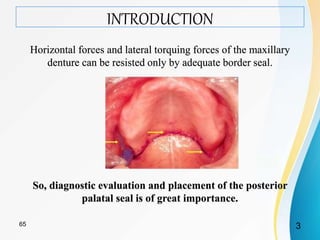
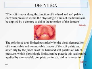
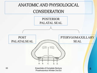
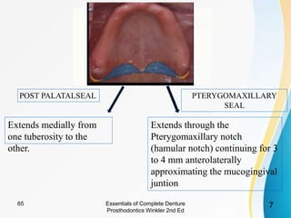
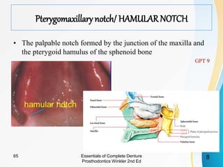
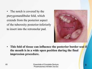
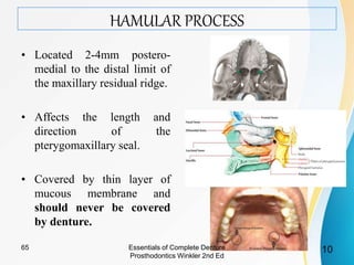
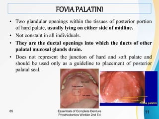
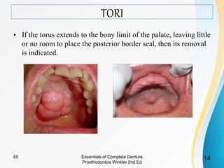
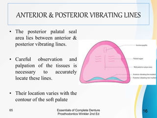
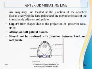
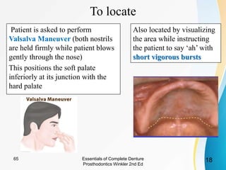
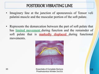
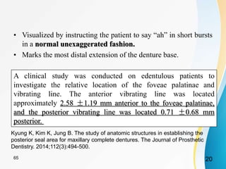
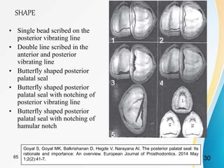
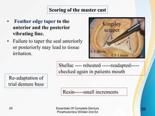
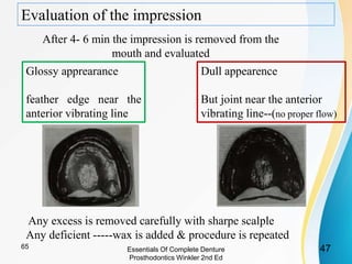
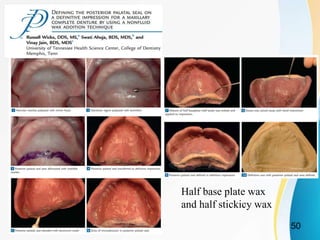
2. CONTENTS
• Introduction
• Definition
• Functions of posterior palatal seal
• Anatomical and physiological considerations
• Anterior & Posterior vibrating lines
• Classification of soft palate
• Parameters of posterior palatal seal
• Rationale behind recording posterior palatal seal.
• The techniques used to mark posterior palatal seal
• Failure in recording posterior palatal seal
• Adding posterior palatal seal to the existing denture
• Conclusion
265
3. INTRODUCTION
Horizontal forces and lateral torquing forces of the maxillary
denture can be resisted only by adequate border seal.
So, diagnostic evaluation and placement of the posterior
palatal seal is of great importance.
365
4. DEFINITION
“The soft tissues along the junction of the hard and soft palates
on which pressure within the physiologic limits of the tissues can
be applied by a denture to aid in the retention of the denture”
The soft tissue area limited posteriorly by the distal demarcation
of the movable and nonmovable tissues of the soft palate and
anteriorly by the junction of the hard and soft palates on which
pressure, within physiologic limits, can be placed; this seal can be
applied by a removable complete denture to aid in its retention
465
5. FUNCTIONS OF POSTERIOR PALATAL SEAL
1. Retention of the maxillary denture base by resisting the
horizontal forces and lateral torquing of the maxillary denture.
2. Maintains contact of the denture with the anterior portion of
soft palate during functional movements.
3. Reduces patient’s awareness of the denture and reduction
in the gag reflex as there is no separation of denture base and
soft palate during normal functional movements.
4. Reduces food accumulation beneath the posterior aspect of
the denture due to proper utilization of tissue compressibility.
when contact occurs between the
tongue and the posterior end of the denture base as the
posterior denture will closely approximate the soft palatal
tissues.
6. Compensate for the volumetric shrinkage that occurs during
the polymerization of methylmethacrylate resin.
565
7. Extends medially from
one tuberosity to the
other.
Extends through the
Pterygomaxillary notch
(hamular notch) continuing for 3
to 4 mm anterolaterally
approximating the mucogingival
juntion
POST PALATALSEAL PTERYGOMAXILLARY
SEAL
Essentials of Complete Denture
Prosthodontics Winkler 2nd Ed
765
8. Pterygomaxillary notch/ HAMULARNOTCH
• The palpable notch formed by the junction of the maxilla and
the pterygoid hamulus of the sphenoid bone
Essentials of Complete Denture
Prosthodontics Winkler 2nd Ed
865
9. • The notch is covered by the
pterygomandibular fold, which
extends from the posterior aspect
of the tuberosity posterior-inferiorly
to insert into the retromolar pad.
• This fold of tissue can influence the posterior border seal if
the mouth is in a wide open position during the final
impression procedure.
Essentials of Complete Denture
Prosthodontics Winkler 2nd Ed
965
10. HAMULAR PROCESS
• Located 2-4mm postero-
medial to the distal limit of
the maxillary residual ridge.
• Affects the length and
direction of the
pterygomaxillary seal.
• Covered by thin layer of
mucous membrane and
should never be covered
by denture.
Essentials of Complete Denture
Prosthodontics Winkler 2nd Ed
1065
11. FOVIA PALATINI
• Two glandular openings within the tissues of posterior portion
of hard palate, usually lying on either side of midline.
• Not constant in all individuals.
• They are the ductal openings into which the ducts of other
palatal mucosal glands drain.
• Does not represent the junction of hard and soft palate and
should be used only as a guideline to placement of posterior
palatal seal.
Essentials of Complete Denture
Prosthodontics Winkler 2nd Ed
1165
12. Disagreements regarding position-
• Swenson (1970) :- Vibrating line 2mm in front of fovea
palatini.
• Lye (1975) :- 1.31mm anterior to the anterior vibrating line.
• Chen (1981) :- Located either on or behind the anterior
vibrating line.
• Boucher stated that the vibrating line is usually located 2mm
anterior to the fovea palatinae
1265
13. MID PALATAL RAPHAE
• This overlies the medial palatal suture, contains little or no
submucosa and will tolerate little compression.
• According to Heartwell and Rahn, this band of tissues is not
meant to be compressed, rather should be relieved if prominent
when prominent
midpalatal fissure
present, extends
onto the soft
palate.
carefully
reproduced in the
master cast
posterior seal should
be extended into this
fissure to ensure
proper peripheral
seal.
Essentials of Complete Denture
Prosthodontics Winkler 2nd Ed
1365
14. TORI
• If the torus extends to the bony limit of the palate, leaving little
or no room to place the posterior border seal, then its removal
is indicated.
Essentials of Complete Denture
Prosthodontics Winkler 2nd Ed
1465
15. CONSISTENCY OF THE SALIVA.
• Thick saliva can create hydrostatic pressure in the area
anterior to the posterior palatal seal, resulting in a downward
dislodging force exerted upon the denture base.
According to Watt and MacGregor,
• A fine line or Cupid’s bow can be scribed on the master cast,
anterior to the cluster of palatal mucous glands (and distal to
any torus that is present).
• This extension of the posterior palatal seal line will contain the
thick mucus in the posterior part of the denture to provide a seal
even if the posterior portion of the denture base is slightly out
of contact with the palatal tissues.
Essentials of Complete Denture
Prosthodontics Winkler 2nd Ed
1565
16. ANTERIOR & POSTERIOR VIBRATING LINES
• The posterior palatal seal
area lies between anterior &
posterior vibrating lines.
• Careful observation and
palpation of the tissues is
necessary to accurately
locate these lines.
• Their location varies with the
contour of the soft palate
Essentials of Complete Denture
Prosthodontics Winkler 2nd Ed
1665
17. ANTERIOR VIBRATING LINE
• An imaginary line located at the junction of the attached
tissues overlying the hard palate and the movable tissues of the
immediately adjacent soft palate.
• Cupid's bow shaped due to the projection of posterior nasal
spine.
• Always on soft palatal tissues.
• Should not be confused with junction between hard and
soft palate.
Essentials of Complete Denture
Prosthodontics Winkler 2nd Ed
1765
18. To locate
Patient is asked to perform
Valsalva Maneuver (both nostrils
are held firmly while patient blows
gently through the nose)
This positions the soft palate
inferiorly at its junction with the
hard palate
Also located by visualizing
the area while instructing
the patient to say ‘ah’ with
short vigorous bursts
Essentials of Complete Denture
Prosthodontics Winkler 2nd Ed
1865
19. POSTERIOR VIBRATING LINE
• Imaginary line at the junction of aponeurosis of Tensor veli
palatini muscle and the muscular portion of the soft palate.
• Represents the demarcation between the part of soft palate that
has limited movement during function and the remainder of
soft palate that is markedly displaced during functional
movements.
Essentials of Complete Denture
Prosthodontics Winkler 2nd Ed
1965
20. • Visualized by instructing the patient to say “ah” in short bursts
in a normal unexaggerated fashion.
• Marks the most distal extension of the denture base.
A clinical study was conducted on edentulous patients to
investigate the relative location of the foveae palatinae and
vibrating line. The anterior vibrating line was located
approximately 2.58 ±1.19 mm anterior to the foveae palatinae,
and the posterior vibrating line was located 0.71 ±0.68 mm
posterior.
Kyung K, Kim K, Jung B. The study of anatomic structures in establishing the
posterior seal area for maxillary complete dentures. The Journal of Prosthetic
Dentistry. 2014;112(3):494-500.
2065
21. SOFT PALATE
Part of a dual valve system which separates the oropharynx from
the oral space and the nasopharynx from the nasal space.
The function of the soft palate in these dual valving actions
requires freedom of movement in three dimensions or planes of
space, i.e., superoinferiorly, mediolaterally and anteroposteriorly.
An impression should be made when the soft palate is placed at a
desired denture border position.
The functional position is achieved when patient is seated in
upright position, with head flexed 30 degrees forward and placing
the tongue under tension against either on the handle of
impression tray or dentist’s fingers, and should not protrude
beyond lips.
dimensions and displacement patterns of posterior palatal seal, Silverman, j prosthet dent, may 19712165
23. Clinical Significance
• Tensor Veli Palatini - When taut, can influence the denture
contour in the hamular notch area.
• Levator Veli Palatini - Closing of the oropharynx from the
nasopharynx during swallowing and determining the position
of the vibrating line.
• Palatoglossus – On contraction, draw the tongue and soft
palate towards each other and causes lateral pressure to the
lingual extension of the mandibular denture.
Arthur O. Rahn & Charles M. Heartwell, Elsevier, Syllabus of complete dentures,4th edition
2365
24. CLASSIFICATION OF SOFTPALATE
• Based upon the angle the soft palate makes with the hard
palate.
– The more acute the angle of the soft palate in relation to
the hard palate, more muscular activity will be necessary
to effect velopharyngeal closure (closing of the
nasopharynx).
• So the more the soft palate is markedly displaced in
function, the less that can be covered by the denture base.
• Determined when the patient is in upright position with the
head held erect.
Essentials of Complete Denture
Prosthodontics Winkler 2nd Ed
2465
25. CLASS I
• A soft palate that is rather horizontal as it extends posteriorly
with minimal muscular activity.
• Wide posterior palatal seal
• Most favorable configuration as more tissue surface can be
covered,yielding potentilaly more retentive denture base
Essentials of Complete Denture
Prosthodontics Winkler 2nd Ed
2565
26. • CLASS II
Palatal contours between a class I and class II.
• CLASS III
• Most acute contour in relation to the hard palate
• Marked elevation of the musculature to create velopharyngeal closure
• Seen along with a high V-shaped palatal vault usually.
• Smaller in width but deeper posterior palatal seal area
Essentials of Complete Denture
Prosthodontics Winkler 2nd Ed
2665
27. PALATAL THROAT FORM CLASSIFICATION ACCORDING TO
• Class I :- Large and normal in form
with a relatively immovable band of
resilient tissue 5-12 mm distal to a line
drawn across distal edge of the
tuberosities.
• Class II :- Medium size and normal
in form with relatively immovable
resilient band of tissue 3-5 mm distal to
a line drawn across distal edge of the
tuberosities.
• Class III :- Usually accompanies a
small maxilla. The curtain of soft
tissues turns down abruptly 3-5 mm
anterior to a line drawn across distal
edge of the tuberosities.
House MM. The relationship of oral examination to dental diagnosis. J Prosthet Dent 1958;8:208-19. 2768
28. PARAMETERS OF POSTERIOR PALATAL SEAL
• Posterior palatal seal has specific characteristics with different
parameters,
• Its variable in its
– size,
– shape
– location
• Depends on anatomical configuration of soft and hard palatal,
their relationship, muscle coordination, and amount of tissue
displaceability.
Goyal S, Goyal MK, Balkrishanan D, Hegde V, Narayana AI. The posterior palatal seal: Its
rationale and importance: An overview. European Journal of Prosthodontics. 2014 May
1;2(2):41-7.
2865
29. SIZE:
• According to Hardy and
Kapur (1958) , the dimension
of PPS was 2 mm at the
midpalatal region and
hamular notch and 4mm at the
greatest curvature region of
PPS.
• Silverman performed a study
on 92 patients & found the
following –
The greatest mean
anteroposterior width of PPS
is 8.0 mm (with 5-12 mm of
range) Goyal S, Goyal MK, Balkrishanan D, Hegde V, Narayana AI. The posterior
palatal seal: Its rationale and importance: An overview. European Journal of
Prosthodontics. 2014 May 1;2(2):41-7.
2965
30. SHAPE
• Single bead scribed on the
posterior vibrating line
• Double line scribed in the
anterior and posterior
vibrating line
• Butterfly shaped posterior
palatal seal
• Butterfly shaped posterior
palatal seal with notching of
posterior vibrating line
• Butterfly shaped posterior
palatal seal with notching of
hamular notch
Goyal S, Goyal MK, Balkrishanan D, Hegde V, Narayana AI. The posterior palatal seal: Its
rationale and importance: An overview. European Journal of Prosthodontics. 2014 May
1;2(2):41-7. 3065
31. Variations used with different shaped soft palate based
on the classification.
• Class 1: A butterfly
shaped posterior palatal
seal with 3-4 mm width
• Class 2: Posterior
palatal seal is narrow
with 2-3 mm of width
• Class 3: A single
beading made on the
posterior vibrating line.
Goyal S, Goyal MK, Balkrishanan D, Hegde V, Narayana AI. The posterior palatal seal: Its
rationale and importance: An overview. European Journal of Prosthodontics. 2014 May
1;2(2):41-7.
3165
32. LOCATION
• Location of posterior palatal seal is not consistent and show lot
of variation but on an average anterior vibrating line is 1.31
mm distal to fovea palatine.
Goyal S, Goyal MK, Balkrishanan D, Hegde V, Narayana AI. The posterior palatal seal: Its
rationale and importance: An overview. European Journal of Prosthodontics. 2014 May
1;2(2):41-7.
3265
33. To establish positive contact posteriorly to prevent the final
impression material from sliding down the pharynx.
To serve as a guide for positioning the impression tray,
especially if a shim has been used within the tray to
establish the borders.
To create slight displacement of the soft palate.
To determine if adequate retention and seal of the potential
denture border is present.
Essentials of Complete Denture
Prosthodontics Winkler 2nd Ed
3365
34. POINTS TO BE NOTED
• Determine the type of soft palate during initial examination.
• Transfer the posterior extent onto the custom tray.
• 1-2 mm distal to the expected denture border should be present
in the impression tray to protect against any overtrimming of
the processed denture base.
3465
35. Classification of techniques of recording PPS-
Hardy and Kapur (1958) –
• Functional :- Final impression is border molded in PPS area
with soft stick modeling compound / wax by sucking
movements performed by the patient.
• Semi functional :- Border molding is done by the dentist.
• Empirical :- Developed on the cast by grooving the cast to the
desired depth.
Natarajan R, Ramesh P, Selvaraj S, Mohan J. Evolution of Techniques in Recording Posterior Palatal
Seal–A Review. Journal of Academy of Dental Education.;13:17.
3565
36. The techniques used to mark posterior palatal seal
1. Conventional technique
2. Fluid wax technique
3. Arbitrary scraping of the master cast
3665 Essentials Of Complete Denture
Prosthodontics Winkler 2nd Ed
37. CONVENTIONAL TECHNIQUE
(winkler)
After accurate & fully extended final impression has been
made,boxed and poured, master cast is obtained
Adaption of trial denture base
Autopolymerising
resin
Shellac
Patient in upright position Astringent mouth
wash------remove
stringy saliva
drying with gauze
piece
37
65
38. Transfer the marking onto the denture base and then onto the cast
Trimming of the trial base
Returning to the patient's mouth,palatal tissue anterior to the
posterior vibrating line is palpated to check for
compressibility in terms of width & depth
Locating anterior vibrating line and
transfer of the same
short vigorous
burst of “ah”
valsalva
maneuver
Anterior vibrating line-----cupid's bow shape
38
65
39. • Feather edge taper to the
anterior and the posterior
vibrating line.
• Failure to taper the seal anteriorly
or posteriorly may lead to tissue
irritation.
Scoring of the master cast
3965 Essentials Of Complete Denture
Prosthodontics Winkler 2nd Ed
1-1.5mm.
0.5-1.0mm
Kingsley
scraper
Re-adaptation of
trial denture base
Shellac ---- reheated -----readapted-----
checked again in patients mouth
Resin-----small increments
40. • Evaluation of the seal
while patient says “ah” in short unexaggerated manner.
• No space adequate seal
• Presence of space further increase the depth.
CONVENTIONALTECHNIQUE
(WINKLER)
4065 Essentials Of Complete Denture
Prosthodontics Winkler 2nd Ed
41. CONVENTIAL TECHNIQUE
Rententive Record base--->
Accurate maxillomandibular
relation
Pyschological security to patient
The dentist is able to understand
the retentive qualities of the
finished denture.
Ease adjustment period
Not a physiologic method
depends on accurate
transfer of the vibrating
lines.
Tissue overcompression
Essentials Of Complete Denture
Prosthodontics Winkler 2nd Ed
4165
42. FLUID WAX TECHNIQUE
(functional technique or physiological technique)
• All the procedures for location and transfer marking of the
anterior and posterior vibrating lines are same as for the
conventional approach.
• Indelible transfer markings are recorded on the final wash
impression.
• Zinc oxide eugenol /plaster are preferred over the elastic
impression material, as they set rigid.
• Chances of distortion of relation between added wax on the
posterior border and rest of the denture bearing surface
Essentials Of Complete Denture
Prosthodontics Winkler 2nd Ed
4265
43. • 4 types of wax –
1. Iowa Wax (White) – Dr. Earl S. Smith
2. Korecta Wax no.4 (Orange) – Dr. O.C. Applegate
3. H-L physiologic paste (Yellow-White) – Dr. C.S.
Harkins
4. Adaptol (Green) – Dr. Nathan G. Kyne
• Designed to flow at mouth temperature.
Essentials Of Complete Denture
Prosthodontics Winkler 2nd Ed
4365
45. PROCEDURE
Melted wax in excess of the estimated
depth is painted onto the outlined seal area
on impression
allowed to cool and carried to the mouth
Impression held under gentle pressure for
4- 6 mintues
Position of head and tongue
Essentials Of Complete Denture
Prosthodontics Winkler 2nd Ed
4565
46. POSITION OF HEAD AND TONGUE
Soft palate Functionally
depressed possition
Prevents progression of material
down the pharynx
Patient should not protrude
tongue beyond the
approximated position of
incisors ---------tray handle
The patient is asked to periodically rotate the head so that all
functional positions of the soft palate are recorded.
30
degrees
Essentials Of Complete Denture
Prosthodontics Winkler 2nd Ed
4665
47. Evaluation of the impression
After 4- 6 min the impression is removed from the
mouth and evaluated
Glossy apprearance
feather edge near the
anterior vibrating line
Dull appearence
But joint near the anterior
vibrating line--(no proper flow)
Any excess is removed carefully with sharpe scalple
Any deficient -----wax is added & procedure is repeated
Essentials Of Complete Denture
Prosthodontics Winkler 2nd Ed
4765
48. PRECAUTIONS:
• The patient should not protrude his tongue beyond the
approximated position of the incisal edge as this may shorten
the posterior border of the final impression.
• The patient should be cautioned against rinsing with cold
water as this may contract the tissues and reduce the flow
properties of wax.
Essentials Of Complete Denture
Prosthodontics Winkler 2nd Ed
4865
49. FLUID WAX TECHNIQUE
Physiological technique
overcompression is
avoided
Added Retention
Mechanical scraping is
avoided
Time consuming
Difficulty in handling
material and extra care
during boxing procedure
Essentials Of Complete Denture
Prosthodontics Winkler 2nd Ed
4965
51. MODIFIED BORDER MOULDING TECHNIQUE
• In an Indian set-up, we usually follow this modified version
of the fluid wax technique.
• Here before making the definitive impression, PPS Area is
recorded in the Low-fusing impression compound.
• Followed by Definitive Impression.
Posterior Palatal Seal (PPS): A brief review Journal of Scientific and Innovative Research
2014; 3(6): 602-605
5165
52. ARBITRARY SCRAPING OF THE MASTER CAST
• Anterior and the posterior vibrating lines are visualized by
examining the patient’s mouth and approximately marked and
scraped on the master cast.
• Least accurate and leaves the most to chance at insertion
appointment since it relies on dentist’s recollection of palatal
configuration and tissue compressibility.
Essentials Of Complete Denture
Prosthodontics Winkler 2nd Ed
5265
53. Other techniques
• Boucher’s Technique
• Bernard Levin’s Technique
• Swenson’s Technique
• Pound’s Technique
• Silverman’s Technique
• Hardy and KapurTechnique
Natarajan R, Ramesh P, Selvaraj S, Mohan J. Evolution of Techniques in Recording Posterior
Palatal Seal–A Review. Journal of Academy of Dental Education.;13:17.
5365
Emperical
methods
54.
55. TROUBLESHOOTING
• Under extension
• Over extension
• Under post damming
• Over post damming
5565 Essentials Of Complete Denture
Prosthodontics Winkler 2nd Ed
56. Source : Natarajan R, Ramesh P, Selvaraj S, Mohan J. Evolution of Techniques in
Recording Posterior Palatal Seal–A Review. Journal of Academy of Dental
Education.;13:17.
5665
57. • Moghadam and Scandrett suggest a procedure that utilizes the
fluid wax technique.
• After the wax has had an adequate chance to flow, the denture is
removed from the mouth and the anterior extent of the seal on
the denture is outlined.
ADDING POSTERIOR PALATAL SEAL TO THE EXISTING
DENTURE
Moghadam BScandrett F. A technique for adding the posterior palatal seal. The Journal of Prosthetic
Dentistry. 1974;32(4):443-447.
5765
58. • Utility wax is placed vertically across the palate, separating the
posterior two thirds from the anterior region, and extended
around the posterior portion of the denture.
• Stone is vibrated into the denture-wax surface outlined by the
utility wax. After the stone has set, the wax is eliminated and
the denture cleaned. The denture base is ground distal to the
anterior vibrating line that has been delineated by the indelible
pencil.
Moghadam BScandrett F. A technique for adding the posterior palatal seal. The
Journal of Prosthetic Dentistry. 1974;32(4):443-447.
5865
59. • A separating medium is applied to the stone cast
and the denture is then replaced on the stone
cast and held firmly with rubber bands.
• Autopolymerizing acrylic powder is sprinkled
between the denture base and the cast while held
on a vibrator and monomer is then added
dropwise.
• The cast and denture are placed in an upright
position until the initial set has taken place.
• They are then placed in a pressure pot with
water (140°F) for 20 minutes under 30 psi
pressure.
• After the cast and denture are separated, the
excess acrylic is trimmed and the border
polished lightly.
• Denture should be stored in water for 24 to 36
hours to avoid tissue irritation due to excess
monomer.
Moghadam BScandrett F. A technique for adding the
posterior palatal seal. The Journal of Prosthetic Dentistry.
1974;32(4):443-447.
5965
60. ADDING POSTERIOR PALATAL SEAL TO THE EXISTING
DENTURE
• Mark the vibrating line in the mouth with an indelible marker.
• Form the desired thickness and extension of the PPS on the denture in the
patient’s mouth with softened green modeling compound
• Transfer the locations of the vibrating line to the denture
• Make a cast of the intaglio surface of the denture with putty material; the cast
must include all of PPS addition and extend 5 to 6 mm posteriorly
• After putty material has set, use a scalpel to cut channels which will allow
excess autopolymerizing acrylic resin to escape.
• Remove the green stick compound and replace with autopolymerizing resin
in a pressure pot.
Ansari IH. A procedure for adding posterior palatal seal to an existing denture in dental office. J
Prosthet Dent 1994;72:449.
6065
61. Arthur Nimmo - Suggested correction of
posterior palatal seal by using a visible
light cured resin.
Identify and mark the vibrating line in the
mouth with an indelible marking stick
• Roughen the denture surface in the
posterior palatal seal area with a carbide
bur.
• Adapt the VLC resin
• Place the denture in the mouth and allow
it to remain in place for approximately
3 minutes. During this time the material
will flow.
• Position a hand-held visible light source
near the border of the denture and apply
light directly to the region for several
minutes.
• Remove any excess resin with a carbide
bur and smooth the junction between the
seal and the polished surface of the
denture.
Nimmo A. Correction of the posterior palatal seal by
using a visible light-cured resin: A clinical report. The
Journal of Prosthetic Dentistry. 1988;59(5):529-531.
ADDING POSTERIOR PALATAL SEAL TO THE EXISTING
DENTURE
6165
62. ADVANTAGES
1. No exothermic reaction to irritate the oral tissues.
2. Minimal volumetric shrinkage during curing.
3. More closely approximates a physiologic technique.
4. Can be performed with relatively little chair time.
DISADVANTAGE:
Cost of the curing unit
Nimmo A. Correction of the posterior palatal seal by
using a visible light-cured resin: A clinical report. The
Journal of Prosthetic Dentistry. 1988;59(5):529-531.
ADDING POSTERIOR PALATAL SEAL TO THE EXISTING
DENTURE
6265
63. CONCLUSION
• The recording of PPS is of great significance because it is vital
factor in establishing the peripheral seal which enhances
retention by utilizing the atmospheric pressure.
• The PPS of a maxillary complete denture can be established
during the making of the preliminary impression, during the
making of final impression, by scoring the final cast or by
incorporating the seal in the final denture.
• It is not a difficult procedure once you have an intimate
knowledge of the anatomy and physiology of the tissues of the
region.
6365
64. REFERENCES & cross references
• Sheldon Winkler ,A.I.T.B.S. Publishers,Essentials of complete denture
Prosthodontics,2nd edition
• Zarb Bolender, Mosby,Prosthodontic treatment for edentulous patients,12th
edition
• Arthur O. Rahn & Charles M. Heartwell, Elsevier,Textbook of complete
dentures,5th edition
• Hardy I R, Posterior border seal –its rationale and importance, J Prosthet
Dent 1958:8;386-97
• Silverman S.L. “Dimensions and displacement patterns of the posterior
palatal seal”. J Prosthet Dent 1971:25;470-88
• Winland RD, Young JM, Maxillary complete denture posterior palatal seal:
Variations in size, shape & location , J Prosthet Dent 1973:29;256-61
• Lye TL, The significance of the fovea palatine in complete denture
prosthodontics. J Prosthet Dent 1975:33;504-10
• Wicks R, Ahuja S, Jain V. Defining the posterior palatal seal on a definitive
impression for a maxillary complete denture by using a non fluid wax
addition technique. J Prosthet Dent 2014;112:1597-600
6465
65. • E Abd AlAziz O, A Baraka O, Y Farahat M. An In-Vivo Comparative
Study of Retention of Heat Cured and Thermoplastic Acrylic Resins in
Maxillary Complete Denture Bases Made with Different Posterior
Palatal Sealing Techniques. Al-Azhar Journal of Dental Science. 2018
Jul 1;21(3):237-43.
• Nikoukari H, A study of posterior palatal seals with varying palatal
forms, J Prosthet Dent 1975:34;605-13
• Nimmo A.,Correction of the posterior palatal seal by using a visible-
light cure resin : A clinical report J Prosthet Dent 1988:59;529-30
• Izharul Haque Ansari , A procedure for adding posterior palatal seal to
an existing denture in dental office, J Prosthet Dent 1994:72;449
• Izharul Haque Ansari “Establishing the posterior palatal seal during
the final impression stage”. J Prosthet Dent 1997:78;324-26
6565
Editor's Notes
The dentist who observes the fovea and utilizes these anatomic landmarks as the posterior extent of the denture base can deprive
his patients of from several millimetersThis, in turn, will have a direct effect upon the retentive potential of the denture base.
Sicher (1952) :- Posterior to the location of hard and soft palate.
Nagle and Sears (1958) :- Posterior limit of hard palate.
Fenn and associates (1961) :- Glandular region of soft palate.
a narrow, cordlike band of tissue extending between the posterior nasal spine and the aponeurosis of the tensor veli palatini muscle.
POST PALATAL SEAL:- Area between the anterior and posterior vibrating line
(Sharry’s Method)
The head and tongue position translate the mandible anteriorly The soft palate will then be passively brought downward and forward due to the indirect attachment of the soft palatal tissues to the body of the mandible and the insertion of the palatoglossus muscle into the side of the tongue.
of the seal are located on the either side of the midline, one third the distance anteriorly from the post vibrating line.
The tissue covering the median palatal raphe has little submucosa and cannot withstand the same compressive force on the tissues lateral to it.