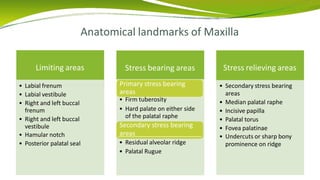
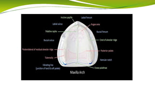
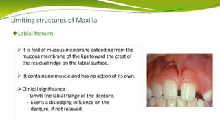
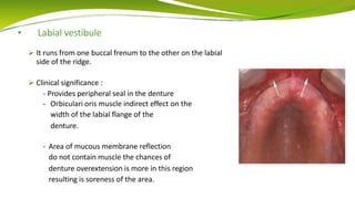
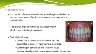
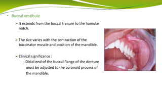
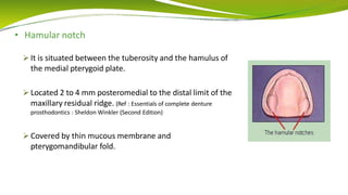
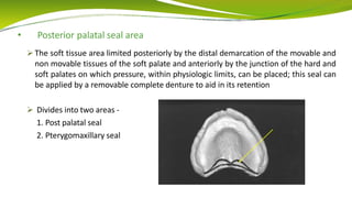
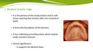
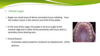
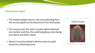
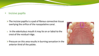
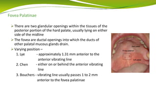
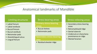
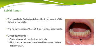
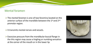
BY -
DR. POOJA LANGOTE
MDS 1st YEAR
Contents
• Definition
• Mucous membrane
• Bone
• Anatomical landmarks in maxilla –
- Lining areas
- Stress Bearing areas
- Stress Relieving areas
• Anatomical landmarks in mandible –
- Limiting areas
- Stress bearing areas
- Stress relieving areas
• References
What is Anatomical Landmark?
⚫According to the Glossary of Prosthodontic Terms, anatomical landmark is a
recognizable anatomic structure used as a point of reference.
⚫Sound knowledge of anatomical landmark is important, if one has to achieve the
retention, stability and support in complete denture.
Anatomical
landmarks
Limiting
structures
Supporting
structures
Relief areas
• Limiting structures or the peripheral structures
1. Determine the peripheral seal for the denture’s border.
2. Provide optimum extension of the denture border.
• Supporting structures or stress bearing areas
1. Acts as a foundation for the denture base.
2. Tolerate the stresses produced by masticatory forces when the denture is in
function.
• Relief areas or the stress relieving areas
1. Resorbs under constant load.
2. Have fragile structures within.
Mucous membrane
⚫Mucous membrane serves as a cushion between the denture base and the
supporting bone.
⚫It comprises stratified squamous epithelium.
⚫Consist of two layers
⚫The mucous membrane is attached to bone, the attachment occurs between the
submucosa and the periosteal covering of the bone.
⚫The submucosa is formed by connective tissue that varies in character from dense to
loose areolar tissue and also varies considerably in thickness.
⚫Thickness and the consistency of the submucous layer is responsible for providing
the support to the denture.
⚫Healthy residual ridge has the adequate thickness of the submucosa and it is firmly
attached to the underlying bone, hence withstands the pressure exerted by the
denture base.
⚫In case of very thin submucous layer, the mucous membrane will be non resilient and
can be easily traumazied under the pressure.
Osteology
⚫Osseous structures beneath the denture provides the support.
Bone
CORTICAL CANCELLOUS
⚫Cancellous bone :
- Trabecular pattern
- Can not withstand high amount of stresses
- Undergoes resorption under constant load
⚫Cortical bone :
- Compact in nature
- Can withstand the high amount of stresses
- Do not resorb readily
• Depending upon the type of underlying bone the stress bearing areas can be easily
marked out.
Anatomical landmarks of Maxilla
Limiting areas
• Labial frenum
• Labial vestibule
• Right and left buccal
frenum
• Right and left buccal
vestibule
• Hamular notch
• Posterior palatal seal
Stress bearing areas Stress relieving areas
• Secondary stress bearing
areas
• Median palatal raphe
• Incisive papilla
• Palatal torus
• Fovea palatinae
• Undercuts or sharp bony
prominence on ridge
Primary stress bearing
areas
• Firm tuberosity
• Hard palate on either side
of the palatal raphe
Secondary stress bearing
areas
• Residual alveolar ridge
• Palatal Rugue
Limiting structures of Maxilla
⚫Labial frenum
It is fold of mucous membrane extending from the
mucous membrane of the lips toward the crest of
the residual ridge on the labial surface.
 It contains no muscle and has no action of its own.
Clinical significance :
- Limits the labial flange of the denture.
- Exerts a dislodging influence on the
denture, if not relieved.
• Labial vestibule
 It runs from one buccal frenum to the other on the labial
side of the ridge.
 Clinical significance :
- Provides peripheral seal in the denture
- Orbiculari oris muscle indirect effect on the
width of the labial flange of the
denture.
- Area of mucous membrane reflection
do not contain muscle the chances of
denture overextension is more in this region
resulting is soreness of the area.
• Buccal frenum
 It is the fold of mucous membrane, extending from the buccal
mucous membrane reflection area towards the slope of the
residual ridge.
 The levator anguli oris muscle attaches beneath
the frenum, affecting its position.
 Clinical significance :
- Due to the action of orbicularis oris and the
buccinator muscles it requires more clearance.
- Overriding of denture on the frenum causes
denture dislodgement, soreness and pain in the region.
• Buccal vestibule
 It extends from the buccal frenum to the hamular
notch.
 The size varies with the contraction of the
buccinator muscle and position of the mandible.
 Clinical significance :
- Distal end of the buccal flange of the denture
must be adjusted to the coronoid process of
the mandible.
• Hamular notch
 It is situated between the tuberosity and the hamulus of
the medial pterygoid plate.
 Located 2 to 4 mm posteromedial to the distal limit of the
maxillary residual ridge. (Ref : Essentials of complete denture
prosthodontics : Sheldon Winkler (Second Edition)
 Covered by thin mucous membrane and
pterygomandibular fold.
⚫Clinical significance :
- Forms the posterior boundary of maxillary denture
- Retention
- Overextension : traumatized the mucous membrane covering
the pterygomandibular raphe and causes pain.
- Under extension : hampers denture retention.
• Posterior palatal seal area
 The soft tissue area limited posteriorly by the distal demarcation of the movable and
non movable tissues of the soft palate and anteriorly by the junction of the hard and
soft palates on which pressure, within physiologic limits, can be placed; this seal can
be applied by a removable complete denture to aid in its retention
 Divides into two areas -
1. Post palatal seal
2. Pterygomaxillary seal
 Clinical significance :
1. Retention
2. Reduction in the gag reflex
3. Reduce food accumulation
4. Compensate for the volumetric shrinkage of resin
Stress bearing areas of maxilla
Stress bearing area
Primary stress bearing area Secondary stress bearing area
Thicker mucosa
Underlying bone is cortical
bone that is less subject to
resorption.
Comparatively thinner
mucosa
Underlying bone is cancellous
bone which subject to
resorption with long-term
denture wear.
• Hard palate
 Anteriorly hard palate is formed by palatal shelves
of two maxillary bone. Posteriorly it has underlying
palatal bone.
 In anterolateral surface of hard palate, the
submucosa contains adipose tissue and
posterolaterally it contains glandular tissue.
 Clinical significance :
- Acts as a primary stress bearing area
• Maxillary tuberosity
 The posterior convexity of the maxillary body is termed
the maxillary tuberosity or tuber.
 Bulbous extension of residual ridge in 2nd or 3rd molar
region, terminating in hamular notch.
 Clinical significance :
- The alveolar tubercle supports the denture.
- It acts as the stress bearing area.
• Residual alveolar ridge
 It is the portion of the residual bone and its soft
tissue covering that remains after the removal of
teeth.
 It forms the foundation of the denture.
 It has underlying cancellous bone which resorbs
under constant stresses.
 Clinical significance :
- It supports the denture base.
• Palatal rugae
 Rugae are raised areas of dense connective tissue radiating
the median suture in the anterior one third of the palate.
from
 In the area of the rugae, the palate is set at an angle to the
residual ridge and is rather thinly covered by soft tissue and is a
secondary stress-bearing area.
 Clinical feature :
- It provides anterio-posterior resistance to displacement of the
denture.
Mid palatal raphe
 The median palatal suture is the area extending from
the incisive papilla to the distal end of the hard palate.
 The mucosa over this area is usually tightly attached,
non resilient and thin, the underlying bony union being
very dense and often raised.
 Hence it must be relived in denture base to avoid
trauma to underlying tissue.
• Incisive papilla
 The incisive papilla is a pad of fibrous connective tissue
overlying the orifice of the nasopalatine canal.
 In the edentulous mouth it may lie on or labial to the
crest of the residual ridge.
 Pressure on this area results in burning sensation in the
anterior third of the palate.
Fovea Palatinae
 There are two glandular openings within the tissues of the
posterior portion of the hard palate, usually lying on either
side of the midline
 The fovea are ductal openings into which the ducts of
other palatal mucous glands drain.
 Varying position –
1. Lye - approximately 1.31 mm anterior to the
anterior vibrating line
- either on or behind the anterior vibrating
line
2. Chen
3. Bouchers - vibrating line usually passes 1 to 2 mm
anterior to the fovea palatinae
 Clinical significance -
- Provide a guide to the posterior extent of the denture base. This inturn, will
have a direct effect upon the retentive potential of the denture base
- Thick ropey saliva layer can create the hydrostatic pressure in fluid and leads
to denture displacement
• Palatal torus
 Hyperplastic over growth of bone, called the palatal torus, is
seen in mid palatal area.
 They are covered by thin mucous membrane.
 For tori extending on the posterior most area of the denture
base, hampering the retention, relief in the denture base is
indicated.
 Surgical removal is indicated for the less extensive.
Anatomical landmarks of Mandible
Limiting structures
• Labial frenum
• Labial vestibule
• Buccal frenum
• Buccal vestibule
• Retromolar pads
• Alveololingual sulcus
• Lingual frenum
Stress bearing areas Stress relieving areas
• Secondary stress bearing
areas
• Retromylohyoid ridge
• Genial tubercle
• Undercuts or sharp bony
prominence on ridge
• Mental foramen
Primary stress bearing
areas
• Buccal shelves
• Retromolar pads
Secondary stress bearing
area
• Residual alveolar ridge
Labial frenum
 The mucolabial fold extends from the inner aspect of the
lip to the mandible.
 The frenum contains fibers of the orbicularis oris muscle.
 Clinical significance :
- Gives idea about the denture extension
- Notch in the denture base should be made to relieve
labial frenum.
Labial vestibule
 Mandibular labial vestibule extends from right buccal
frenum to the left.
 Two soft elevations, one on each side of the frenum.
These pads mark the origin of the mentalis muscle.
 Laterally, has arrangement of fibres of orbicularis oris.
 Clinical significance –
- Marks the extension of the denture border in this
region.
- Overextension on the mentalis muscle, leads to
displacement of mandibular denture.
Buccal frenum
 It is the fold or folds of mucous membrane, extending from
the buccal mucous membrane reflection area towards the
slope of the residual ridge.
 It is the dividing line between the labial and buccal
vestibules.
 It overlies depressor anguli oris muscle.
 Should be relived in denture to avoid displacement of
denture.
Buccal vestibule
 The buccal vestibule extends posteriorly from the buccal frenum to
the posterior lateral aspect of the retromolar pad.
 The extent of the buccal vestibule is influenced by the buccinator
muscle, which extends from the modiolus anteriorly to the
pterygomandibular raphe posteriorly.
 The distobuccal border of the buccal vestibule is bordered by the
vertical fibers of the masseter muscle.
 On activation masseter muscle cause a bulge in the buccinators
muscle, creating the masseteric notch in the posterior lateral
denture border.
Retromolar pads
 The retromolar pad is a triangular pad of tissue at the distal
end of the residual ridge.
 The anterior portion of the triangle is keratinized tissue of
the remnant gingiva of the third molar called the pear-
shaped.
 The posterior aspect of the triangle is thin, nonkeratinized
epithelium; loose connective tissue; glandular tissue; fibers
of the temporalis tendon and of the buccinator and superior
constrictor muscles; and the pterygomandibular raphe.
 The underlying bone is dense cortical bone because of the muscle attachments and
is resistant to resorption.
 Clinical significance :
- Primary stress bearing area
- Provides the peripheral posterior seal of the denture
- Soft tissue distal to the last molar approximates the retromolar pad at the
established occlusal vertical dimension
Alveololingual sulcus
 The alveololingual sulcus, which is the space between the residual ridge and
the tongue, extends from the lingual frenum to the retromylohyoid curtain.
 The lingual border is defined by the mylohyoid muscle along the entire
length of the mandible.
 The value of the border’s maximum lingual border extension is lateral
stability of the denture and reduction of food collection under the denture.
 The lingual border of the denture is studied in three
regions -
1. The anterior region : This extends from the
lingual frenum back to where the mylohyoid
ridge curves above the level of the sulcus
2. The middle region : This part extends from the
premylohyoid fossa to the distal end of the
mylohyoid ridge
3. The posterior region : Here the flange passes into
the retromylohyoid fossa
Lingual frenum
 It is a fold of mucous membrane seen when the tip of
the tongue is elevated.
 It is either cord like or fan-shaped and requires
functional freedom.
 It overlies genioglossus muscle.
 Clinical significance :
- Marks lingual extension of the denture border.
- Short frenum is corrected a it affects the stability of
denture.
Buccal shelf area
 Boundaries of buccal shelf area –
Anteriorly - Mandibular buccal frenum
Posteriorly - Anterior edge of the masseter muscle.
Medially
Laterally
Distally
- crest of the ridge
- boney external oblique ridge
- retromolar pad.
 The buccinator muscle fibers attach horizontally along the boney
oblique ridge.
 It is composed of dense cortical bone.
 It is at right angle to the direction of vertical occlusal force.
 Clinical significance : Acts as a primary stress bearing area.
Residual alveolar ridge
 The crest of the residual alveolar ridge is covered by fibrous
connective tissue
 The slopes of the ridge have cancellous bone and without a
good cortical bony plate covering it.
 Clinical significance
- Acts as a secondary stress bearing area.
Genial tubercle
 The genial tubercles are a dense cortical prominence at
the inferior border of the mandible at the lingual midline.
 They represent the muscle attachment of the
genioglossus and geniohyoid muscles.
 When there is extensive resorption of the residual ridge,
they often lie on the ridge and need to be relieved.
Mental foramen
 The mental foramen is one of two foramina located on the
anterior surface of the mandible between the 1st and 2nd
premolar region.
 It transmits mental nerves and vessels.
 Excessive pressure from the mandibular buccal flange in
the this region may cause a tingling or numbing sensation
at the corner of the mouth or in the lower lip
Retromyelohyoid ridge
 The mylohyoid ridge is a boney prominence along the lingual
aspect of the mandible.
 Gives attachment to mylohyoid muscle. Anteriorly, the muscle
is attached to the inferior border of the mandible, but
continuing posteriorly it is attached to this ridge and is nearer
the level of the alveolar ridge.
 This posterior prominence often requires relief in the denture
and may require surgical reduction.
Mandibular tori
 The torus mandibularis is found on the lingual cortical
surface of the mandible located in the premolar area.
 They are usually covered with keratinized, nonmobile
tissue.
 Require removal as they will severely limit the
extension of the denture in the floor of mouth.
 Smaller tori may only require relief in the denture.
References
⚫The glossary of prosthodontics terms (Ninth Edition)
⚫Boucher’s prosthodontics treatment for edentulous patients (Thirteen
Edition)
⚫Prosthodontic treatment for edentulous patients : Zarb-Bolender (Twelth
Edition)
⚫Syllabus of complete dentures : Charles Heartwell (Fourth Edition)
⚫Essentials of complete denture prosthodontics : Sheldon Winkler (Second
Edition)
Thank you!

anatomicallandmarksofmaxillaandmandibleautosaved-200820132830.pptx

  • 1.
    BY - DR. POOJALANGOTE MDS 1st YEAR
  • 2.
    Contents • Definition • Mucousmembrane • Bone • Anatomical landmarks in maxilla – - Lining areas - Stress Bearing areas - Stress Relieving areas • Anatomical landmarks in mandible – - Limiting areas - Stress bearing areas - Stress relieving areas • References
  • 3.
    What is AnatomicalLandmark? ⚫According to the Glossary of Prosthodontic Terms, anatomical landmark is a recognizable anatomic structure used as a point of reference. ⚫Sound knowledge of anatomical landmark is important, if one has to achieve the retention, stability and support in complete denture. Anatomical landmarks Limiting structures Supporting structures Relief areas
  • 4.
    • Limiting structuresor the peripheral structures 1. Determine the peripheral seal for the denture’s border. 2. Provide optimum extension of the denture border. • Supporting structures or stress bearing areas 1. Acts as a foundation for the denture base. 2. Tolerate the stresses produced by masticatory forces when the denture is in function. • Relief areas or the stress relieving areas 1. Resorbs under constant load. 2. Have fragile structures within.
  • 5.
    Mucous membrane ⚫Mucous membraneserves as a cushion between the denture base and the supporting bone. ⚫It comprises stratified squamous epithelium. ⚫Consist of two layers
  • 6.
    ⚫The mucous membraneis attached to bone, the attachment occurs between the submucosa and the periosteal covering of the bone. ⚫The submucosa is formed by connective tissue that varies in character from dense to loose areolar tissue and also varies considerably in thickness.
  • 7.
    ⚫Thickness and theconsistency of the submucous layer is responsible for providing the support to the denture. ⚫Healthy residual ridge has the adequate thickness of the submucosa and it is firmly attached to the underlying bone, hence withstands the pressure exerted by the denture base. ⚫In case of very thin submucous layer, the mucous membrane will be non resilient and can be easily traumazied under the pressure.
  • 9.
    Osteology ⚫Osseous structures beneaththe denture provides the support. Bone CORTICAL CANCELLOUS ⚫Cancellous bone : - Trabecular pattern - Can not withstand high amount of stresses - Undergoes resorption under constant load
  • 10.
    ⚫Cortical bone : -Compact in nature - Can withstand the high amount of stresses - Do not resorb readily • Depending upon the type of underlying bone the stress bearing areas can be easily marked out.
  • 11.
    Anatomical landmarks ofMaxilla Limiting areas • Labial frenum • Labial vestibule • Right and left buccal frenum • Right and left buccal vestibule • Hamular notch • Posterior palatal seal Stress bearing areas Stress relieving areas • Secondary stress bearing areas • Median palatal raphe • Incisive papilla • Palatal torus • Fovea palatinae • Undercuts or sharp bony prominence on ridge Primary stress bearing areas • Firm tuberosity • Hard palate on either side of the palatal raphe Secondary stress bearing areas • Residual alveolar ridge • Palatal Rugue
  • 13.
    Limiting structures ofMaxilla ⚫Labial frenum It is fold of mucous membrane extending from the mucous membrane of the lips toward the crest of the residual ridge on the labial surface.  It contains no muscle and has no action of its own. Clinical significance : - Limits the labial flange of the denture. - Exerts a dislodging influence on the denture, if not relieved.
  • 14.
    • Labial vestibule It runs from one buccal frenum to the other on the labial side of the ridge.  Clinical significance : - Provides peripheral seal in the denture - Orbiculari oris muscle indirect effect on the width of the labial flange of the denture. - Area of mucous membrane reflection do not contain muscle the chances of denture overextension is more in this region resulting is soreness of the area.
  • 15.
    • Buccal frenum It is the fold of mucous membrane, extending from the buccal mucous membrane reflection area towards the slope of the residual ridge.  The levator anguli oris muscle attaches beneath the frenum, affecting its position.  Clinical significance : - Due to the action of orbicularis oris and the buccinator muscles it requires more clearance. - Overriding of denture on the frenum causes denture dislodgement, soreness and pain in the region.
  • 16.
    • Buccal vestibule It extends from the buccal frenum to the hamular notch.  The size varies with the contraction of the buccinator muscle and position of the mandible.  Clinical significance : - Distal end of the buccal flange of the denture must be adjusted to the coronoid process of the mandible.
  • 17.
    • Hamular notch It is situated between the tuberosity and the hamulus of the medial pterygoid plate.  Located 2 to 4 mm posteromedial to the distal limit of the maxillary residual ridge. (Ref : Essentials of complete denture prosthodontics : Sheldon Winkler (Second Edition)  Covered by thin mucous membrane and pterygomandibular fold.
  • 18.
    ⚫Clinical significance : -Forms the posterior boundary of maxillary denture - Retention - Overextension : traumatized the mucous membrane covering the pterygomandibular raphe and causes pain. - Under extension : hampers denture retention.
  • 19.
    • Posterior palatalseal area  The soft tissue area limited posteriorly by the distal demarcation of the movable and non movable tissues of the soft palate and anteriorly by the junction of the hard and soft palates on which pressure, within physiologic limits, can be placed; this seal can be applied by a removable complete denture to aid in its retention  Divides into two areas - 1. Post palatal seal 2. Pterygomaxillary seal
  • 20.
     Clinical significance: 1. Retention 2. Reduction in the gag reflex 3. Reduce food accumulation 4. Compensate for the volumetric shrinkage of resin
  • 21.
    Stress bearing areasof maxilla Stress bearing area Primary stress bearing area Secondary stress bearing area Thicker mucosa Underlying bone is cortical bone that is less subject to resorption. Comparatively thinner mucosa Underlying bone is cancellous bone which subject to resorption with long-term denture wear.
  • 22.
    • Hard palate Anteriorly hard palate is formed by palatal shelves of two maxillary bone. Posteriorly it has underlying palatal bone.  In anterolateral surface of hard palate, the submucosa contains adipose tissue and posterolaterally it contains glandular tissue.  Clinical significance : - Acts as a primary stress bearing area
  • 23.
    • Maxillary tuberosity The posterior convexity of the maxillary body is termed the maxillary tuberosity or tuber.  Bulbous extension of residual ridge in 2nd or 3rd molar region, terminating in hamular notch.  Clinical significance : - The alveolar tubercle supports the denture. - It acts as the stress bearing area.
  • 24.
    • Residual alveolarridge  It is the portion of the residual bone and its soft tissue covering that remains after the removal of teeth.  It forms the foundation of the denture.  It has underlying cancellous bone which resorbs under constant stresses.  Clinical significance : - It supports the denture base.
  • 25.
    • Palatal rugae Rugae are raised areas of dense connective tissue radiating the median suture in the anterior one third of the palate. from  In the area of the rugae, the palate is set at an angle to the residual ridge and is rather thinly covered by soft tissue and is a secondary stress-bearing area.  Clinical feature : - It provides anterio-posterior resistance to displacement of the denture.
  • 26.
    Mid palatal raphe The median palatal suture is the area extending from the incisive papilla to the distal end of the hard palate.  The mucosa over this area is usually tightly attached, non resilient and thin, the underlying bony union being very dense and often raised.  Hence it must be relived in denture base to avoid trauma to underlying tissue.
  • 27.
    • Incisive papilla The incisive papilla is a pad of fibrous connective tissue overlying the orifice of the nasopalatine canal.  In the edentulous mouth it may lie on or labial to the crest of the residual ridge.  Pressure on this area results in burning sensation in the anterior third of the palate.
  • 28.
    Fovea Palatinae  Thereare two glandular openings within the tissues of the posterior portion of the hard palate, usually lying on either side of the midline  The fovea are ductal openings into which the ducts of other palatal mucous glands drain.  Varying position – 1. Lye - approximately 1.31 mm anterior to the anterior vibrating line - either on or behind the anterior vibrating line 2. Chen 3. Bouchers - vibrating line usually passes 1 to 2 mm anterior to the fovea palatinae
  • 29.
     Clinical significance- - Provide a guide to the posterior extent of the denture base. This inturn, will have a direct effect upon the retentive potential of the denture base - Thick ropey saliva layer can create the hydrostatic pressure in fluid and leads to denture displacement
  • 30.
    • Palatal torus Hyperplastic over growth of bone, called the palatal torus, is seen in mid palatal area.  They are covered by thin mucous membrane.  For tori extending on the posterior most area of the denture base, hampering the retention, relief in the denture base is indicated.  Surgical removal is indicated for the less extensive.
  • 31.
    Anatomical landmarks ofMandible Limiting structures • Labial frenum • Labial vestibule • Buccal frenum • Buccal vestibule • Retromolar pads • Alveololingual sulcus • Lingual frenum Stress bearing areas Stress relieving areas • Secondary stress bearing areas • Retromylohyoid ridge • Genial tubercle • Undercuts or sharp bony prominence on ridge • Mental foramen Primary stress bearing areas • Buccal shelves • Retromolar pads Secondary stress bearing area • Residual alveolar ridge
  • 33.
    Labial frenum  Themucolabial fold extends from the inner aspect of the lip to the mandible.  The frenum contains fibers of the orbicularis oris muscle.  Clinical significance : - Gives idea about the denture extension - Notch in the denture base should be made to relieve labial frenum.
  • 34.
    Labial vestibule  Mandibularlabial vestibule extends from right buccal frenum to the left.  Two soft elevations, one on each side of the frenum. These pads mark the origin of the mentalis muscle.  Laterally, has arrangement of fibres of orbicularis oris.  Clinical significance – - Marks the extension of the denture border in this region. - Overextension on the mentalis muscle, leads to displacement of mandibular denture.
  • 35.
    Buccal frenum  Itis the fold or folds of mucous membrane, extending from the buccal mucous membrane reflection area towards the slope of the residual ridge.  It is the dividing line between the labial and buccal vestibules.  It overlies depressor anguli oris muscle.  Should be relived in denture to avoid displacement of denture.
  • 36.
    Buccal vestibule  Thebuccal vestibule extends posteriorly from the buccal frenum to the posterior lateral aspect of the retromolar pad.  The extent of the buccal vestibule is influenced by the buccinator muscle, which extends from the modiolus anteriorly to the pterygomandibular raphe posteriorly.  The distobuccal border of the buccal vestibule is bordered by the vertical fibers of the masseter muscle.  On activation masseter muscle cause a bulge in the buccinators muscle, creating the masseteric notch in the posterior lateral denture border.
  • 37.
    Retromolar pads  Theretromolar pad is a triangular pad of tissue at the distal end of the residual ridge.  The anterior portion of the triangle is keratinized tissue of the remnant gingiva of the third molar called the pear- shaped.  The posterior aspect of the triangle is thin, nonkeratinized epithelium; loose connective tissue; glandular tissue; fibers of the temporalis tendon and of the buccinator and superior constrictor muscles; and the pterygomandibular raphe.
  • 38.
     The underlyingbone is dense cortical bone because of the muscle attachments and is resistant to resorption.  Clinical significance : - Primary stress bearing area - Provides the peripheral posterior seal of the denture - Soft tissue distal to the last molar approximates the retromolar pad at the established occlusal vertical dimension
  • 39.
    Alveololingual sulcus  Thealveololingual sulcus, which is the space between the residual ridge and the tongue, extends from the lingual frenum to the retromylohyoid curtain.  The lingual border is defined by the mylohyoid muscle along the entire length of the mandible.  The value of the border’s maximum lingual border extension is lateral stability of the denture and reduction of food collection under the denture.
  • 40.
     The lingualborder of the denture is studied in three regions - 1. The anterior region : This extends from the lingual frenum back to where the mylohyoid ridge curves above the level of the sulcus 2. The middle region : This part extends from the premylohyoid fossa to the distal end of the mylohyoid ridge 3. The posterior region : Here the flange passes into the retromylohyoid fossa
  • 41.
    Lingual frenum  Itis a fold of mucous membrane seen when the tip of the tongue is elevated.  It is either cord like or fan-shaped and requires functional freedom.  It overlies genioglossus muscle.  Clinical significance : - Marks lingual extension of the denture border. - Short frenum is corrected a it affects the stability of denture.
  • 42.
    Buccal shelf area Boundaries of buccal shelf area – Anteriorly - Mandibular buccal frenum Posteriorly - Anterior edge of the masseter muscle. Medially Laterally Distally - crest of the ridge - boney external oblique ridge - retromolar pad.  The buccinator muscle fibers attach horizontally along the boney oblique ridge.  It is composed of dense cortical bone.  It is at right angle to the direction of vertical occlusal force.  Clinical significance : Acts as a primary stress bearing area.
  • 43.
    Residual alveolar ridge The crest of the residual alveolar ridge is covered by fibrous connective tissue  The slopes of the ridge have cancellous bone and without a good cortical bony plate covering it.  Clinical significance - Acts as a secondary stress bearing area.
  • 44.
    Genial tubercle  Thegenial tubercles are a dense cortical prominence at the inferior border of the mandible at the lingual midline.  They represent the muscle attachment of the genioglossus and geniohyoid muscles.  When there is extensive resorption of the residual ridge, they often lie on the ridge and need to be relieved.
  • 45.
    Mental foramen  Themental foramen is one of two foramina located on the anterior surface of the mandible between the 1st and 2nd premolar region.  It transmits mental nerves and vessels.  Excessive pressure from the mandibular buccal flange in the this region may cause a tingling or numbing sensation at the corner of the mouth or in the lower lip
  • 46.
    Retromyelohyoid ridge  Themylohyoid ridge is a boney prominence along the lingual aspect of the mandible.  Gives attachment to mylohyoid muscle. Anteriorly, the muscle is attached to the inferior border of the mandible, but continuing posteriorly it is attached to this ridge and is nearer the level of the alveolar ridge.  This posterior prominence often requires relief in the denture and may require surgical reduction.
  • 47.
    Mandibular tori  Thetorus mandibularis is found on the lingual cortical surface of the mandible located in the premolar area.  They are usually covered with keratinized, nonmobile tissue.  Require removal as they will severely limit the extension of the denture in the floor of mouth.  Smaller tori may only require relief in the denture.
  • 48.
    References ⚫The glossary ofprosthodontics terms (Ninth Edition) ⚫Boucher’s prosthodontics treatment for edentulous patients (Thirteen Edition) ⚫Prosthodontic treatment for edentulous patients : Zarb-Bolender (Twelth Edition) ⚫Syllabus of complete dentures : Charles Heartwell (Fourth Edition) ⚫Essentials of complete denture prosthodontics : Sheldon Winkler (Second Edition)
  • 49.